Click image to see more details
-

-
-
-
-
+1
Product Info Summary
| SKU: | PA2242 |
|---|---|
| Size: | 100 μg/vial |
| Reactive Species: | Human |
| Host: | Rabbit |
| Application: | WB |
Customers Who Bought This Also Bought

Product info
Product Name
Anti-RIP3/RIPK3 Antibody Picoband®
SKU/Catalog Number
PA2242
BA3486-2 is an alternative SKU for this antibody, used in previous lots.
Size
100 μg/vial
Form
Lyophilized
Description
Boster Bio Anti-RIP3/RIPK3 Antibody catalog # PA2242. Tested in WB applications. This antibody reacts with Human. The brand Picoband indicates this is a premium antibody that guarantees superior quality, high affinity, and strong signals with minimal background in Western blot applications. Only our best-performing antibodies are designated as Picoband, ensuring unmatched performance.
Storage & Handling
Store at -20˚C for one year from date of receipt. After reconstitution, at 4˚C for one month. It can also be aliquotted and stored frozen at -20˚C for six months. Avoid repeated freeze-thaw cycles.
Cite This Product
Anti-RIP3/RIPK3 Antibody Picoband® (Boster Biological Technology, Pleasanton CA, USA, Catalog # PA2242)
Host
Rabbit
Contents
Each vial contains antibody formulated with stabilizing components, 0.9mg NaCl, 0.2mg Na2HPO4, 0.05mg Thimerosal, 0.05mg NaN3.
*This antibody is supplied in a stabilized formulation.
Compatibility with conjugation reactions depends on the chemistry of the conjugation method used.
For conjugation methods that are not compatible with the stabilizing components present in this formulation, a carrier-free antibody format is required.
Clonality
Polyclonal
Isotype
Rabbit IgG
Immunogen
A synthetic peptide corresponding to a sequence at the N-terminus of human RIP3.
Cross-reactivity
No cross-reactivity with other proteins
Reactive Species
PA2242 is reactive to RIPK3 in Human
Observed Molecular Weight
56 kDa, 77 kDa
Calculated molecular weight
56.9 kDa
Background of RIPK3
Receptor-interacting serine/threonine-protein kinase 3 (RIPK3), also known as RIP3 is an enzyme that in humans is encoded by the RIPK3 gene. This gene is mapped to 14q12. The product of this gene is a member of the receptor-interacting protein (RIP) family of serine/threonine protein kinases, and contains a C-terminal domain unique from other RIP family members. The encoded protein is predominantly localized to the cytoplasm, and can undergo nucleocytoplasmic shuttling dependent on novel nuclear localization and export signals. It is a component of the tumor necrosis factor (TNF) receptor-I signaling complex, and can induce apoptosis and weakly activate the NF-kappaB transcription factor.
Antibody Validation
Boster validates all antibodies on WB, IHC, ICC, Immunofluorescence, and ELISA with known positive control and negative samples to ensure specificity and high affinity, including thorough antibody incubations.
Application & Images
Applications
PA2242 is guaranteed for WB Boster Guarantee
Recommend Dilution
| Application | Dilution | Species |
|---|---|---|
| Western blot | 0.1-0.5μg/ml | Human |
Tested application
Suggested blocking solution with 5% non-fat milk or BSA; (*)Recommended protein loading: 20-40 µg per lane
Validation Images & Assay Conditions
Click image to see more details
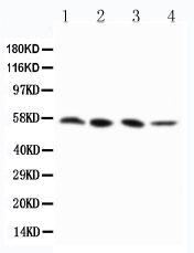
Anti-RIP3 antibody, PA2242, Western blotting
Lane 1: PANC Cell Lysate
Lane 2: SW620 Cell Lysate
Lane 3: SKOV-3 Cell Lysate
Lane 4: M231 Cell Lysate

Click image to see more details
Conditional knockout of TGF-βRII prevented cisplatin-induced renal injury and apoptotic signaling in vivo . A: Periodic acid-Schiff (PAS) staining and quantitative analysis show conditional knockout of TGF-βRII reduced renal injury in cisplatin nephropathy. B: Creatinine assay. C: BUN assay. Serum creatinine and BUN show conditional knockout of TGF-βRII prevented decline of renal function in cisplatin nephropathy. D. Real-time PCR data show conditional knockout of TGF-βRII reduced TGF-β mRNA level in cisplatin-induced nephropathy. E. Western blot analysis of phospho-Smad2 and phospho-Smad3. F. Immunohistochemistry and quantitative data show conditional knockout of TGF-βRII reduced KIM-1 protein and F4/80+ macrophages infiltration in cisplatin nephropathy. G. Western blot analysis of KIM-1, RIPK1, RIPK3, cleaved caspase-3. Data represent mean ± SEM for 6-8 mice. **P<0.01, ***P<0.001 versus normal; ## P<0.01, ### P<0.001 versus TGF-βRII FF+ cisplatin group. TGF-βRII FF: TGF-βRII flox/flox mouse; TGF-βRII KspCre: conditional TGF-βRII knockout mice; Magnification: 100X.
Index in PubMed under a CC BY license. PMID: 31754396

Click image to see more details
Conditional knockout of Smad2 prevented cisplatin-induced renal injury, decline of renal function and attenuated signaling molecules regulating programmed cell death in vivo . A: PAS staining and quantitative analysis show conditional knockout of Smad2 reduced renal injury in cisplatin-induced AKI mice. B: Creatinine assay. C: BUN assay. Serum creatinine and BUN show conditional deletion of Smad2 prevented decline of renal function in cisplatin nephropathy. D: Immunohistochemistry and quantitative data show conditional knockout of Smad2 reduced KIM-1 in cisplatin-induced nephropathy. E and F: Western blot and Real-time PCR analysis of KIM-1. G: Western blot of P-p53, p53, RIPK1, RIPK3 and cleaved caspase-3. Data represent mean ± SEM for 6-8 mice. **P<0.01, ***P<0.001 versus normal; # P<0.05, ## P<0.01, ### P<0.001 versus Smad2FF+cisplatin group. S2FF: Smad2 flox/flox mouse; S2 KspCre: conditional Smad2 knockout mice; KIM-1: kidney injury molecule-1. Magnification: 100X.
Index in PubMed under a CC BY license. PMID: 31754396

Click image to see more details
Knockdown of Smad2 attenuates cisplatin-induced renal injury, inflammation response, and programmed cell death in established nephrotoxic AKI model. A: PAS staining and quantitative analysis show knockdown of Smad2 reduced renal injury in cisplatin nephropathy. B: Creatinine assay. C: BUN assay. Serum creatinine and BUN show knockdown of Smad2 prevented decline of renal function in cisplatin nephropathy. D: Real-time PCR data show knockdown of Smad2 reduced mRNA of TNF-α, IL-1β, MCP-1 and KIM-1. E: Immunohistochemistry and quantitative data show knockdown of Smad2 reduced KIM-1 protein and F4/80+ macrophages in cisplatin-induced nephropathy. F: TUNEL assay. Knockdown of Smad2 reduced apoptosis in injured kidney. G: Western blot analysis of KIM-1, RIPK1, RIPK3, cleaved caspase-3. Data represent mean ± SEM for 6 mice. *P<0.05, **P<0.01, ***P<0.001 versus control; # P<0.05, ## P<0.01, ### P<0.001 versus Smad2 vector control+cisplatin. KD: knockdown. Magnification: 100X.
Index in PubMed under a CC BY license. PMID: 31754396

Click image to see more details
Knockdown of Smad2 attenuates renal injury, inflammation response, and programmed cell death in established ischemic AKI model. A: PAS staining and quantitative analysis show knockdown of Smad2 reduced renal injury in I/R-induced AKI model. B: Creatinine assay. C: BUN assay. Serum creatinine and BUN show knockdown of Smad2 prevented decline of renal function. D: Real-time PCR data show knockdown of Smad2 reduced mRNA of TNF-α, IL-1β, MCP-1 and KIM-1. E: Immunohistochemistry and quantitative data show knockdown of Smad2 reduced KIM-1 protein and F4/80+ macrophages infiltration in injured kidney. F: TUNEL assay. G: Western blot analysis of KIM-1, RIPK1, RIPK3, cleaved caspase-3. Data represent mean ± SEM for 6 mice. *P<0.05, **P<0.01, ***P<0.001 versus control; # P<0.05, ## P<0.01, ### P<0.001 versus Smad2 vector control+IRI. KD: knockdown. Magnification: 100X.
Index in PubMed under a CC BY license. PMID: 31754396
Specific Publications For Anti-RIP3/RIPK3 Antibody Picoband® (PA2242)
Loading publications
Recommended Resources
Here are featured tools and databases that you might find useful.
- Boster's Pathways Library
- Protein Databases
- Bioscience Research Protocol Resources
- Data Processing & Analysis Software
- Photo Editing Software
- Scientific Literature Resources
- Research Paper Management Tools
- Molecular Biology Software
- Primer Design Tools
- Bioinformatics Tools
- Phylogenetic Tree Analysis
Customer Reviews
Have you used Anti-RIP3/RIPK3 Antibody Picoband®?
Share your experimental results or join a short interview to earn up to $1,000 in product credits or other rewards.
0 Reviews For Anti-RIP3/RIPK3 Antibody Picoband®
Customer Q&As
Have a question?
Find answers in Q&As, reviews.
Can't find your answer?
Submit your question
4 Customer Q&As for Anti-RIP3/RIPK3 Antibody Picoband®
Question
We are currently using anti-RIP3/RIPK3 antibody PA2242 for human tissue, and we are satisfied with the WB results. The species of reactivity given in the datasheet says human. Is it true that the antibody can work on feline tissues as well?
Verified Customer
Verified customer
Asked: 2019-12-23
Answer
The anti-RIP3/RIPK3 antibody (PA2242) has not been tested for cross reactivity specifically with feline tissues, but there is a good chance of cross reactivity. We have an innovator award program that if you test this antibody and show it works in feline you can get your next antibody for free. Please contact me if I can help you with anything.
Boster Scientific Support
Answered: 2019-12-23
Question
Our team were happy with the WB result of your anti-RIP3/RIPK3 antibody. However we have observed positive staining in aortic endothelium fetal brain cytosol using this antibody. Is that expected? Could you tell me where is RIPK3 supposed to be expressed?
L. Miller
Verified customer
Asked: 2019-08-05
Answer
Based on literature, aortic endothelium fetal brain does express RIPK3. Generally RIPK3 expresses in cytoplasm, cytosol. Regarding which tissues have RIPK3 expression, here are a few articles citing expression in various tissues:
Aortic endothelium, and Fetal brain, Pubmed ID: 10358032
Blood, Pubmed ID: 15489334
Cervix carcinoma, and Lymphocyte, Pubmed ID: 10339433
Thalamus, Pubmed ID: 14702039
Boster Scientific Support
Answered: 2019-08-05
Question
We have been able to see staining in human blood. Any tips? Is anti-RIP3/RIPK3 antibody supposed to stain blood positively?
Verified Customer
Verified customer
Asked: 2019-05-09
Answer
According to literature blood does express RIPK3. According to Uniprot.org, RIPK3 is expressed in blood, cervix carcinoma lymphocyte, aortic endothelium fetal brain, thalamus, among other tissues. Regarding which tissues have RIPK3 expression, here are a few articles citing expression in various tissues:
Aortic endothelium, and Fetal brain, Pubmed ID: 10358032
Blood, Pubmed ID: 15489334
Cervix carcinoma, and Lymphocyte, Pubmed ID: 10339433
Thalamus, Pubmed ID: 14702039
Boster Scientific Support
Answered: 2019-05-09
Question
We need using your anti-RIP3/RIPK3 antibody for thymus development studies. Has this antibody been tested with western blotting on sw620 cell lysate? We would like to see some validation images before ordering.
Verified Customer
Verified customer
Asked: 2017-08-25
Answer
Thanks for your inquiry. This PA2242 anti-RIP3/RIPK3 antibody is validated on panc cell lysate, sw620 cell lysate, m231 cell lysate. It is guaranteed to work for WB in human. Our Boster guarantee will cover your intended experiment even if the sample type has not been be directly tested.
Boster Scientific Support
Answered: 2017-08-25
