
Click image to see more details
-

-
-
-
-
+1
Product Info Summary
| SKU: | PB9387 |
|---|---|
| Size: | 100 μg/vial |
| Reactive Species: | Human, Mouse, Rat |
| Host: | Rabbit |
| Application: | IHC, WB |
Customers Who Bought This Also Bought

Product info
Product Name
Anti-ROCK2 Antibody Picoband®
SKU/Catalog Number
PB9387
Size
100 μg/vial
Form
Lyophilized
Description
Boster Bio Anti-ROCK2 Antibody Picoband® catalog # PB9387. Tested in IHC, WB applications. This antibody reacts with Human, Mouse, Rat. The brand Picoband indicates this is a premium antibody that guarantees superior quality, high affinity, and strong signals with minimal background in Western blot applications. Only our best-performing antibodies are designated as Picoband, ensuring unmatched performance.
Storage & Handling
Store at -20˚C for one year from date of receipt. After reconstitution, at 4˚C for one month. It can also be aliquotted and stored frozen at -20˚C for six months. Avoid repeated freeze-thaw cycles.
Cite This Product
Anti-ROCK2 Antibody Picoband® (Boster Biological Technology, Pleasanton CA, USA, Catalog # PB9387)
Host
Rabbit
Contents
Each vial contains antibody formulated with stabilizing components, 0.9 mg NaCl, 0.2 mg Na2HPO4, and 0.05 mg NaN3.
*This antibody is supplied in a stabilized formulation.
Compatibility with conjugation reactions depends on the chemistry of the conjugation method used.
For conjugation methods that are not compatible with the stabilizing components present in this formulation, a carrier-free antibody format is required.
Clonality
Polyclonal
Isotype
Rabbit IgG
Immunogen
E.coli-derived human ROCK2 recombinant protein (Position: E652-D923). Human ROCK2 shares 94.9% and 95.2% amino acid (aa) sequence identity with mouse and rat ROCK2, respectively.
Cross-reactivity
No cross-reactivity with other proteins.
Reactive Species
PB9387 is reactive to ROCK2 in Human, Mouse, Rat
Observed Molecular Weight
161 kDa
Calculated molecular weight
160.9 kDa
Background of ROCK2
Rho-associated kinase (ROCK), including the ROCK-I and ROCK-II isoforms, is a protein kinase involved in signaling from Rho to actin cytoskeleton. Serine/threonine kinase ROCK II/Rho kinase, which is an isozyme of ROCK I, is one of the targets for the small GTPase Rho. ROCK II regulates the formation of actin stress fibers and focal adhesions, cytokinesis, smooth muscle contraction, and the activation of c-fos serum response element. Sequencing analysis has shown that human ROCK II contains 1388 amino acid residues with a calculated molecular mass of approximately 161 kDa. Fluorescence in situ hybridization analysis showed that the human ROCK II gene is located on chromosome 2p24. Thumkeo et al. concluded that ROCK-II is essential in inhibiting blood coagulation and maintaining blood flow in the endothelium-free labyrinth layer and that loss of ROCK-II leads to thrombus formation, placental dysfunction, intrauterine growth retardation, and fetal death.
Antibody Validation
Boster validates all antibodies on WB, IHC, ICC, Immunofluorescence, and ELISA with known positive control and negative samples to ensure specificity and high affinity, including thorough antibody incubations.
Application & Images
Applications
PB9387 is guaranteed for IHC, WB Boster Guarantee
Recommend Dilution
| Application | Dilution | Species |
|---|---|---|
| Immunohistochemistry (Paraffin-embedded Section) | 0.5-1μg/ml | Human, Mouse, Rat |
| Western blot | 0.1-0.5μg/ml | Human, Mouse, Rat |
Tested application
Suggested blocking solution with 5% non-fat milk or BSA; (*)Recommended protein loading: 20-40 µg per lane
Use TE buffer pH 9.0 for antigen retrieval; (*) citrate buffer pH 6.0 is an alternative.
Validation Images & Assay Conditions
Click image to see more details
Western blot analysis of ROCK2 using anti-ROCK2 antibody (PB9387).
Electrophoresis was performed on a 5-20% SDS-PAGE gel at 70V (Stacking gel) / 90V (Resolving gel) for 2-3 hours. The sample well of each lane was loaded with 30 ug of sample under reducing conditions.
Lane 1: human Hela whole cell lysates,
Lane 2: rat liver tissue lysates,
Lane 3: mouse brain tissue lysates,
Lane 4: mouse liver tissue lysates.
After electrophoresis, proteins were transferred to a nitrocellulose membrane at 150 mA for 50-90 minutes. Blocked the membrane with 5% non-fat milk/TBS for 1.5 hour at RT. The membrane was incubated with rabbit anti-ROCK2 antigen affinity purified polyclonal antibody (Catalog # PB9387) at 0.5 μg/mL overnight at 4°C, then washed with TBS-0.1%Tween 3 times with 5 minutes each and probed with a goat anti-rabbit IgG-HRP secondary antibody at a dilution of 1:5000 for 1.5 hour at RT. The signal is developed using an Enhanced Chemiluminescent detection (ECL) kit (Catalog # EK1002) with Tanon 5200 system. A specific band was detected for ROCK2 at approximately 161 kDa. The expected band size for ROCK2 is at 161 kDa.

Click image to see more details
IHC analysis of ROCK2 using anti-ROCK2 antibody (PB9387).
ROCK2 was detected in a paraffin-embedded section of Mouse Cardiac Muscle tissue. Heat mediated antigen retrieval was performed in EDTA buffer (pH 8.0, epitope retrieval solution). The tissue section was blocked with 10% goat serum. The tissue section was then incubated with 1 μg/ml rabbit anti-ROCK2 Antibody (PB9387) overnight at 4°C. Biotinylated goat anti-rabbit IgG was used as secondary antibody and incubated for 30 minutes at 37°C. The tissue section was developed using Strepavidin-Biotin-Complex (SABC) (Catalog # SA1022) with DAB as the chromogen.

Click image to see more details
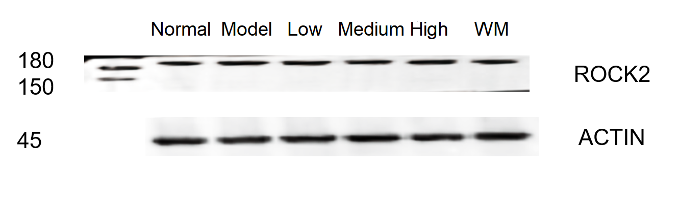
Western blot analysis of ROCK2 using anti-ROCK2 antibody (PB9387).
Electrophoresis was performed on a 5-20% SDS-PAGE gel at 70V (Stacking gel) / 90V (Resolving gel) for 2-3 hours. The sample well of each lane was loaded with 30 ug of sample under reducing conditions.
Lane 1: Normal group-rat colon tissue lysates,
Lane 2: Model group-rat colon tissue lysates,
Lane 3: Triditional Chinese medicine treatment (low dose)-rat colon tissue lysates,
Lane 4: Triditional Chinese medicine treatment (medium dose)-rat colon tissue lysates,
Lane 5: Triditional Chinese medicine treatment(high dose)-rat colon tissue lysates,
Lane 6: Western medicine treatment-rat colon tissue lysates.
After electrophoresis, proteins were transferred to a nitrocellulose membrane at 150 mA for 50-90 minutes. Blocked the membrane with 5% non-fat milk/TBS for 1.5 hour at RT. The membrane was incubated with rabbit anti-ROCK2 antigen affinity purified polyclonal antibody (Catalog # PB9387) at 1:1000 overnight at 4°C, then washed with TBS-0.1%Tween 3 times with 5 minutes each and probed with a HRP Conjugated AffiniPure Goat Anti-rabbit IgG (H+L)secondary antibody at a dilution of 1:5000 for 1 hour at RT. The signal is developed using an Enhanced Chemiluminescent detection (ECL) kit (Catalog # EK1002) with ChemiDoc MP system. A specific band was detected for ROCK2 at approximately 180 kDa. The expected band size for ROCK2 is at 161 kDa.

Click image to see more details
IHC analysis of ROCK2 using anti-ROCK2 antibody (PB9387).
ROCK2 was detected in a paraffin-embedded section of Rat Brain tissue. Heat mediated antigen retrieval was performed in EDTA buffer (pH 8.0, epitope retrieval solution). The tissue section was blocked with 10% goat serum. The tissue section was then incubated with 1 μg/ml rabbit anti-ROCK2 Antibody (PB9387) overnight at 4°C. Biotinylated goat anti-rabbit IgG was used as secondary antibody and incubated for 30 minutes at 37°C. The tissue section was developed using Strepavidin-Biotin-Complex (SABC) (Catalog # SA1022) with DAB as the chromogen.

Click image to see more details
IHC analysis of ROCK2 using anti-ROCK2 antibody (PB9387).
ROCK2 was detected in a paraffin-embedded section of Human Mammary Cancer tissue. Heat mediated antigen retrieval was performed in EDTA buffer (pH 8.0, epitope retrieval solution). The tissue section was blocked with 10% goat serum. The tissue section was then incubated with 1 μg/ml rabbit anti-ROCK2 Antibody (PB9387) overnight at 4°C. Biotinylated goat anti-rabbit IgG was used as secondary antibody and incubated for 30 minutes at 37°C. The tissue section was developed using Strepavidin-Biotin-Complex (SABC) (Catalog # SA1022) with DAB as the chromogen.
Specific Publications For Anti-ROCK2 Antibody Picoband® (PB9387)
Loading publications
Recommended Resources
Here are featured tools and databases that you might find useful.
- Boster's Pathways Library
- Protein Databases
- Bioscience Research Protocol Resources
- Data Processing & Analysis Software
- Photo Editing Software
- Scientific Literature Resources
- Research Paper Management Tools
- Molecular Biology Software
- Primer Design Tools
- Bioinformatics Tools
- Phylogenetic Tree Analysis
Customer Reviews
Have you used Anti-ROCK2 Antibody Picoband®?
Share your experimental results or join a short interview to earn up to $1,000 in product credits or other rewards.
1 Reviews For Anti-ROCK2 Antibody Picoband®
The antibody is highly specific and efficient, suitable for detecting ROCK2 protein in rat colon by Western blot, with only minimal non-specific bands.
Excellent
| SKU | PB9387 |
|---|---|
| Application | Western Blot |
| Sample | rat colon tissue |
| Sample Processing Description | RIPA lysis buffer with protease inhibitor PMSF (100:1) was used to lyse the sample for 10 minutes, followed by centrifugation at 12,000 rpm for 15 minutes. The supernatant was mixed with 5× loading buffer, denatured at 100°C for 10 minutes, and then loaded onto SDS-PAGE. |
| Other Reagents | Blocking buffer |
| Primary Antibody | ROCK2 Antibody Picoband® |
| Primary Incubation | 1:1000, overnight at 4 ℃ |
| Secondary Antibody | HRP Conjugated AffiniPure Goat Anti-Rabbit IgG (H+L) |
| Secondary Incubation | 1:5000, 1 hour in room temperature |
| Detection | Substrate: ECL, Imaging system:ChemiDoc MP |
| Results Summary | The figure shows the Western blot results of the target protein ROCK2 and the internal control Actin in rat colon across the following groups: normal, disease model, low/middle/high dose traditional Chinese medicine treatment, and Western medicine treatment. Expression was increased in the model group. Among the traditional Chinese medicine treatments, the high-dose group showed the best effect. The target bands are clear and distinct, and the experimental results are satisfactory. |
Shiyu Zhang, LUTCM
Verified customer
Submitted 2026-01-06
Customer Q&As
Have a question?
Find answers in Q&As, reviews.
Can't find your answer?
Submit your question
1 Customer Q&As for Anti-ROCK2 Antibody Picoband®
Question
We are currently using anti-ROCK2 antibody PB9387 for mouse tissue, and we are well pleased with the IHC results. The species of reactivity given in the datasheet says human, mouse, rat. Is it true that the antibody can work on goat tissues as well?
A. Parker
Verified customer
Asked: 2015-03-11
Answer
The anti-ROCK2 antibody (PB9387) has not been tested for cross reactivity specifically with goat tissues, but there is a good chance of cross reactivity. We have an innovator award program that if you test this antibody and show it works in goat you can get your next antibody for free. Please contact me if I can help you with anything.
Boster Scientific Support
Answered: 2015-03-11
