Click image to see more details
-

-
-
-
-
+11
Product Info Summary
| SKU: | A00177-2 |
|---|---|
| Size: | 100 μg/vial |
| Reactive Species: | Human, Mouse, Rat |
| Host: | Rabbit |
| Application: | IHC, WB |
Customers Who Bought This Also Bought

Product info
Product Name
Anti-SOX9 Antibody Picoband®
SKU/Catalog Number
A00177-2
Size
100 μg/vial
Form
Lyophilized
Description
Boster Bio Anti-SOX9 Antibody Picoband® catalog # A00177-2. Tested in IHC, WB applications. This antibody reacts with Human, Mouse, Rat. The brand Picoband indicates this is a premium antibody that guarantees superior quality, high affinity, and strong signals with minimal background in Western blot applications. Only our best-performing antibodies are designated as Picoband, ensuring unmatched performance.
Storage & Handling
Store at -20˚C for one year from date of receipt. After reconstitution, at 4˚C for one month. It can also be aliquotted and stored frozen at -20˚C for six months. Avoid repeated freeze-thaw cycles.
Cite This Product
Anti-SOX9 Antibody Picoband® (Boster Biological Technology, Pleasanton CA, USA, Catalog # A00177-2)
Host
Rabbit
Contents
Each vial contains 4mg Trehalose, 0.9mg NaCl, 0.2mg Na2HPO4, 0.05mg NaN3.
Clonality
Polyclonal
Isotype
Rabbit IgG
Immunogen
A synthetic peptide corresponding to a sequence in the middle region of human SOX9, identical to the related mouse and rat sequences.
Cross-reactivity
No cross-reactivity with other proteins.
Reactive Species
A00177-2 is reactive to SOX9 in Human, Mouse, Rat
Observed Molecular Weight
70 kDa
Calculated molecular weight
56.1 kDa
Background of SOX9
Transcription factor SOX-9 is a protein that in humans is encoded by the SOX9 gene. The protein encoded by this gene recognizes the sequence CCTTGAG along with other members of the HMG-box class DNA-binding proteins. It acts during chondrocyte differentiation and, with steroidogenic factor 1, regulates transcription of the anti-Muellerian hormone (AMH) gene. Deficiencies lead to the skeletal malformation syndrome campomelic dysplasia, frequently with sex reversal.
Antibody Validation
Boster validates all antibodies on WB, IHC, ICC, Immunofluorescence, and ELISA with known positive control and negative samples to ensure specificity and high affinity, including thorough antibody incubations.
Application & Images
Applications
A00177-2 is guaranteed for IHC, WB Boster Guarantee
Recommend Dilution
| Application | Dilution | Species |
|---|---|---|
| Western blot | 0.1-0.5μg/ml | |
| Immunohistochemistry (Paraffin-embedded Section) | 0.5-1μg/ml |
Tested application
Suggested blocking solution with 5% non-fat milk or BSA; (*)Recommended protein loading: 20-40 µg per lane
Use TE buffer pH 9.0 for antigen retrieval; (*) citrate buffer pH 6.0 is an alternative.
Validation Images & Assay Conditions
Click image to see more details
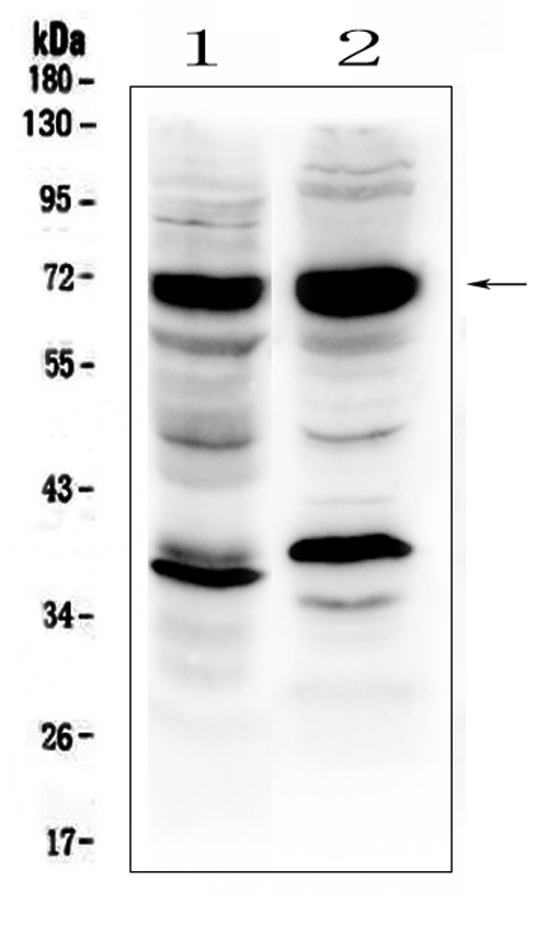
Western blot analysis of SOX9 using anti-SOX9 antibody (A00177-2).
Electrophoresis was performed on a 5-20% SDS-PAGE gel at 70V (Stacking gel) / 90V (Resolving gel) for 2-3 hours. The sample well of each lane was loaded with 50ug of sample under reducing conditions.
Lane 1: human HepG2 whole cell lysates
Lane 2: human PC-3 whole cell lysates
After Electrophoresis, proteins were transferred to a Nitrocellulose membrane at 150mA for 50-90 minutes. Blocked the membrane with 5% Non-fat Milk/ TBS for 1.5 hour at RT. The membrane was incubated with rabbit anti-SOX9 antigen affinity purified polyclonal antibody (Catalog # A00177-2) at 0.5 μg/mL overnight at 4°C, then washed with TBS-0.1%Tween 3 times with 5 minutes each and probed with a goat anti-rabbit IgG-HRP secondary antibody at a dilution of 1:10000 for 1.5 hour at RT. The signal is developed using an Enhanced Chemiluminescent detection (ECL) kit (Catalog # EK1002) with Tanon 5200 system. A specific band was detected for SOX9 at approximately 70KD. The expected band size for SOX9 is at 56KD.

Click image to see more details
Hoxc10 exists in mesodermal derived callus. The immunofluorescence of Sox9 and CD44 in the mandible homotopic grafting (A), femoral heterotopic grafting (B) and femoral homotopic grafting(C). Sox9 represents cartilage (red), CD44 is a BMSCs marker (green), and DAPI marks the nucleus. (Scale bars, 500 μm) (D–F) represent the immunofluorescence of CD105 in mandible homotopic grafting, femoral heterotopic grafting, and femoral homotopic grafting, respectively. The white dotted line shows the edge of the femoral graft and the mandibular graft, and the middle of the dotted line is the callus. (Scale bars, 200 μm) (G) Localization of Hoxc10 at callus in femoral heterotopic grafting. (Scale bars, 100 μm; Scale bars, 20 μm) The data are presented as the mean ± SD ( n = 6). * p < 0.05.
Index in PubMed under a CC BY license. PMID: 39434203

Click image to see more details
Hoxc10 is retained in L/M‐BMSCs in vitro. (A) Schematic of Transwell co‐culture model of L‐BMSCs and M‐BMSCs. (B) Schematic of the limb bones and mandibles from different embryonic origins. The mandible is of neural crest origin (blue) and the limb bone is of mesodermal origin (orange) (C) qPCR verified the gene expression levels of Sox9 and Col2a1 before and after co‐culture of L‐BMSCs and M‐BMSCs. (D) The proliferation, osteogenic and chondrogenic genes of femoral homotopic grafting, femoral heterotopic grafting and mandibles homotopic grafting. (E) After 21 days of chondrogenic induction in the upper layer cells of Transwell model before and after co cultivation with L‐BMSCs and M‐BMSCs, blue stained proteoglycans were observed using Alcian blue. (F) Quantitative analysis of Alizarin blue staining before and after co culture of L‐BMSCs and M‐BMSCs. The data are presented as the mean ± SD ( n = 3). * p < 0.05.
Index in PubMed under a CC BY license. PMID: 39434203

Click image to see more details
Hoxc10 is positively correlated with cartilage. n (C) q‐PCR validated the expression levels of Sox9 gene after overexpression and knockout of Hoxc10. (D) q‐PCR validated the expression levels of the Col2a1 gene after overexpression and knockout of Hoxc10. (E) q‐PCR validated the expression levels of Aggrecan gene after overexpression and knockout of Hoxc10. (F) The proteoglycan of BMSCs after overexpression and knockdown Hoxc10 was observed by Alcian blue staining 21 days after chondrogenic induction. (G) Quantitative analysis of Alcian blue staining. (H) Schematic diagram of co‐culture of L‐BMSCs and M‐BMSCs with and without Hoxc10 knockout. (I) Col2a1 gene expression in L‐BMSCs after Hoxc10 knockout and co‐culture with M‐BMSCs compared to control. (J) ChIP experiment of Sox9 and Hoxc10 protein binding. The data are presented as the mean ± SD ( n = 3).* p < 0.05.
Index in PubMed under a CC BY license. PMID: 39434203

Click image to see more details
LRRK2-IN-1 suppresses the IL-1β-induced inflammation and catabolism and induces anabolism without causing the inhibition of chondrocyte viability. A Schematic diagram of cell treatment and experimental procedures. B Cell viability assessed by CCK8 assay. No obvious inhibition of chondrocyte proliferation was observed when treated with 0.5, 1.0, 2.5, and 5.0 µM LRRK2-IN-1 for 24 h. Data represent mean ± SD; N = 6/group; one-way ANOVA; ns, not significant. C Western blot analyses of the protein levels of anabolic, catabolic, inflammatory factors in the IL-1β-induced chondrocytes treated with 0.5, 1.0, 2.5, and 5.0 µM LRRK2-IN-1 for 24 h. LRRK2-IN-1 suppressed MMP3, MMP13, iNOS, and COX2 and induced COL2 and SOX9 in a dose-dependent manner. D Quantitative analyses of the western blot results. Data represent mean ± SD; N = 3/group; *P < 0.05; **P < 0.01 by one-way ANOVA. E Immunofluorescence of iNOS, MMP13, and aggrecan expression in the IL-1β-induced chondrocytes treated with 5.0 µM LRRK2-IN-1 for 24 h. Scar bar: 400 μm
Index in PubMed under a CC BY license. PMID: 37605203

Click image to see more details
W-Exo-L@GelMA exhibits a strong chondrocyte-targeting effect and a pronounced action on promoting anabolism and suppressing catabolism and inflammation without causing the inhibition of chondrocyte viability. A Cell viability assessed by CCK8 assay. No obvious cytotoxicity on chondrocytes was observed when treated with W-Exo-L@GelMA loaded with 0.5, 1.0, 2.5, and 5.0 µM LRRK2-IN-1 for 48 h. Data represent mean ± SD; N = 6/group; one-way ANOVA; ns, not significant. B Immunofluorescence of Dil-labeled exosomes. The uptake of exosomes was observed in the chondrocytes when treated with Exo-L, Exo-L@GelMA or W-Exo-L@GelMA for 48 h. Dil was used for labeling exosomes (red), DAPI to label nuclei (blue), and Phalloidin to label the cytoskeleton (green). Scar bar: 200 μm. C Western blot analyses of the protein levels of anabolic, catabolic, and inflammatory factors in the IL-1β-induced chondrocytes treated with W-Exo-L@GelMA loaded with 0.5, 1.0, 2.5, and 5.0 µM LRRK2-IN-1 for 48 h. W-Exo-L@GelMA promoted COL2 and SOX9 and inhibited iNOS, COX2, MMP3, and MMP13 protein levels in a dose-dependent manner. D Quantitative analysis of the western blot results. Data represent mean ± SD; N = 3/group; *P < 0.05; **P < 0.01 by one-way ANOVA
Index in PubMed under a CC BY license. PMID: 37605203

Click image to see more details
Antioxidants preserve ADSC cell stemness and multidirectional differentiation potential during long-term in vitro expansion. After treatment with 10 μM GSH or melatonin, the ADSCs cultured for passage 3 (P3), passage 6 (P6), and passage 9 (P9) were used in the following analysis. a Osteogenesis differentiation of passaged ADSCs (Alizarin Red S staining; scale bar, 50 μm). b Adipogenesis differentiation of passaged ADSCs (Oil Red O staining; scale bar, 50 μm). c Western blot analysis for RUNX-2 in osteogenic cells. d Western blot analysis for perilipin A in adipogenic cells. e Chondrogenesis differentiation of passaged ADSCs (Alcian blue staining; scale bar, 50 μm). f Western blot analysis for SOX-9 in chondrogenic cells. g Western blot analysis for SOX-2, OCT-4, and β-actin in ADSCs. ADSCs, adipose tissue-derived stem cells; GSH, reduced glutathione
Index in PubMed under a CC BY license. PMID: 31623678

Click image to see more details
IHC analysis of SOX9 using anti-SOX9 antibody (A00177-2).
SOX9 was detected in paraffin-embedded section of mouse lung tissues. Heat mediated antigen retrieval was performed in citrate buffer (pH6, epitope retrieval solution) for 20 mins. The tissue section was blocked with 10% goat serum. The tissue section was then incubated with 1μg/ml rabbit anti-SOX9 Antibody (A00177-2) overnight at 4°C. Biotinylated goat anti-rabbit IgG was used as secondary antibody and incubated for 30 minutes at 37°C. The tissue section was developed using Strepavidin-Biotin-Complex (SABC)(Catalog # SA1022) with DAB as the chromogen.

Click image to see more details
IHC analysis of SOX9 using anti-SOX9 antibody (A00177-2).
SOX9 was detected in paraffin-embedded section of mouse testis tissues. Heat mediated antigen retrieval was performed in citrate buffer (pH6, epitope retrieval solution) for 20 mins. The tissue section was blocked with 10% goat serum. The tissue section was then incubated with 1μg/ml rabbit anti-SOX9 Antibody (A00177-2) overnight at 4°C. Biotinylated goat anti-rabbit IgG was used as secondary antibody and incubated for 30 minutes at 37°C. The tissue section was developed using Strepavidin-Biotin-Complex (SABC)(Catalog # SA1022) with DAB as the chromogen.

Click image to see more details
IHC analysis of SOX9 using anti-SOX9 antibody (A00177-2).
SOX9 was detected in paraffin-embedded section of rat lung tissues. Heat mediated antigen retrieval was performed in citrate buffer (pH6, epitope retrieval solution) for 20 mins. The tissue section was blocked with 10% goat serum. The tissue section was then incubated with 1μg/ml rabbit anti-SOX9 Antibody (A00177-2) overnight at 4°C. Biotinylated goat anti-rabbit IgG was used as secondary antibody and incubated for 30 minutes at 37°C. The tissue section was developed using Strepavidin-Biotin-Complex (SABC)(Catalog # SA1022) with DAB as the chromogen.

Click image to see more details
IHC analysis of SOX9 using anti-SOX9 antibody (A00177-2).
SOX9 was detected in paraffin-embedded section of rat testis tissues. Heat mediated antigen retrieval was performed in citrate buffer (pH6, epitope retrieval solution) for 20 mins. The tissue section was blocked with 10% goat serum. The tissue section was then incubated with 1μg/ml rabbit anti-SOX9 Antibody (A00177-2) overnight at 4°C. Biotinylated goat anti-rabbit IgG was used as secondary antibody and incubated for 30 minutes at 37°C. The tissue section was developed using Strepavidin-Biotin-Complex (SABC)(Catalog # SA1022) with DAB as the chromogen.

Click image to see more details
IHC analysis of SOX9 using anti-SOX9 antibody (A00177-2).
SOX9 was detected in paraffin-embedded section of human intestinal cancer tissues. Heat mediated antigen retrieval was performed in citrate buffer (pH6, epitope retrieval solution) for 20 mins. The tissue section was blocked with 10% goat serum. The tissue section was then incubated with 1μg/ml rabbit anti-SOX9 Antibody (A00177-2) overnight at 4°C. Biotinylated goat anti-rabbit IgG was used as secondary antibody and incubated for 30 minutes at 37°C. The tissue section was developed using Strepavidin-Biotin-Complex (SABC)(Catalog # SA1022) with DAB as the chromogen.

Click image to see more details
IHC analysis of SOX9 using anti-SOX9 antibody (A00177-2).
SOX9 was detected in paraffin-embedded section of human mammary cancer tissues. Heat mediated antigen retrieval was performed in citrate buffer (pH6, epitope retrieval solution) for 20 mins. The tissue section was blocked with 10% goat serum. The tissue section was then incubated with 1μg/ml rabbit anti-SOX9 Antibody (A00177-2) overnight at 4°C. Biotinylated goat anti-rabbit IgG was used as secondary antibody and incubated for 30 minutes at 37°C. The tissue section was developed using Strepavidin-Biotin-Complex (SABC)(Catalog # SA1022) with DAB as the chromogen.
Specific Publications For Anti-SOX9 Antibody Picoband® (A00177-2)
Loading publications
Recommended Resources
Here are featured tools and databases that you might find useful.
- Boster's Pathways Library
- Protein Databases
- Bioscience Research Protocol Resources
- Data Processing & Analysis Software
- Photo Editing Software
- Scientific Literature Resources
- Research Paper Management Tools
- Molecular Biology Software
- Primer Design Tools
- Bioinformatics Tools
- Phylogenetic Tree Analysis
Customer Reviews
Have you used Anti-SOX9 Antibody Picoband®?
Share your experimental results or join a short interview to earn up to $1,000 in product credits or other rewards.
0 Reviews For Anti-SOX9 Antibody Picoband®
Customer Q&As
Have a question?
Find answers in Q&As, reviews.
Can't find your answer?
Submit your question
16 Customer Q&As for Anti-SOX9 Antibody Picoband®
Question
Our lab were well pleased with the WB result of your anti-SOX9 antibody. However we have seen positive staining in testis nucleus using this antibody. Is that expected? Could you tell me where is SOX9 supposed to be expressed?
Z. Mangal
Verified customer
Asked: 2020-04-02
Answer
According to literature, testis does express SOX9. Generally SOX9 expresses in nucleus. Regarding which tissues have SOX9 expression, here are a few articles citing expression in various tissues:
Eye, and PNS, Pubmed ID: 15489334
Testis, Pubmed ID: 7990924
Boster Scientific Support
Answered: 2020-04-02
Question
My question regards to test anti-SOX9 antibody A00177-2 on rat testis for research purposes, then I may be interested in using anti-SOX9 antibody A00177-2 for diagnostic purposes as well. Is the antibody suitable for diagnostic purposes?
Verified Customer
Verified customer
Asked: 2020-03-04
Answer
The products we sell, including anti-SOX9 antibody A00177-2, are only intended for research use. They would not be suitable for use in diagnostic work. If you have the means to develop a product into diagnostic use, and are interested in collaborating with us and develop our product into an IVD product, please contact us for more discussions.
Boster Scientific Support
Answered: 2020-03-04
Question
We purchased anti-SOX9 antibody for WB on eye pns a few months ago. I am using human, and We intend to use the antibody for IHC-P next. My question regards examining eye pns as well as testis in our next experiment. Do you have any suggestion on which antibody would work the best for IHC-P?
D. Miller
Verified customer
Asked: 2020-02-26
Answer
I viewed the website and datasheets of our anti-SOX9 antibody and I see that A00177-2 has been tested on human in both WB and IHC-P. Thus A00177-2 should work for your application. Our Boster satisfaction guarantee will cover this product for IHC-P in human even if the specific tissue type has not been validated. We do have a comprehensive range of products for IHC-P detection and you can check out our website bosterbio.com to find out more information about them.
Boster Scientific Support
Answered: 2020-02-26
Question
We appreciate helping with my inquiry over the phone. Here are the WB image, lot number and protocol we used for testis using anti-SOX9 antibody A00177-2. Let me know if you need anything else.
Verified Customer
Verified customer
Asked: 2020-01-03
Answer
We appreciate the data. You have provided everything we needed. Our lab team are working to resolve your inquiry as quickly as possible, and we appreciate your patience and understanding! Please let me know if there is anything you need in the meantime.
Boster Scientific Support
Answered: 2020-01-03
Question
Will anti-SOX9 antibody A00177-2 work for IHC-P with testis?
Verified Customer
Verified customer
Asked: 2019-11-28
Answer
According to the expression profile of testis, SOX9 is highly expressed in testis. So, it is likely that anti-SOX9 antibody A00177-2 will work for IHC-P with testis.
Boster Scientific Support
Answered: 2019-11-28
Question
We are currently using anti-SOX9 antibody A00177-2 for human tissue, and we are happy with the WB results. The species of reactivity given in the datasheet says human, mouse, rat. Is it possible that the antibody can work on monkey tissues as well?
Verified Customer
Verified customer
Asked: 2019-08-01
Answer
The anti-SOX9 antibody (A00177-2) has not been validated for cross reactivity specifically with monkey tissues, but there is a good chance of cross reactivity. We have an innovator award program that if you test this antibody and show it works in monkey you can get your next antibody for free. Please contact me if I can help you with anything.
Boster Scientific Support
Answered: 2019-08-01
Question
I was wanting to use your anti-SOX9 antibody for IHC-P for rat testis on frozen tissues, but I want to know if it has been validated for this particular application. Has this antibody been validated and is this antibody a good choice for rat testis identification?
Verified Customer
Verified customer
Asked: 2019-06-07
Answer
You can see on the product datasheet, A00177-2 anti-SOX9 antibody has been tested for IHC-P, WB on human, mouse, rat tissues. We have an innovator award program that if you test this antibody and show it works in rat testis in IHC-frozen, you can get your next antibody for free.
Boster Scientific Support
Answered: 2019-06-07
Question
Is this A00177-2 anti-SOX9 antibody reactive to the isotypes of SOX9?
Verified Customer
Verified customer
Asked: 2019-05-06
Answer
The immunogen of A00177-2 anti-SOX9 antibody is A synthetic peptide corresponding to a sequence of human SOX9 (QPRRRKSVKNGQAEAEEATEQTHISPNAIFKALQAD). Could you tell me which isotype you are interested in so I can help see if the immunogen is part of this isotype?
Boster Scientific Support
Answered: 2019-05-06
Question
Is a blocking peptide available for product anti-SOX9 antibody (A00177-2)?
Verified Customer
Verified customer
Asked: 2018-08-27
Answer
We do provide the blocking peptide for product anti-SOX9 antibody (A00177-2). If you would like to place an order for it please contact support@bosterbio.com and make a special request.
Boster Scientific Support
Answered: 2018-08-27
Question
My lab would like using your anti-SOX9 antibody for protein-containing complex assembly studies. Has this antibody been tested with western blotting on mouse lung? We would like to see some validation images before ordering.
Verified Customer
Verified customer
Asked: 2018-06-11
Answer
I appreciate your inquiry. This A00177-2 anti-SOX9 antibody is validated on human hepg2 whole cell lysates, mouse lung. It is guaranteed to work for IHC-P, WB in human, mouse, rat. Our Boster guarantee will cover your intended experiment even if the sample type has not been be directly tested.
Boster Scientific Support
Answered: 2018-06-11
Question
Is there a BSA free version of anti-SOX9 antibody A00177-2 available?
R. Parker
Verified customer
Asked: 2018-05-21
Answer
Thank you for your recent telephone inquiry. I can confirm that some lots of this anti-SOX9 antibody A00177-2 are BSA free. For now, these lots are available and we can make a BSA free formula for you free of charge. It will take 3 extra days to prepare. If you require this antibody BSA free again in future, please do not hesitate to contact me and I will be pleased to check which lots we have in stock that are BSA free.
Boster Scientific Support
Answered: 2018-05-21
Question
We have been able to see staining in rat testis. What should we do? Is anti-SOX9 antibody supposed to stain testis positively?
C. Krishna
Verified customer
Asked: 2016-05-03
Answer
According to literature testis does express SOX9. According to Uniprot.org, SOX9 is expressed in testis, eye pns, among other tissues. Regarding which tissues have SOX9 expression, here are a few articles citing expression in various tissues:
Eye, and PNS, Pubmed ID: 15489334
Testis, Pubmed ID: 7990924
Boster Scientific Support
Answered: 2016-05-03
Question
Please see the WB image, lot number and protocol we used for testis using anti-SOX9 antibody A00177-2. Please let me know if you require anything else.
J. Huang
Verified customer
Asked: 2015-09-24
Answer
Thank you very much for the data. Our lab team are working to resolve this as quickly as possible, and we appreciate your patience and understanding! You have provided everything we needed. Please let me know if there is anything you need in the meantime.
Boster Scientific Support
Answered: 2015-09-24
Question
I have a question about product A00177-2, anti-SOX9 antibody. I was wondering if it would be possible to conjugate this antibody with biotin. I would need it to be without BSA or sodium azide. I am planning on using a buffer exchange of sodium azide with PBS only. Would there be problems for me to conjugate the antibody and store it in -20 degrees in small aliquots?
M. Williams
Verified customer
Asked: 2013-12-17
Answer
We do not recommend storing this antibody with PBS buffer only in -20 degrees. If you want to store it in -20 degrees it is best to add some cryoprotectant like glycerol. If you want carrier free A00177-2 anti-SOX9 antibody, we can provide it to you in a special formula with trehalose and/or glycerol. These molecules will not interfere with conjugation chemistry and provide a good level of protection for the antibody from degradation. Please be sure to specify this in your purchase order.
Boster Scientific Support
Answered: 2013-12-17
Question
I see that the anti-SOX9 antibody A00177-2 works with IHC-P, what is the protocol used to produce the result images on the product page?
B. Banerjee
Verified customer
Asked: 2013-08-30
Answer
You can find protocols for IHC-P on the "support/technical resources" section of our navigation menu. If you have any further questions, please send an email to support@bosterbio.com
Boster Scientific Support
Answered: 2013-08-30
Question
Does A00177-2 anti-SOX9 antibody work on parafin embedded sections? If so, which fixation method do you recommend we use (PFA, paraformaldehyde, other)?
M. Jha
Verified customer
Asked: 2013-03-19
Answer
It shows on the product datasheet, A00177-2 anti-SOX9 antibody as been tested on IHC-P. It is best to use PFA for fixation because it has better tissue penetration ability. PFA needs to be prepared fresh before use. Long term stored PFA turns into formalin, as the PFA molecules congregate and become formalin.
Boster Scientific Support
Answered: 2013-03-19
