Click image to see more details
-

-
-
-
-
+9
Product Info Summary
| SKU: | A00312-1 |
|---|---|
| Size: | 100 µg/vial |
| Reactive Species: | Human, Mouse, Rat |
| Host: | Rabbit |
| Application: | ELISA, Flow Cytometry, IF, IHC, WB |
Customers Who Bought This Also Bought

Product info
Product Name
Anti-STIM1 Antibody Picoband®
SKU/Catalog Number
A00312-1
Size
100 µg/vial
Form
Lyophilized
Description
Boster Bio Anti-STIM1 Antibody Picoband® catalog # A00312-1. Tested in ELISA, IHC, IF, WB, Flow Cytometry applications. This antibody reacts with Human, Mouse, Rat. The brand Picoband indicates this is a premium antibody that guarantees superior quality, high affinity, and strong signals with minimal background in Western blot applications. Only our best-performing antibodies are designated as Picoband, ensuring unmatched performance.
Storage & Handling
At -20°C for one year from date of receipt. After reconstitution, at 4°C for one month. It can also be aliquotted and stored frozen at -20°C for six months. Avoid repeated freezing and thawing.
Cite This Product
Anti-STIM1 Antibody Picoband® (Boster Biological Technology, Pleasanton CA, USA, Catalog # A00312-1)
Host
Rabbit
Contents
Each vial contains 4 mg Trehalose, 0.9 mg NaCl, 0.2 mg Na2HPO4.
Clonality
Polyclonal
Isotype
IgG
Immunogen
E.coli-derived human STIM1 recombinant protein (Position: E42-L599). Human STIM1 shares 98.2% and 98% amino acid (aa) sequence identity with mouse and rat STIM1, respectively.
Cross-reactivity
No cross reactivity with other proteins.
Reactive Species
A00312-1 is reactive to STIM1 in Human, Mouse, Rat
Observed Molecular Weight
85 kDa
Calculated molecular weight
77.4 kDa
Background of STIM1
Stromal interaction molecule 1 is a protein that in humans is encoded by the STIM1 gene. STIM1 has a single transmembranedomain, and is localized to the endoplasmic reticulum, and to a lesser extent to the plasma membrane. This gene encodes a type 1 transmembrane protein that mediates Ca2+ influx after depletion of intracellular Ca2+ stores by gating of store-operated Ca2+ influx channels (SOCs). It is one of several genes located in the imprinted gene domain of 11p15.5, an important tumor-suppressor gene region. Alterations in this region have been associated with the Beckwith-Wiedemann syndrome, Wilms tumor, rhabdomyosarcoma, adrenocrotical carcinoma, and lung, ovarian, and breast cancer. This gene may play a role in malignancies and disease that involve this region, as well as early hematopoiesis, by mediating attachment to stromal cells. Mutations in this gene are associated with fatal classic Kaposi sarcoma, immunodeficiency due to defects in store-operated calcium entry (SOCE) in fibroblasts, ectodermal dysplasia and tubular aggregate myopathy. This gene is oriented in a head-to-tail configuration with the ribonucleotide reductase 1 gene (RRM1), with the 3' end of this gene situated 1.6 kb from the 5' end of the RRM1 gene. Alternative splicing of this gene results in multiple transcript variants.
Antibody Validation
Boster validates all antibodies on WB, IHC, ICC, Immunofluorescence, and ELISA with known positive control and negative samples to ensure specificity and high affinity, including thorough antibody incubations.
Application & Images
Applications
A00312-1 is guaranteed for ELISA, Flow Cytometry, IF, IHC, WB Boster Guarantee
Assay Dilutions Recommendation
The recommendations below provide a starting point for assay optimization. The actual working concentration varies and should be decided by the user.
Western blot, 0.25-0.5 μg/ml, Human, Mouse, Rat
Immunohistochemistry(Paraffin-embedded Section), 2-5 μg/ml, Human, Mouse, Rat
Immunofluorescence, 5 μg/ml, Human, Mouse, Rat
Flow Cytometry (Fixed), 1-3 μg/1x106 cells, Human
ELISA, 0.1-0.5 μg/ml, -
Positive Control
WB: human K562 whole cell, human HepG2 whole cell, human Hela whole cell, rat PC-12 whole cell, mouse liver tissue, mouse NIH/3T3 whole cell
IHC: human breast cancer tissue, human breast cancer tissue, human colon adenocarcinoma tissue, human placenta tissue, human thyroid papillary carcinoma tissue, human thyroid papillary carcinoma tissue, mouse testis tissue, rat testis tissue
IF: human placenta tissue, mouse testis tissue, rat testis tissue
FCM: K562 cell
Validation Images & Assay Conditions
Click image to see more details
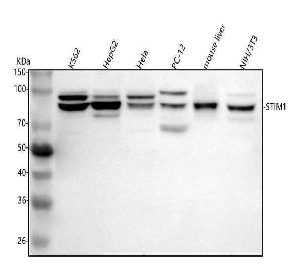
Western blot analysis of STIM1 using anti-STIM1 antibody (A00312-1).
Electrophoresis was performed on a 5-20% SDS-PAGE gel at 70V (Stacking gel) / 90V (Resolving gel) for 2-3 hours. The sample well of each lane was loaded with 30 ug of sample under reducing conditions.
Lane 1: human K562 whole cell lysates,
Lane 2: human HepG2 whole cell lysates,
Lane 3: human Hela whole cell lysates,
Lane 4: rat PC-12 whole cell lysates,
Lane 5: mouse liver tissue lysates,
Lane 6: mouse NIH/3T3 whole cell lysates.
After electrophoresis, proteins were transferred to a nitrocellulose membrane at 150 mA for 50-90 minutes. Blocked the membrane with 5% non-fat milk/TBS for 1.5 hour at RT. The membrane was incubated with rabbit anti-STIM1 antigen affinity purified polyclonal antibody (Catalog # A00312-1) at 0.5 μg/mL overnight at 4°C, then washed with TBS-0.1%Tween 3 times with 5 minutes each and probed with a goat anti-rabbit IgG-HRP secondary antibody at a dilution of 1:5000 for 1.5 hour at RT. The signal is developed using an Enhanced Chemiluminescent detection (ECL) kit (Catalog # EK1002) with Tanon 5200 system. A specific band was detected for STIM1 at approximately 85 kDa. The expected band size for STIM1 is at 77 kDa.

Click image to see more details
IHC analysis of STIM1 using anti-STIM1 antibody (A00312-1).
STIM1 was detected in a paraffin-embedded section of human breast cancer tissue. Heat mediated antigen retrieval was performed in EDTA buffer (pH 8.0, epitope retrieval solution). The tissue section was blocked with 10% goat serum. The tissue section was then incubated with 2 μg/ml rabbit anti-STIM1 Antibody (A00312-1) overnight at 4°C. Peroxidase Conjugated Goat Anti-rabbit IgG was used as secondary antibody and incubated for 30 minutes at 37°C. The tissue section was developed using HRP Conjugated Rabbit IgG Super Vision Assay Kit (Catalog # SV0002) with DAB as the chromogen.

Click image to see more details
IHC analysis of STIM1 using anti-STIM1 antibody (A00312-1).
STIM1 was detected in a paraffin-embedded section of human colon adenocarcinoma tissue. Heat mediated antigen retrieval was performed in EDTA buffer (pH 8.0, epitope retrieval solution). The tissue section was blocked with 10% goat serum. The tissue section was then incubated with 2 μg/ml rabbit anti-STIM1 Antibody (A00312-1) overnight at 4°C. Peroxidase Conjugated Goat Anti-rabbit IgG was used as secondary antibody and incubated for 30 minutes at 37°C. The tissue section was developed using HRP Conjugated Rabbit IgG Super Vision Assay Kit (Catalog # SV0002) with DAB as the chromogen.

Click image to see more details
IHC analysis of STIM1 using anti-STIM1 antibody (A00312-1).
STIM1 was detected in a paraffin-embedded section of human placenta tissue. Heat mediated antigen retrieval was performed in EDTA buffer (pH 8.0, epitope retrieval solution). The tissue section was blocked with 10% goat serum. The tissue section was then incubated with 2 μg/ml rabbit anti-STIM1 Antibody (A00312-1) overnight at 4°C. Peroxidase Conjugated Goat Anti-rabbit IgG was used as secondary antibody and incubated for 30 minutes at 37°C. The tissue section was developed using HRP Conjugated Rabbit IgG Super Vision Assay Kit (Catalog # SV0002) with DAB as the chromogen.

Click image to see more details
IHC analysis of STIM1 using anti-STIM1 antibody (A00312-1).
STIM1 was detected in a paraffin-embedded section of human thyroid papillary carcinoma tissue. Heat mediated antigen retrieval was performed in EDTA buffer (pH 8.0, epitope retrieval solution). The tissue section was blocked with 10% goat serum. The tissue section was then incubated with 2 μg/ml rabbit anti-STIM1 Antibody (A00312-1) overnight at 4°C. Peroxidase Conjugated Goat Anti-rabbit IgG was used as secondary antibody and incubated for 30 minutes at 37°C. The tissue section was developed using HRP Conjugated Rabbit IgG Super Vision Assay Kit (Catalog # SV0002) with DAB as the chromogen.

Click image to see more details
IHC analysis of STIM1 using anti-STIM1 antibody (A00312-1).
STIM1 was detected in a paraffin-embedded section of mouse testis tissue. Heat mediated antigen retrieval was performed in EDTA buffer (pH 8.0, epitope retrieval solution). The tissue section was blocked with 10% goat serum. The tissue section was then incubated with 2 μg/ml rabbit anti-STIM1 Antibody (A00312-1) overnight at 4°C. Peroxidase Conjugated Goat Anti-rabbit IgG was used as secondary antibody and incubated for 30 minutes at 37°C. The tissue section was developed using HRP Conjugated Rabbit IgG Super Vision Assay Kit (Catalog # SV0002) with DAB as the chromogen.

Click image to see more details
IHC analysis of STIM1 using anti-STIM1 antibody (A00312-1).
STIM1 was detected in a paraffin-embedded section of rat testis tissue. Heat mediated antigen retrieval was performed in EDTA buffer (pH 8.0, epitope retrieval solution). The tissue section was blocked with 10% goat serum. The tissue section was then incubated with 2 μg/ml rabbit anti-STIM1 Antibody (A00312-1) overnight at 4°C. Peroxidase Conjugated Goat Anti-rabbit IgG was used as secondary antibody and incubated for 30 minutes at 37°C. The tissue section was developed using HRP Conjugated Rabbit IgG Super Vision Assay Kit (Catalog # SV0002) with DAB as the chromogen.

Click image to see more details
Flow Cytometry analysis of K562 cells using anti-STIM1 antibody (A00312-1).
Overlay histogram showing K562 cells stained with A00312-1 (Blue line). To facilitate intracellular staining, cells were fixed with 4% paraformaldehyde and permeabilized with permeabilization buffer. The cells were blocked with 10% normal goat serum. And then incubated with rabbit anti-STIM1 Antibody (A00312-1, 1 μg/1x106 cells) for 30 min at 20°C. DyLight®488 conjugated goat anti-rabbit IgG (BA1127, 5-10 μg/1x106 cells) was used as secondary antibody for 30 minutes at 20°C. Isotype control antibody (Green line) was rabbit IgG (1 μg/1x106) used under the same conditions. Unlabelled sample (Red line) was also used as a control.

Click image to see more details
IF analysis of STIM1 using anti-STIM1 antibody (A00312-1).
STIM1 was detected in a paraffin-embedded section of human placenta tissue. Heat mediated antigen retrieval was performed in EDTA buffer (pH 8.0, epitope retrieval solution). The tissue section was blocked with 10% goat serum. The tissue section was then incubated with 5 μg/mL rabbit anti-STIM1 Antibody (A00312-1) overnight at 4°C. DyLight®594 Conjugated Goat Anti-Rabbit IgG (BA1142) was used as secondary antibody at 1:500 dilution and incubated for 30 minutes at 37°C. The section was counterstained with DAPI. Visualize using a fluorescence microscope and filter sets appropriate for the label used.

Click image to see more details
IF analysis of STIM1 using anti-STIM1 antibody (A00312-1).
STIM1 was detected in a paraffin-embedded section of mouse testis tissue. Heat mediated antigen retrieval was performed in EDTA buffer (pH 8.0, epitope retrieval solution). The tissue section was blocked with 10% goat serum. The tissue section was then incubated with 5 μg/mL rabbit anti-STIM1 Antibody (A00312-1) overnight at 4°C. DyLight®594 Conjugated Goat Anti-Rabbit IgG (BA1142) was used as secondary antibody at 1:500 dilution and incubated for 30 minutes at 37°C. The section was counterstained with DAPI. Visualize using a fluorescence microscope and filter sets appropriate for the label used.

Click image to see more details
IF analysis of STIM1 using anti-STIM1 antibody (A00312-1).
STIM1 was detected in a paraffin-embedded section of rat testis tissue. Heat mediated antigen retrieval was performed in EDTA buffer (pH 8.0, epitope retrieval solution). The tissue section was blocked with 10% goat serum. The tissue section was then incubated with 5 μg/mL rabbit anti-STIM1 Antibody (A00312-1) overnight at 4°C. DyLight®594 Conjugated Goat Anti-Rabbit IgG (BA1142) was used as secondary antibody at 1:500 dilution and incubated for 30 minutes at 37°C. The section was counterstained with DAPI. Visualize using a fluorescence microscope and filter sets appropriate for the label used.
Specific Publications For Anti-STIM1 Antibody Picoband® (A00312-1)
Loading publications
Recommended Resources
Here are featured tools and databases that you might find useful.
- Boster's Pathways Library
- Protein Databases
- Bioscience Research Protocol Resources
- Data Processing & Analysis Software
- Photo Editing Software
- Scientific Literature Resources
- Research Paper Management Tools
- Molecular Biology Software
- Primer Design Tools
- Bioinformatics Tools
- Phylogenetic Tree Analysis
Customer Reviews
Have you used Anti-STIM1 Antibody Picoband®?
Share your experimental results or join a short interview to earn up to $1,000 in product credits or other rewards.
0 Reviews For Anti-STIM1 Antibody Picoband®
Customer Q&As
Have a question?
Find answers in Q&As, reviews.
Can't find your answer?
Submit your question
