Click image to see more details
-

-
-
-
-
+1
Product Info Summary
| SKU: | PB10100 |
|---|---|
| Size: | 100 μg/vial |
| Reactive Species: | Human, Mouse, Pig, Rat |
| Host: | Rabbit |
| Application: | IF, IHC, ICC, WB |
Customers Who Bought This Also Bought

Product info
Product Name
Anti-Synapsin II/SYN2 Antibody Picoband®
SKU/Catalog Number
PB10100
PB1153 is an alternative SKU for this antibody, used in previous lots.
Size
100 μg/vial
Form
Lyophilized
Description
Boster Bio Anti-Synapsin II/SYN2 Antibody Picoband® catalog # PB10100. Tested in ICC/IF, IHC, WB applications. This antibody reacts with Human, Mouse, Rat, Pig. The brand Picoband indicates this is a premium antibody that guarantees superior quality, high affinity, and strong signals with minimal background in Western blot applications. Only our best-performing antibodies are designated as Picoband, ensuring unmatched performance.
Storage & Handling
Store at -20˚C for one year from date of receipt. After reconstitution, at 4˚C for one month. It can also be aliquotted and stored frozen at -20˚C for six months. Avoid repeated freeze-thaw cycles.
Cite This Product
Anti-Synapsin II/SYN2 Antibody Picoband® (Boster Biological Technology, Pleasanton CA, USA, Catalog # PB10100)
Host
Rabbit
Contents
Each vial contains 4 mg Trehalose, 0.9 mg NaCl and 0.2 mg Na2HPO4.
Clonality
Polyclonal
Isotype
Rabbit IgG
Immunogen
E. coli-derived human Synapsin II recombinant protein (Position: A497-D582). Human Synapsin II shares 93.1% and 89.7% amino acid (aa) sequence identity with mouse and rat Synapsin II, respectively.
Cross-reactivity
No cross-reactivity with other proteins.
Reactive Species
PB10100 is reactive to SYN2 in Human, Mouse, Pig, Rat
Observed Molecular Weight
55, 80 kDa
Calculated molecular weight
63.0 kDa
Background of SYN2
Synapsin II is the collective name for synapsin IIa and synapsin IIb, two nearly identical phosphoproteins in the synapsin family that in humans are encoded by the SYN2 gene. Synapsins encode neuronal phosphoproteins which associate with the cytoplasmic surface of synaptic vesicles. Family members are characterized by common protein domains, and they are implicated in synaptogenesis and the modulation of neurotransmitter release, suggesting a potential role in several neuropsychiatric diseases. This member of the synapsin family encodes a neuron-specific phosphoprotein that selectively binds to small synaptic vesicles in the presynaptic nerve terminal. Polymorphisms in this gene are associated with abnormal presynaptic function and related neuronal disorders, including autism, epilepsy, bipolar disorder and schizophrenia. Alternative splicing of this gene results in multiple transcript variants. The tissue inhibitor of metalloproteinase 4 gene is located within an intron of this gene and is transcribed in the opposite direction.
Antibody Validation
Boster validates all antibodies on WB, IHC, ICC, Immunofluorescence, and ELISA with known positive control and negative samples to ensure specificity and high affinity, including thorough antibody incubations.
Application & Images
Applications
PB10100 is guaranteed for IF, IHC, ICC, WB Boster Guarantee
Recommend Dilution
| Application | Dilution | Species |
|---|---|---|
| Western blot | 0.1-0.5μg/ml | Human, Mouse, Rat |
| Immunohistochemistry (Paraffin-embedded Section) | 2-5μg/ml | Pig, Rat |
| Immunocytochemistry/Immunofluorescence | 5 μg/ml | Human |
Tested application
Suggested blocking solution with 5% non-fat milk or BSA; (*)Recommended protein loading: 20-40 µg per lane
Use TE buffer pH 9.0 for antigen retrieval; (*) citrate buffer pH 6.0 is an alternative.
Validation Images & Assay Conditions
Click image to see more details
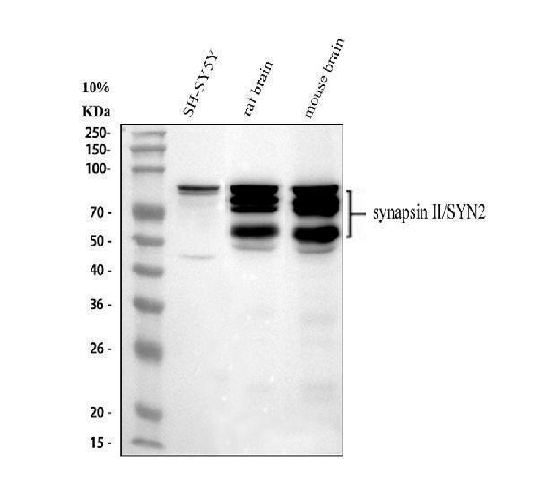
Western blot analysis of Synapsin II using anti-Synapsin II antibody (PB10100).
Electrophoresis was performed on a 10% SDS-PAGE gel at 80V (Stacking gel) / 120V (Resolving gel) for 2 hours. The sample well of each lane was loaded with 30 ug of sample under reducing conditions.
Lane 1: human SH-SY5Y whole cell lysates,
Lane 2: rat brain tissue lysates,
Lane 3: mouse brain tissue lysates.
After electrophoresis, proteins were transferred to a nitrocellulose membrane at 150 mA for 50-90 minutes. Blocked the membrane with 5% non-fat milk/TBS for 1.5 hour at RT. The membrane was incubated with rabbit anti-Synapsin II antigen affinity purified polyclonal antibody (Catalog # PB10100) at 0.5 μg/mL overnight at 4°C, then washed with TBS-0.1%Tween 3 times with 5 minutes each and probed with a goat anti-rabbit IgG-HRP secondary antibody at a dilution of 1:5000 for 1.5 hour at RT. The signal is developed using an ECL Plus Western Blotting Substrate (Catalog # AR1196-200) with Tanon 5200 system. A specific band was detected for Synapsin II at approximately 55, 80 kDa. The expected band size for Synapsin II is at 63 kDa.

Click image to see more details
IHC analysis of Synapsin II using anti-Synapsin II antibody (PB10100).
Synapsin II was detected in a paraffin-embedded section of pig brain tissue. Heat mediated antigen retrieval was performed in EDTA buffer (pH 8.0, epitope retrieval solution). The tissue section was blocked with 10% goat serum. The tissue section was then incubated with 2 μg/ml rabbit anti-Synapsin II Antibody (PB10100) overnight at 4°C. Peroxidase Conjugated Goat Anti-rabbit IgG was used as secondary antibody and incubated for 30 minutes at 37°C. The tissue section was developed using HRP Conjugated Rabbit IgG Super Vision Assay Kit (Catalog # SV0002) with DAB as the chromogen.

Click image to see more details
IHC analysis of Synapsin II using anti-Synapsin II antibody (PB10100).
Synapsin II was detected in a paraffin-embedded section of rat brain tissue. Heat mediated antigen retrieval was performed in EDTA buffer (pH 8.0, epitope retrieval solution). The tissue section was blocked with 10% goat serum. The tissue section was then incubated with 2 μg/ml rabbit anti-Synapsin II Antibody (PB10100) overnight at 4°C. Peroxidase Conjugated Goat Anti-rabbit IgG was used as secondary antibody and incubated for 30 minutes at 37°C. The tissue section was developed using HRP Conjugated Rabbit IgG Super Vision Assay Kit (Catalog # SV0002) with DAB as the chromogen.

Click image to see more details
IF analysis of Synapsin II using anti-Synapsin II antibody (PB10100) and anti-Tubulin Alpha antibody (M03989-3).
Synapsin II was detected in immunocytochemical section of U2OS cell. Enzyme antigen retrieval was performed using IHC enzyme antigen retrieval reagent (AR0022) for 15 mins. The cells were blocked with 10% goat serum. And then incubated with 5 μg/mL rabbit anti-Synapsin II Antibody (PB10100) and mouse anti-Tubulin Alpha antibody (M03989-3) overnight at 4°C. DyLight®488 Conjugated Goat Anti-Rabbit IgG (BA1127) and Cy3 Conjugated Goat Anti-Mouse IgG (BA1031) were used as secondary antibody at 1:500 dilution and incubated for 30 minutes at 37°C. Visualize using a fluorescence microscope and filter sets appropriate for the label used.
Specific Publications For Anti-Synapsin II/SYN2 Antibody Picoband® (PB10100)
Loading publications
Recommended Resources
Here are featured tools and databases that you might find useful.
- Boster's Pathways Library
- Protein Databases
- Bioscience Research Protocol Resources
- Data Processing & Analysis Software
- Photo Editing Software
- Scientific Literature Resources
- Research Paper Management Tools
- Molecular Biology Software
- Primer Design Tools
- Bioinformatics Tools
- Phylogenetic Tree Analysis
Customer Reviews
Have you used Anti-Synapsin II/SYN2 Antibody Picoband®?
Share your experimental results or join a short interview to earn up to $1,000 in product credits or other rewards.
0 Reviews For Anti-Synapsin II/SYN2 Antibody Picoband®
Customer Q&As
Have a question?
Find answers in Q&As, reviews.
Can't find your answer?
Submit your question
5 Customer Q&As for Anti-Synapsin II/SYN2 Antibody Picoband®
Question
We are currently using anti-Synapsin II/SYN2 antibody PB10100 for rat tissue, and we are content with the WB results. The species of reactivity given in the datasheet says human, mouse, rat. Is it true that the antibody can work on pig tissues as well?
Verified Customer
Verified customer
Asked: 2019-07-11
Answer
The anti-Synapsin II/SYN2 antibody (PB10100) has not been validated for cross reactivity specifically with pig tissues, but there is a good chance of cross reactivity. We have an innovator award program that if you test this antibody and show it works in pig you can get your next antibody for free. Please contact me if I can help you with anything.
Boster Scientific Support
Answered: 2019-07-11
Question
Is there a BSA free version of anti-Synapsin II/SYN2 antibody PB10100 available?
Verified Customer
Verified customer
Asked: 2018-11-05
Answer
I appreciate your recent telephone inquiry. I can confirm that some lots of this anti-Synapsin II/SYN2 antibody PB10100 are BSA free. For now, these lots are available and we can make a BSA free formula for you free of charge. It will take 3 extra days to prepare. If you require this antibody BSA free again in future, please do not hesitate to contact me and I will be pleased to check which lots we have in stock that are BSA free.
Boster Scientific Support
Answered: 2018-11-05
Question
I see that the anti-Synapsin II/SYN2 antibody PB10100 works with WB, what is the protocol used to produce the result images on the product page?
Verified Customer
Verified customer
Asked: 2018-04-09
Answer
You can find protocols for WB on the "support/technical resources" section of our navigation menu. If you have any further questions, please send an email to support@bosterbio.com
Boster Scientific Support
Answered: 2018-04-09
Question
Does anti-Synapsin II/SYN2 antibody PB10100 work for WB with middle temporal gyrus?
Verified Customer
Verified customer
Asked: 2017-06-06
Answer
According to the expression profile of middle temporal gyrus, SYN2 is highly expressed in middle temporal gyrus. So, it is likely that anti-Synapsin II/SYN2 antibody PB10100 will work for WB with middle temporal gyrus.
Boster Scientific Support
Answered: 2017-06-06
Question
My question regarding product PB10100, anti-Synapsin II/SYN2 antibody. I was wondering if it would be possible to conjugate this antibody with biotin. I would need it to be without BSA or sodium azide. I am planning on using a buffer exchange of sodium azide with PBS only. Would there be problems for me to conjugate the antibody and store it in -20 degrees in small aliquots?
Z. Carter
Verified customer
Asked: 2015-05-25
Answer
We suggest not storing this antibody with PBS buffer only in -20 degrees. If you want to store it in -20 degrees it is best to add some cryoprotectant like glycerol. If you want carrier free PB10100 anti-Synapsin II/SYN2 antibody, we can provide it to you in a special formula with trehalose and/or glycerol. These molecules will not interfere with conjugation chemistry and provide a good level of protection for the antibody from degradation. Please be sure to specify this in your purchase order.
Boster Scientific Support
Answered: 2015-05-25
