Click image to see more details
Product Info Summary
| SKU: | M00097-1 |
|---|---|
| Size: | 100 μl |
| Reactive Species: | Human, Mouse, Rat |
| Host: | Rabbit |
| Application: | IP, IHC, WB |
Customers Who Bought This Also Bought

Product info
Product Name
Anti-Tau MAPT Rabbit Monoclonal Antibody
SKU/Catalog Number
M00097-1
BM3928 is an alternative SKU for this antibody, used in previous lots.
Size
100 μl
Form
Liquid
Description
Boster Bio Anti-Tau MAPT Rabbit Monoclonal Antibody catalog # M00097-1. Tested in WB, IHC, IP applications. This antibody reacts with Human, Mouse, Rat.
Storage & Handling
Store at -20°C for one year. For short term storage and frequent use, store at 4°C for up to one month. Avoid repeated freeze-thaw cycles.
Cite This Product
Anti-Tau MAPT Rabbit Monoclonal Antibody (Boster Biological Technology, Pleasanton CA, USA, Catalog # M00097-1)
Host
Rabbit
Contents
Rabbit IgG in stabilizing components, phosphate buffered saline, pH 7.4, 150mM NaCl, 0.02% sodium azide and 50% glycerol.
*This antibody is supplied in a stabilized formulation.
Compatibility with conjugation reactions depends on the chemistry of the conjugation method used.
For conjugation methods that are not compatible with the stabilizing components present in this formulation, a carrier-free antibody format is required.
Clonality
Monoclonal
Clone Number
GF-13
Isotype
Rabbit IgG
Immunogen
A synthesized peptide derived from human Tau
Reactive Species
M00097-1 is reactive to MAPT in Human, Mouse, Rat
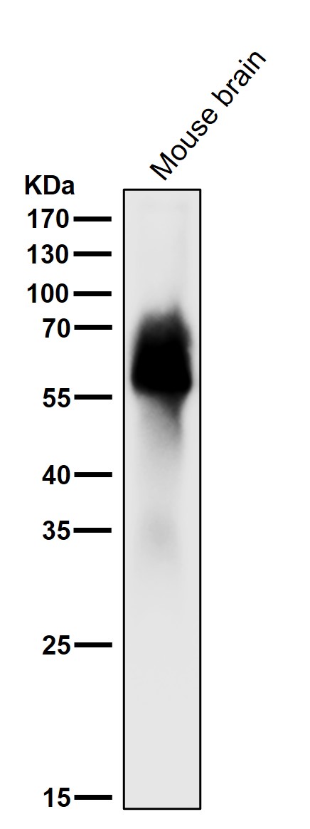
Observed Molecular Weight
50-80 kDa
Calculated molecular weight
78.9 kDa
Antibody Validation
Boster validates all antibodies on WB, IHC, ICC, Immunofluorescence, and ELISA with known positive control and negative samples to ensure specificity and high affinity, including thorough antibody incubations.
Application & Images
Applications
M00097-1 is guaranteed for IP, IHC, WB Boster Guarantee
Recommend Dilution
WB 1:500-2000
IHC 1:50-200
IP 1:20
Validation Images & Assay Conditions
Click image to see more details
All lanes use the Antibody at 1:1k dilution for 1 hour at room temperature.

Click image to see more details
Western blot analysis of Tau expression in SH-SY5Y cell lysate.
Specific Publications For Anti-Tau MAPT Rabbit Monoclonal Antibody (M00097-1)
Loading publications
Recommended Resources
Here are featured tools and databases that you might find useful.
- Boster's Pathways Library
- Protein Databases
- Bioscience Research Protocol Resources
- Data Processing & Analysis Software
- Photo Editing Software
- Scientific Literature Resources
- Research Paper Management Tools
- Molecular Biology Software
- Primer Design Tools
- Bioinformatics Tools
- Phylogenetic Tree Analysis
Customer Reviews
Have you used Anti-Tau MAPT Rabbit Monoclonal Antibody?
Share your experimental results or join a short interview to earn up to $1,000 in product credits or other rewards.
0 Reviews For Anti-Tau MAPT Rabbit Monoclonal Antibody
Customer Q&As
Have a question?
Find answers in Q&As, reviews.
Can't find your answer?
Submit your question
5 Customer Q&As for Anti-Tau MAPT Rabbit Monoclonal Antibody
Question
We ordered your anti-Tau Rabbit Monoclonal antibody for IP on liver last year. I am using rat, and I plan to use the antibody for IF next. I would like examining liver as well as fetal brain cortex in our next experiment. Do you have any suggestion on which antibody would work the best for IF?
Verified Customer
Verified customer
Asked: 2020-04-23
Answer
I have checked the website and datasheets of our anti-Tau Rabbit Monoclonal antibody and I see that M00097-1 has been validated on rat in both IP and IF. Thus M00097-1 should work for your application. Our Boster satisfaction guarantee will cover this product for IF in rat even if the specific tissue type has not been validated. We do have a comprehensive range of products for IF detection and you can check out our website bosterbio.com to find out more information about them.
Boster Scientific Support
Answered: 2020-04-23
Question
We have observed staining in rat cervix carcinoma. Do you have any suggestions? Is anti-Tau Rabbit Monoclonal antibody supposed to stain cervix carcinoma positively?
Verified Customer
Verified customer
Asked: 2020-01-15
Answer
According to literature cervix carcinoma does express MAPT. According to Uniprot.org, MAPT is expressed in parietal lobe, brain, fetal brain, fetal brain cortex, cervix carcinoma, leukemic t-cell, cervix carcinoma erythroleukemia, liver, among other tissues. Regarding which tissues have MAPT expression, here are a few articles citing expression in various tissues:
Brain, Pubmed ID: 1512244, 2484340, 2495000, 2498079, 3131773, 15489334
Cervix carcinoma, Pubmed ID: 16964243, 18220336, 20068231
Cervix carcinoma, and Erythroleukemia, Pubmed ID: 23186163
Fetal brain, Pubmed ID: 2516729
Fetal brain cortex, Pubmed ID: 16443603
Leukemic T-cell, Pubmed ID: 19690332
Liver, Pubmed ID: 24275569
Boster Scientific Support
Answered: 2020-01-15
Question
What is the difference between Anti-Tau Rabbit Monoclonal antibodies M00097 and M00097-1?
Verified customer
Asked: 2019-10-10
Answer
The Anti-Tau MAPT Rabbit Monoclonal Antibody (M00097) and Anti-Tau MAPT Rabbit Monoclonal Antibody (M00097-1) differ in the immunogen sequence and application.
Boster Scientific Support
Answered: 2019-10-11
Question
We are currently using anti-Tau Rabbit Monoclonal antibody M00097-1 for rat tissue, and we are satisfied with the WB results. The species of reactivity given in the datasheet says human, mouse, rat. Is it true that the antibody can work on bovine tissues as well?
Verified Customer
Verified customer
Asked: 2018-08-09
Answer
The anti-Tau Rabbit Monoclonal antibody (M00097-1) has not been validated for cross reactivity specifically with bovine tissues, though there is a good chance of cross reactivity. We have an innovator award program that if you test this antibody and show it works in bovine you can get your next antibody for free. Please contact me if I can help you with anything.
Boster Scientific Support
Answered: 2018-08-09
Question
My colleagues were happy with the WB result of your anti-Tau Rabbit Monoclonal antibody. However we have been able to see positive staining in brain cytosol using this antibody. Is that expected? Could you tell me where is MAPT supposed to be expressed?
C. Li
Verified customer
Asked: 2015-05-12
Answer
From literature, brain does express MAPT. Generally MAPT expresses in cytoplasm, cytosol. Regarding which tissues have MAPT expression, here are a few articles citing expression in various tissues:
Brain, Pubmed ID: 1512244, 2484340, 2495000, 2498079, 3131773, 15489334
Cervix carcinoma, Pubmed ID: 16964243, 18220336, 20068231
Cervix carcinoma, and Erythroleukemia, Pubmed ID: 23186163
Fetal brain, Pubmed ID: 2516729
Fetal brain cortex, Pubmed ID: 16443603
Leukemic T-cell, Pubmed ID: 19690332
Liver, Pubmed ID: 24275569
Boster Scientific Support
Answered: 2015-05-12
