Click image to see more details
-

-
-
-
-
+2
Product Info Summary
| SKU: | M02389 |
|---|---|
| Size: | 100 μg/vial |
| Reactive Species: | Human |
| Host: | Mouse |
| Application: | Flow Cytometry, IF, IHC, ICC, WB |
Customers Who Bought This Also Bought

Product info
Product Name
Anti-TCP1 alpha Antibody Picoband® (monoclonal, 2E7)
SKU/Catalog Number
M02389
Size
100 μg/vial
Form
Lyophilized
Description
Boster Bio Anti-TCP1 alpha Antibody Picoband® (monoclonal, 2E7) catalog # M02389. Tested in Flow Cytometry, IF, IHC, ICC, WB applications. This antibody reacts with Human. The brand Picoband indicates this is a premium antibody that guarantees superior quality, high affinity, and strong signals with minimal background in Western blot applications. Only our best-performing antibodies are designated as Picoband, ensuring unmatched performance.
Storage & Handling
Store at -20˚C for one year from date of receipt. After reconstitution, at 4˚C for one month. It can also be aliquotted and stored frozen at -20˚C for six months. Avoid repeated freeze-thaw cycles.
Cite This Product
Anti-TCP1 alpha Antibody Picoband® (monoclonal, 2E7) (Boster Biological Technology, Pleasanton CA, USA, Catalog # M02389)
Host
Mouse
Contents
Each vial contains 4mg Trehalose, 0.9mg NaCl, 0.2mg Na2HPO4, 0.05mg NaN3.
Clonality
Monoclonal
Clone Number
2E7
Isotype
Mouse IgG1
Immunogen
A synthetic peptide corresponding to a sequence at the C-terminus of human TCP1 alpha, different from the related mouse sequence by one amino acid, and from the related rat sequence by two amino acids.
Cross-reactivity
No cross-reactivity with other proteins.
Reactive Species
M02389 is reactive to TCP1 in Human
Observed Molecular Weight
60 kDa
Calculated molecular weight
60.3 kDa
Background of TCP1
T-complex protein 1 subunit alpha is a protein that in humans is encoded by the TCP1 gene. The protein encoded by this gene is a molecular chaperone that is a member of the chaperonin containing TCP1 complex (CCT), also known as the TCP1 ring complex (TRiC). This complex consists of two identical stacked rings, each containing eight different proteins. Unfolded polypeptides enter the central cavity of the complex and are folded in an ATP-dependent manner. The complex folds various proteins, including actin and tubulin. Alternate transcriptional splice variants of this gene, encoding different isoforms, have been characterized. In addition, three pseudogenes that appear to be derived from this gene have been found.
Antibody Validation
Boster validates all antibodies on WB, IHC, ICC, Immunofluorescence, and ELISA with known positive control and negative samples to ensure specificity and high affinity, including thorough antibody incubations.
Application & Images
Applications
M02389 is guaranteed for Flow Cytometry, IF, IHC, ICC, WB Boster Guarantee
Recommend Dilution
| Application | Dilution | Species |
|---|---|---|
| Western blot | 0.1-0.5μg/ml | |
| Immunohistochemistry (Paraffin-embedded Section) | 0.5-1μg/ml | |
| Immunocytochemistry/Immunofluorescence | 2μg/ml | |
| Flow Cytometry (Fixed) | 1-3μg/1x106 cells |
Tested application
Suggested blocking solution with 5% non-fat milk or BSA; (*)Recommended protein loading: 20-40 µg per lane
Use TE buffer pH 9.0 for antigen retrieval; (*) citrate buffer pH 6.0 is an alternative.
Validation Images & Assay Conditions
Click image to see more details
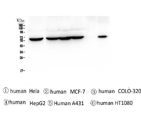
Western blot analysis of TCP1 alpha using anti-TCP1 alpha antibody (M02389).
Electrophoresis was performed on a 5-20% SDS-PAGE gel at 70V (Stacking gel) / 90V (Resolving gel) for 2-3 hours. The sample well of each lane was loaded with 50ug of sample under reducing conditions.
Lane 1: human Hela whole cell lysates,
Lane 2: human MCF-7 whole cell lysates,
Lane 3: human COLO-320 whole cell lysates,
Lane 4: human HepG2 whole cell lysates,
Lane 5: human A431 whole cell lysates,
Lane 6: human HT1080 whole cell lysates.
After Electrophoresis, proteins were transferred to a Nitrocellulose membrane at 150mA for 50-90 minutes. Blocked the membrane with 5% Non-fat Milk/ TBS for 1.5 hour at RT. The membrane was incubated with mouse anti-TCP1 alpha antigen affinity purified monoclonal antibody (Catalog # M02389) at 0.5 μg/mL overnight at 4°C, then washed with TBS-0.1%Tween 3 times with 5 minutes each and probed with a goat anti-mouse IgG-HRP secondary antibody at a dilution of 1:10000 for 1.5 hour at RT. The signal is developed using an Enhanced Chemiluminescent detection (ECL) kit (Catalog # EK1001) with Tanon 5200 system.

Click image to see more details
IHC analysis of TCP1 alpha using anti-TCP1 alpha antibody (M02389).
TCP1 alpha was detected in paraffin-embedded section of human lung cancer tissue. Heat mediated antigen retrieval was performed in citrate buffer (pH6, epitope retrieval solution) for 20 mins. The tissue section was blocked with 10% goat serum. The tissue section was then incubated with 2μg/ml mouse anti-TCP1 alpha Antibody (M02389) overnight at 4°C. Biotinylated goat anti-mouse IgG was used as secondary antibody and incubated for 30 minutes at 37°C. The tissue section was developed using Strepavidin-Biotin-Complex (SABC)(Catalog # SA1021) with DAB as the chromogen.

Click image to see more details
IHC analysis of TCP1 alpha using anti-TCP1 alpha antibody (M02389).
TCP1 alpha was detected in paraffin-embedded section of human placenta tissue. Heat mediated antigen retrieval was performed in citrate buffer (pH6, epitope retrieval solution) for 20 mins. The tissue section was blocked with 10% goat serum. The tissue section was then incubated with 2μg/ml mouse anti-TCP1 alpha Antibody (M02389) overnight at 4°C. Biotinylated goat anti-mouse IgG was used as secondary antibody and incubated for 30 minutes at 37°C. The tissue section was developed using Strepavidin-Biotin-Complex (SABC)(Catalog # SA1021) with DAB as the chromogen.

Click image to see more details
Flow Cytometry analysis of HepG2 cells using anti-TCP1 alpha antibody (M02389).
Overlay histogram showing HepG2 cells stained with M02389 (Blue line). To facilitate intracellular staining, cells were fixed with 4% paraformaldehyde and permeabilized with permeabilization buffer. The cells were blocked with 10% normal goat serum. And then incubated with mouse anti-TCP1 alpha Antibody (M02389,1μg/1x106 cells) for 30 min at 20°C. DyLight®488 conjugated goat anti-mouse IgG (BA1126, 5-10μg/1x106 cells) was used as secondary antibody for 30 minutes at 20°C. Isotype control antibody (Green line) was rabbit IgG (1μg/1x106) used under the same conditions. Unlabelled sample (Red line) was also used as a control.

Click image to see more details
IF analysis of TCP1 alpha using anti-TCP1 alpha antibody (M02389).
TCP1 alpha was detected in immunocytochemical section of MCF7 cells. Enzyme antigen retrieval was performed using IHC enzyme antigen retrieval reagent (AR0022) for 15 mins. The cells were blocked with 10% goat serum. And then incubated with 2μg/mL mouse anti-TCP1 alpha Antibody (M02389) overnight at 4°C. DyLight®488 Conjugated Goat Anti-Mouse IgG (BA1126) was used as secondary antibody at 1:100 dilution and incubated for 30 minutes at 37°C. The section was counterstained with DAPI. Visualize using a fluorescence microscope and filter sets appropriate for the label used.

Click image to see more details
Western blot analysis of TCP1 using anti-TCP1 antibody (M02389).
Electrophoresis was performed on a 5-20% SDS-PAGE gel at 80V (Stacking gel) / 120V (Resolving gel) for 2 hours. The sample well of each lane was loaded with 30 ug of sample under reducing conditions.
Lane 1-6: human U2OS whole cell lysates,
After electrophoresis, proteins were transferred to a nitrocellulose membrane at 150 mA for 50-90 minutes. Blocked the membrane with 5% non-fat milk/TBS for 1.5 hour at RT. The membrane was incubated with mouse anti-TCP1 antigen affinity purified monoclonal antibody (M02389) at 1:1000 overnight at 4°C, then washed with TBS-0.1%Tween 3 times with 5 minutes each and probed with a goat anti-mouse IgG-HRP secondary antibodyat a dilution of 1:5000 for 1.5 hour at RT. The signal is developed using an ECL Plus Western Blotting Substrate (Catalog # AR1196-200) with ChemiDoc MP system. A specific band was detected for TCP1 at approximately 60 kDa. The expected band size for TCP1 is at 60 kDa.
Specific Publications For Anti-TCP1 alpha Antibody Picoband® (monoclonal, 2E7) (M02389)
Loading publications
Recommended Resources
Here are featured tools and databases that you might find useful.
- Boster's Pathways Library
- Protein Databases
- Bioscience Research Protocol Resources
- Data Processing & Analysis Software
- Photo Editing Software
- Scientific Literature Resources
- Research Paper Management Tools
- Molecular Biology Software
- Primer Design Tools
- Bioinformatics Tools
- Phylogenetic Tree Analysis
Customer Reviews
Have you used Anti-TCP1 alpha Antibody Picoband® (monoclonal, 2E7)?
Share your experimental results or join a short interview to earn up to $1,000 in product credits or other rewards.
1 Reviews For Anti-TCP1 alpha Antibody Picoband® (monoclonal, 2E7)
In this study, these two antibodies were primarily used for Western blot experiments. In the Western blot results, the CCT1 antibody showed a strong specific band and maintained a robust signal even after multiple reuses.
Excellent
| SKU | M02389 |
|---|---|
| Application | Western Blot |
| Sample | U2OS cell |
| Sample Processing Description | Cells were lysed with NP-40 lysis buffer to extract proteins. After quantification using the BCA assay, 5× loading buffer was added, and the samples were boiled for denaturation. Then, 20 µg of protein was loaded into each lane. |
| Primary Antibody | Anti-TCP1 alpha Antibody Picoband® (monoclonal, 2E7) |
| Primary Incubation | 1:1000, overnight at 4 ℃ |
| Blocking Agent | BSA |
| Secondary Antibody | Goat Anti-Rabbit IgG (H+L) Secondary Antibody, Unconjugated (BA1039) |
| Secondary Incubation | Incubate at room temperature for 1 hour |
| Detection | Substrate: ECL substrate, Imaging system: ChemiDoc MP (Bio-Rad) |
| Results Summary | I will purchase Boster products again and recommend them to my classmates and colleagues. |
Xinyu Ma, Tsinghua University
Verified customer
Submitted 2025-10-27
Customer Q&As
Have a question?
Find answers in Q&As, reviews.
Can't find your answer?
Submit your question
