Click image to see more details
-

-
-
-
-
+3
Product Info Summary
| SKU: | A00197-2 |
|---|---|
| Size: | 0.1 mg |
| Reactive Species: | Human, Mouse, Rat |
| Host: | Rabbit |
| Application: | ELISA, IF, IHC-P, WB |
Customers Who Bought This Also Bought

Product info
Product Name
Anti-Toll-like receptor 3 TLR3 Antibody
SKU/Catalog Number
A00197-2
Size
0.1 mg
Form
Liquid
Description
Boster Bio Anti-Toll-like receptor 3 TLR3 Antibody (Catalog # A00197-2). Tested in ELISA, WB, IHC-P, IF applications. This antibody reacts with Human, Mouse, Rat.
Storage & Handling
TLR3 antibody can be stored at 4°C up to one year. Antibodies should not be exposed to prolonged high temperatures.
Cite This Product
Anti-Toll-like receptor 3 TLR3 Antibody (Boster Biological Technology, Pleasanton CA, USA, Catalog # A00197-2)
Host
Rabbit
Contents
TLR3 Antibody is supplied in PBS containing 0.02% sodium azide.
Clonality
Polyclonal
Isotype
IgG
Immunogen
TLR3 antibody was raised against a peptide corresponding to 15 amino acids near the carboxy terminus of human TLR3. The immunogen is located within amino acids 780 - 830 of TLR3.
Reactive Species
A00197-2 is reactive to TLR3 in Human, Mouse, Rat
Observed Molecular Weight
104 kDa
Calculated molecular weight
103.8 kDa
Background of TLR3
Toll-like receptors (TLRs) are evolutionarily conserved pattern-recognition molecules resembling the toll proteins that mediate antimicrobial responses in Drosophila. These proteins recognize different microbial products during infection and serve as an important link between the innate and adaptive immune responses. The TLRs act through adaptor molecules such as MyD88 and TIRAP to activate various kinases and transcription factors so the organism can respond to potential infection. TLR3 is known to recognize viral double-stranded (ds) RNA, a molecular pattern associated with viral infection. Recently it has been shown to recognize viruses such as Influenza A and West Nile Virus and can mediate entry of at least West Nile Virus.
Antibody Validation
Boster validates all antibodies on WB, IHC, ICC, Immunofluorescence, and ELISA with known positive control and negative samples to ensure specificity and high affinity, including thorough antibody incubations.
Application & Images
Applications
A00197-2 is guaranteed for ELISA, IF, IHC-P, WB Boster Guarantee
Recommend Dilution
| Application | Dilution | Species |
|---|---|---|
| Antibody validated: Western Blot in human | mouse and rat samples; Immunohistochemistry in mouse | and rat samples; Immunofluorescence in the human and mouse samples. All other applications and species not yet tested. |
Validation Images & Assay Conditions
Click image to see more details
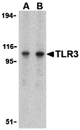
Western blot analysis of TLR3 in Daudi cell lysate with TLR3 antibody at (A) 1 and (B) 2 μg/mL.

Click image to see more details
WB Validation in Human Cells Loading: 15 μg of lysate Antibodies: TLR3, A00197-2, 1 μ g/mL , 1 h incubation at RT in 5% NFDM/TBST. Secondary: Goat Anti-Rabbit IgG HRP conjugate at 1:10000 dilution.

Click image to see more details
WB Validation in Mouse and Rat Brain Loading: 15 μg of lysate Antibodies: TLR3, A00197-2, 1 μg/mL , 1 h incubation at RT in 5% NFDM/TBST. Secondary: Goat Anti-Rabbit IgG HRP conjugate at 1:10000 dilution.

Click image to see more details
Immunofluorescence of TLR3 in mouse spleen tissue with TLR3 antibody at 20 μg/ml.
Green: TLR3 Antibody (A00197-2)
Blue: DAPI staining

Click image to see more details
Immunofluorescence Validation of TLR3 in Human Spleen Immunofluorescent analysis of 4% paraformaldehyde-fixed human spleen tissue labeling TLR3 with A00197-2 at 10 μg/mL, followed by goat anti-rabbit IgG secondary antibody at 1/500 dilution (red) and DAPI staining (blue).

Click image to see more details
Immunohistochemistry Validation of TLR3 in Mouse Brain Immunohistochemical analysis of paraffin-embedded mouse brain tissue using anti-TLR3 antibody (A00197-2) at 1 μg/ml. Tissue was fixed with formaldehyde and blocked with 10% serum for 1 h at RT; antigen retrieval was by heat mediation with a citrate buffer (pH6). Samples were incubated with primary antibody overnight at 4°C. A goat anti-rabbit IgG H&L (HRP) at 1/250 was used as secondary. Counter stained with Hematoxylin.

Click image to see more details
Immunohistochemistry Validation of TLR3 in Rat Brain Immunohistochemical analysis of paraffin-embedded rat brain tissue using anti-TLR3 antibody (A00197-2) at 1 μg/ml. Tissue was fixed with formaldehyde and blocked with 10% serum for 1 h at RT; antigen retrieval was by heat mediation with a citrate buffer (pH6). Samples were incubated with primary antibody overnight at 4°C. A goat anti-rabbit IgG H&L (HRP) at 1/250 was used as secondary. Counter stained with Hematoxylin.
Specific Publications For Anti-Toll-like receptor 3 TLR3 Antibody (A00197-2)
Loading publications
Recommended Resources
Here are featured tools and databases that you might find useful.
- Boster's Pathways Library
- Protein Databases
- Bioscience Research Protocol Resources
- Data Processing & Analysis Software
- Photo Editing Software
- Scientific Literature Resources
- Research Paper Management Tools
- Molecular Biology Software
- Primer Design Tools
- Bioinformatics Tools
- Phylogenetic Tree Analysis
Customer Reviews
Have you used Anti-Toll-like receptor 3 TLR3 Antibody?
Share your experimental results or join a short interview to earn up to $1,000 in product credits or other rewards.
0 Reviews For Anti-Toll-like receptor 3 TLR3 Antibody
Customer Q&As
Have a question?
Find answers in Q&As, reviews.
Can't find your answer?
Submit your question
