Click image to see more details
Product Info Summary
| SKU: | PA1464 |
|---|---|
| Size: | 100 μg/vial |
| Reactive Species: | Human, Mouse, Rat |
| Host: | Rabbit |
| Application: | WB |
Customers Who Bought This Also Bought

Product info
Product Name
Anti-FADD Antibody Picoband®
SKU/Catalog Number
PA1464
BA3618-2 is an alternative SKU for this antibody, used in previous lots.
Size
100 μg/vial
Form
Lyophilized
Description
Boster Bio Anti-FADD Antibody catalog # PA1464. Tested in WB applications. This antibody reacts with Human, Mouse, Rat. The brand Picoband indicates this is a premium antibody that guarantees superior quality, high affinity, and strong signals with minimal background in Western blot applications. Only our best-performing antibodies are designated as Picoband, ensuring unmatched performance.
Storage & Handling
Store at -20˚C for one year from date of receipt. After reconstitution, at 4˚C for one month. It can also be aliquotted and stored frozen at -20˚C for six months. Avoid repeated freeze-thaw cycles.
Cite This Product
Anti-FADD Antibody Picoband® (Boster Biological Technology, Pleasanton CA, USA, Catalog # PA1464)
Host
Rabbit
Contents
Each vial contains antibody formulated with stabilizing components, 0.9mg NaCl, 0.2mg Na2HPO4, 0.05mg Thimerosal, 0.01mg NaN3.
*This antibody is supplied in a stabilized formulation.
Compatibility with conjugation reactions depends on the chemistry of the conjugation method used.
For conjugation methods that are not compatible with the stabilizing components present in this formulation, a carrier-free antibody format is required.
Clonality
Polyclonal
Isotype
Rabbit IgG
Immunogen
A synthetic peptide corresponding to a sequence at the C-terminus of human FADD.
Cross-reactivity
No cross-reactivity with other proteins
Reactive Species
PA1464 is reactive to FADD in Human, Mouse, Rat
Observed Molecular Weight
28 kDa
Calculated molecular weight
23.3 kDa
Background of FADD
FADD, Fas-Associated protein with Death Domain, is a universal adaptor protein in apoptosis that mediates signaling of all known death domain-containing members of the TNF receptor superfamily. The FADD gene contains 2 exons and spans approximately 3.6 kb. By analysis of somatic cell hybrid panels and by fluorescence in situ hybridization, the FADD gene is mapped to 11q13.3. The protein encoded by this gene is an adaptor molecule that interacts with various cell surface receptors and mediates cell apoptotic signals. Through its C-terminal death domain, this protein can be recruited by TNFRSF6/Fas-receptor, tumor necrosis factor receptor, TNFRSF25, and TNFSF10/TRAIL-receptor, thus, it participates in the death signaling initiated by these receptors.
Antibody Validation
Boster validates all antibodies on WB, IHC, ICC, Immunofluorescence, and ELISA with known positive control and negative samples to ensure specificity and high affinity, including thorough antibody incubations.
Application & Images
Applications
PA1464 is guaranteed for WB Boster Guarantee
Recommend Dilution
| Application | Dilution | Species |
|---|---|---|
| Western blot | 0.1-0.5μg/ml | Human, Mouse, Rat |
Tested application
Suggested blocking solution with 5% non-fat milk or BSA; (*)Recommended protein loading: 20-40 µg per lane
Validation Images & Assay Conditions
Click image to see more details
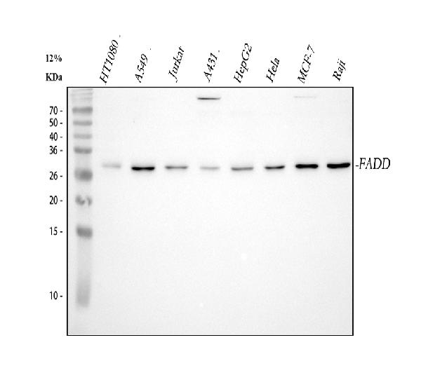
Western blot analysis of FADD using anti-FADD antibody (PA1464).
Electrophoresis was performed on a 5-20% SDS-PAGE gel at 70V (Stacking gel) / 90V (Resolving gel) for 2-3 hours. The sample well of each lane was loaded with 30 ug of sample under reducing conditions.
Lane 1: human HT1080 whole cell lysates,
Lane 2: human A549 whole cell lysates,
Lane 3: human Jurkat whole cell lysates,
Lane 4: human A431 whole cell lysates,
Lane 5: human HepG2 whole cell lysates,
Lane 6: human Hela whole cell lysates,
Lane 7: human MCF-7 whole cell lysates,
Lane 8: human Raji whole cell lysates.
After electrophoresis, proteins were transferred to a nitrocellulose membrane at 150 mA for 50-90 minutes. Blocked the membrane with 5% non-fat milk/TBS for 1.5 hour at RT. The membrane was incubated with rabbit anti-FADD antigen affinity purified polyclonal antibody (Catalog # PA1464) at 0.5 μg/mL overnight at 4°C, then washed with TBS-0.1%Tween 3 times with 5 minutes each and probed with a goat anti-rabbit IgG-HRP secondary antibody at a dilution of 1:5000 for 1.5 hour at RT. The signal is developed using an Enhanced Chemiluminescent detection (ECL) kit (Catalog # EK1002) with Tanon 5200 system. A specific band was detected for FADD at approximately 28 kDa. The expected band size for FADD is at 23 kDa.

Click image to see more details
Western blot analysis of FADD using anti-FADD antibody (PA1464).
Electrophoresis was performed on a 5-20% SDS-PAGE gel at 70V (Stacking gel) / 90V (Resolving gel) for 2-3 hours. The sample well of each lane was loaded with 30 ug of sample under reducing conditions.
Lane 1: rat spleen tissue lysates,
Lane 2: rat PC-12 whole cell lysates,
Lane 3: mouse spleen tissue lysates,
Lane 4: mouse kidney tissue lysates,
Lane 5: mouse RAW264.7 whole cell lysates.
After electrophoresis, proteins were transferred to a nitrocellulose membrane at 150 mA for 50-90 minutes. Blocked the membrane with 5% non-fat milk/TBS for 1.5 hour at RT. The membrane was incubated with rabbit anti-FADD antigen affinity purified polyclonal antibody (Catalog # PA1464) at 0.5 μg/mL overnight at 4°C, then washed with TBS-0.1%Tween 3 times with 5 minutes each and probed with a goat anti-rabbit IgG-HRP secondary antibody at a dilution of 1:5000 for 1.5 hour at RT. The signal is developed using an Enhanced Chemiluminescent detection (ECL) kit (Catalog # EK1002) with Tanon 5200 system. A specific band was detected for FADD at approximately 28 kDa. The expected band size for FADD is at 23 kDa.
Specific Publications For Anti-FADD Antibody Picoband® (PA1464)
Loading publications
Recommended Resources
Here are featured tools and databases that you might find useful.
- Boster's Pathways Library
- Protein Databases
- Bioscience Research Protocol Resources
- Data Processing & Analysis Software
- Photo Editing Software
- Scientific Literature Resources
- Research Paper Management Tools
- Molecular Biology Software
- Primer Design Tools
- Bioinformatics Tools
- Phylogenetic Tree Analysis
Customer Reviews
Have you used Anti-FADD Antibody Picoband®?
Share your experimental results or join a short interview to earn up to $1,000 in product credits or other rewards.
0 Reviews For Anti-FADD Antibody Picoband®
Customer Q&As
Have a question?
Find answers in Q&As, reviews.
Can't find your answer?
Submit your question
1 Customer Q&As for Anti-FADD Antibody Picoband®
Question
We are currently using anti-FADD antibody PA1464 for human tissue, and we are satisfied with the WB results. The species of reactivity given in the datasheet says human. Is it possible that the antibody can work on feline tissues as well?
Verified Customer
Verified customer
Asked: 2019-02-15
Answer
The anti-FADD antibody (PA1464) has not been tested for cross reactivity specifically with feline tissues, though there is a good chance of cross reactivity. We have an innovator award program that if you test this antibody and show it works in feline you can get your next antibody for free. Please contact me if I can help you with anything.
Boster Scientific Support
Answered: 2019-02-15
