Click image to see more details
-

-
-
-
-
+4
Product Info Summary
| SKU: | A01610-1 |
|---|---|
| Size: | 100 μg/vial |
| Reactive Species: | Human, Mouse, Rat |
| Host: | Rabbit |
| Application: | ELISA, WB |
Customers Who Bought This Also Bought

Product info
Product Name
Anti-TPX2 Antibody Picoband®
SKU/Catalog Number
A01610-1
Size
100 μg/vial
Form
Lyophilized
Description
Boster Bio Anti-TPX2 Antibody Picoband® catalog # A01610-1. Tested in ELISA, WB applications. This antibody reacts with Human, Mouse, Rat. The brand Picoband indicates this is a premium antibody that guarantees superior quality, high affinity, and strong signals with minimal background in Western blot applications. Only our best-performing antibodies are designated as Picoband, ensuring unmatched performance.
Storage & Handling
Store at -20˚C for one year from date of receipt. After reconstitution, at 4˚C for one month. It can also be aliquotted and stored frozen at -20˚C for six months. Avoid repeated freeze-thaw cycles.
Cite This Product
Anti-TPX2 Antibody Picoband® (Boster Biological Technology, Pleasanton CA, USA, Catalog # A01610-1)
Host
Rabbit
Contents
Each vial contains 4mg Trehalose, 0.9mg NaCl, 0.2mg Na2HPO4, 0.05mg NaN3.
Clonality
Polyclonal
Isotype
Rabbit IgG
Immunogen
E. coli-derived human TPX2 recombinant protein (Position: D15-Q93).
Cross-reactivity
No cross-reactivity with other proteins.
Reactive Species
A01610-1 is reactive to TPX2 in Human, Mouse, Rat
Observed Molecular Weight
86 kDa, 100 kDa
Calculated molecular weight
85.7 kDa
Background of TPX2
Targeting protein for Xklp2, also known as C20ORF1, is a protein that in humans is encoded by the TPX2 gene. It is mapped to chromosome 20q11.2. It was concluded that even in the presence of duplicated centrosomes, spindle formation requires the function of TPX2 to generate a stable bipolar spindle with overlapping antiparallel microtubule arrays. At the onset of mitosis, GOLGA2 interacts with importin-alpha, liberating TPX2 from importin-alpha, allowing TPX2 to activates AURKA kinase and stimulates local microtubule nucleation.
Antibody Validation
Boster validates all antibodies on WB, IHC, ICC, Immunofluorescence, and ELISA with known positive control and negative samples to ensure specificity and high affinity, including thorough antibody incubations.
Application & Images
Applications
A01610-1 is guaranteed for ELISA, WB Boster Guarantee
Recommend Dilution
| Application | Dilution | Species |
|---|---|---|
| Western blot | 0.1-0.5μg/ml | |
| ELISA | 0.1-0.5μg/ml |
Tested application
Suggested blocking solution with 5% non-fat milk or BSA; (*)Recommended protein loading: 20-40 µg per lane
Validation Images & Assay Conditions
Click image to see more details
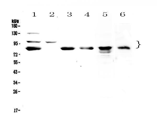
Western blot analysis of TPX2 using anti-TPX2 antibody (A01610-1).
Electrophoresis was performed on a 5-20% SDS-PAGE gel at 70V (Stacking gel) / 90V (Resolving gel) for 2-3 hours. The sample well of each lane was loaded with 50ug of sample under reducing conditions.
Lane 1: human Hela whole cell lysates,
Lane 2: human COLO-320 whole cell lysates,
Lane 3: human U-87MG whole cell lysates,
Lane 4: human SGC-7901 whole cell lysates,
Lane 5: rat PC-12 whole cell lysates,
Lane 6: mouse HEPA1-6 whole cell lysates.
After Electrophoresis, proteins were transferred to a Nitrocellulose membrane at 150mA for 50-90 minutes. Blocked the membrane with 5% Non-fat Milk/ TBS for 1.5 hour at RT. The membrane was incubated with rabbit anti-TPX2 antigen affinity purified polyclonal antibody (Catalog # A01610-1) at 0.5 μg/mL overnight at 4°C, then washed with TBS-0.1%Tween 3 times with 5 minutes each and probed with a goat anti-rabbit IgG-HRP secondary antibody at a dilution of 1:10000 for 1.5 hour at RT. The signal is developed using an Enhanced Chemiluminescent detection (ECL) kit (Catalog # EK1002) with Tanon 5200 system. A specific band was detected for TPX2 at approximately 86KD, 100KD. The expected band size for TPX2 is at 86KD.

Click image to see more details
HMGB3 could inhibit cell survival via TPX2 in NB cells. (A) Nine genes significantly co-expressed with HMGB3 in four datasets of NB. (B) Functional enrichment analysis of nine genes. (C) The mRNA expression of nine genes was detected by qRT-PCR in SK-N-SH cells after HMGB3 knockdown. (D) Pearson correlation of HMGB3 and TPX2 in four NB datasets. (E) Protein expression of TPX2 and HMGB3 was detected by western blotting after HMGB3 knockdown. (F) TPX2 reduction was time-dependent with HMGB3 knockdown in SK-N-SH and SK-N-AS cells. * p < 0.05, ** p < 0.01, and *** p < 0.001.
Index in PubMed under a CC BY license. PMID: 34988076

Click image to see more details
TPX2 overexpression can reverse the inhibition of cell proliferation caused by HMGB3 knockdown in SK-N-SH cells. (A) Protein expression was detected in sh-HMGB3 SK-N-SH cells after TPX2 over-expression by western blotting. (B–D) Colon formation, Transwell assay, and immunocytochemistry assay for sh-HMGB3 SK-N-SH cells after TPX2 over-expression. Scale bars, 50 μm * p < 0.05, ** p < 0.01, and *** p < 0.001.
Index in PubMed under a CC BY license. PMID: 34988076

Click image to see more details
High expression of HMGB3 and TPX2 correlated to inferior NB prognosis in multiple datasets. (A–C) , Overall survival (OS) of NB patients in three independent datasets. (D, E) Progression-free survival (PFS) and Event-free survival (EFS) in NB patients.
Index in PubMed under a CC BY license. PMID: 34988076

Click image to see more details
HMGB3 and TPX2 are closely associated with the survival of patients with NB in GSE49710. (A) HMGB3 and TPX2 can be independent prognostic factors for overall survival. (B–D) Nomogram, random forest, and ROC curves of HMGB3, TPX2 and clinicopathological characteristics for NB patients.
Index in PubMed under a CC BY license. PMID: 34988076

Click image to see more details
CircPOSTN regulated TPX2 expression via sponging miR-361-5p in glioma cells. a The binding sequences between miR-361-5p and TPX2, and matched mutant sites were shown. b , c Luciferase activity was determined by dual-luciferase reporter assay in LN229 and U251 cells following co-transfection with TPX2 3′UTR-WT or TPX2 3′UTR-MUT and miR-361-5p or miR-NC according to the design. d – g The mRNA and protein expression levels of TPX2 were estimated using RT-qPCR and western blot assays in glioma tissues and cells, along with controls. h , i The correlation analysis between TPX2 and miR-361-5p or circPOSTN was conducted by Pearson’s correlation analysis. j , k The RT-qPCR and western blot assays were recruited to evaluate the expression levels of TPX2 level in LN229 and U251 cells transfected with miR-NC, miR-361-5p, miR-361-5p + pcDNA-NC or miR-361-5p + pcDNA-circPOSTN. * P < 0.05
Index in PubMed under a CC BY license. PMID: 32774168

Click image to see more details
TPX2 regulated proliferation, apoptosis, and aerobic glycolysis in glioma cells. a – l LN229 and U251 cells were introduced with si-NC or si-TPX2. a The transfection efficiency of si-TPX2 was checked with RT-qPCR assay in LN229 and U251 cells. b , c The cell viability of LN229 and U251 cells was determined with MTT assay. d The apoptosis rate of transfected LN229 and U251 cells was represented by flow cytometry assay. e The western blot assay was used to assay the expression levels of Bcl-2 and Bax in LN229 and U251 cells. f The activity of caspase-3 was detected with a caspase-3 assay kit. g – i The glucose, lactate, and ATP production levels were shown. j The protein expression levels of HK2 and LDHA were estimated by western blot assay in LN229 and U251 cells. k , l LDHA enzyme activity and ROS content were evaluated in LN229 and U251 cells post-transfection. * P < 0.05
Index in PubMed under a CC BY license. PMID: 32774168

Click image to see more details
Silencing of circPOSTN repressed glioma tumor growth in vivo. a , b Tumor volume and weight were presented. c , d The expression levels of circPOSTN and miR-361-5p in dissected tumor tissues were estimated with RT-qPCR assay. e Western blot assay was executed to test protein level of TPX2 in dissected tumor tissues. f The diagrammatic representation of our conclusion was shown. * P < 0.05
Index in PubMed under a CC BY license. PMID: 32774168
Specific Publications For Anti-TPX2 Antibody Picoband® (A01610-1)
Loading publications
Recommended Resources
Here are featured tools and databases that you might find useful.
- Boster's Pathways Library
- Protein Databases
- Bioscience Research Protocol Resources
- Data Processing & Analysis Software
- Photo Editing Software
- Scientific Literature Resources
- Research Paper Management Tools
- Molecular Biology Software
- Primer Design Tools
- Bioinformatics Tools
- Phylogenetic Tree Analysis
Customer Reviews
Have you used Anti-TPX2 Antibody Picoband®?
Share your experimental results or join a short interview to earn up to $1,000 in product credits or other rewards.
0 Reviews For Anti-TPX2 Antibody Picoband®
Customer Q&As
Have a question?
Find answers in Q&As, reviews.
Can't find your answer?
Submit your question
3 Customer Q&As for Anti-TPX2 Antibody Picoband®
Question
I was wanting to use your anti-TPX2 antibody for ELISA for human brain lung on frozen tissues, but I want to know if it has been tested for this particular application. Has this antibody been tested and is this antibody a good choice for human brain lung identification?
Verified Customer
Verified customer
Asked: 2019-02-06
Answer
It shows on the product datasheet, A01610-1 anti-TPX2 antibody has been tested for ELISA, WB on human, mouse, rat tissues. We have an innovator award program that if you test this antibody and show it works in human brain lung in IHC-frozen, you can get your next antibody for free.
Boster Scientific Support
Answered: 2019-02-06
Question
Will anti-TPX2 antibody A01610-1 work on pig ELISA with brain lung?
C. Moore
Verified customer
Asked: 2018-10-09
Answer
Our lab technicians have not tested anti-TPX2 antibody A01610-1 on pig. You can run a BLAST between pig and the immunogen sequence of anti-TPX2 antibody A01610-1 to see if they may cross-react. If the sequence homology is close, then you can perform a pilot test. Keep in mind that since we have not validated pig samples, this use of the antibody is not covered by our guarantee. However we have an innovator award program that if you test this antibody and show it works in pig brain lung in ELISA, you can get your next antibody for free.
Boster Scientific Support
Answered: 2018-10-09
Question
We are currently using anti-TPX2 antibody A01610-1 for human tissue, and we are well pleased with the WB results. The species of reactivity given in the datasheet says human, mouse, rat. Is it possible that the antibody can work on goat tissues as well?
Verified Customer
Verified customer
Asked: 2017-11-13
Answer
The anti-TPX2 antibody (A01610-1) has not been tested for cross reactivity specifically with goat tissues, though there is a good chance of cross reactivity. We have an innovator award program that if you test this antibody and show it works in goat you can get your next antibody for free. Please contact me if I can help you with anything.
Boster Scientific Support
Answered: 2017-11-13
