Click image to see more details
Product Info Summary
| SKU: | M00182 |
|---|---|
| Size: | 100 μg/vial |
| Reactive Species: | Human, Monkey |
| Host: | Mouse |
| Application: | WB |
Customers Who Bought This Also Bought

Product info
Product Name
Anti-PLK1 Antibody Picoband® (monoclonal, 6D11)
SKU/Catalog Number
M00182
Size
100 μg/vial
Form
Lyophilized
Description
Boster Bio Anti-PLK1 Antibody Picoband® (monoclonal, 6D11) catalog # M00182. Tested in WB applications. This antibody reacts with Human, Monkey. The brand Picoband indicates this is a premium antibody that guarantees superior quality, high affinity, and strong signals with minimal background in Western blot applications. Only our best-performing antibodies are designated as Picoband, ensuring unmatched performance.
Storage & Handling
Store at -20˚C for one year from date of receipt. After reconstitution, at 4˚C for one month. It can also be aliquotted and stored frozen at -20˚C for six months. Avoid repeated freeze-thaw cycles.
Cite This Product
Anti-PLK1 Antibody Picoband® (monoclonal, 6D11) (Boster Biological Technology, Pleasanton CA, USA, Catalog # M00182)
Host
Mouse
Contents
Each vial contains 4mg Trehalose, 0.9mg NaCl, 0.2mg Na2HPO4, 0.05mg NaN3.
Clonality
Monoclonal
Clone Number
6D11
Isotype
Mouse IgG2b
Immunogen
E.coli-derived human PLK1 recombinant protein (Position: K86-N430). Human PLK1 shares 95.4% and 96.2% amino acid (aa) sequence identity with mouse and rat PLK1, respectively.
Cross-reactivity
No cross-reactivity with other proteins.
Reactive Species
M00182 is reactive to PLK1 in Human, Monkey
Observed Molecular Weight
68 kDa
Calculated molecular weight
68.3 kDa
Background of PLK1
Serine/threonine-protein kinase PLK1, also known as polo-like kinase 1 (PLK-1) or serine/threonine-protein kinase 13 (STPK13), is an enzyme that in humans is encoded by the PLK1 (polo-like kinase 1) gene. Plk1 is an early trigger for G2/M transition. It supports the functional maturation of the centrosome in late G2/early prophaseand establishment of the bipolar spindle. Also, Plk1 phosphorylates and activates cdc25C, a phosphatase that dephosphorylates and activates the cyclinB/cdc2 complex. Studies have shown that the loss of PLK1 expression can inducepro-apoptotic pathways and inhibit growth. Based on yeast and murine studies of meiosis, human PLK1 may also have a regulatory function in meiosis.
Antibody Validation
Boster validates all antibodies on WB, IHC, ICC, Immunofluorescence, and ELISA with known positive control and negative samples to ensure specificity and high affinity, including thorough antibody incubations.
Application & Images
Applications
M00182 is guaranteed for WB Boster Guarantee
Recommend Dilution
| Application | Dilution | Species |
|---|---|---|
| Western blot | 0.1-0.5μg/ml |
Tested application
Suggested blocking solution with 5% non-fat milk or BSA; (*)Recommended protein loading: 20-40 µg per lane
Validation Images & Assay Conditions
Click image to see more details
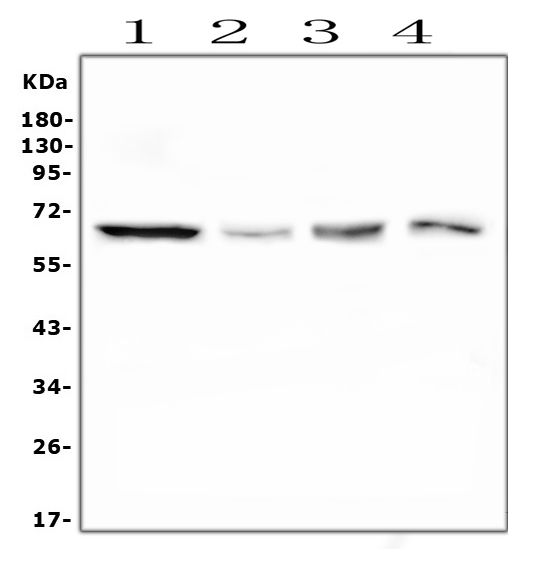
Western blot analysis of PLK1 using anti-PLK1 antibody (M00182).
Electrophoresis was performed on a 5-20% SDS-PAGE gel at 70V (Stacking gel) / 90V (Resolving gel) for 2-3 hours. The sample well of each lane was loaded with 50ug of sample under reducing conditions.
Lane 1: human Hela whole cell lysates
Lane 2: monkey COS-7 whole cell lysates
Lane 3: human Raji whole cell lysates
Lane 4: human U2OS whole cell lysates
After Electrophoresis, proteins were transferred to a Nitrocellulose membrane at 150mA for 50-90 minutes. Blocked the membrane with 5% Non-fat Milk/ TBS for 1.5 hour at RT. The membrane was incubated with mouse anti-PLK1 antigen affinity purified monoclonal antibody (Catalog # M00182) at 0.5 μg/mL overnight at 4°C, then washed with TBS-0.1%Tween 3 times with 5 minutes each and probed with a goat anti-mouse IgG-HRP secondary antibody at a dilution of 1:10000 for 1.5 hour at RT. The signal is developed using an Enhanced Chemiluminescent detection (ECL) kit (Catalog # EK1001) with Tanon 5200 system. A specific band was detected for PLK1 at approximately 68KD. The expected band size for PLK1 is at 68KD.
Specific Publications For Anti-PLK1 Antibody Picoband® (monoclonal, 6D11) (M00182)
Loading publications
Recommended Resources
Here are featured tools and databases that you might find useful.
- Boster's Pathways Library
- Protein Databases
- Bioscience Research Protocol Resources
- Data Processing & Analysis Software
- Photo Editing Software
- Scientific Literature Resources
- Research Paper Management Tools
- Molecular Biology Software
- Primer Design Tools
- Bioinformatics Tools
- Phylogenetic Tree Analysis
Customer Reviews
Have you used Anti-PLK1 Antibody Picoband® (monoclonal, 6D11)?
Share your experimental results or join a short interview to earn up to $1,000 in product credits or other rewards.
0 Reviews For Anti-PLK1 Antibody Picoband® (monoclonal, 6D11)
Customer Q&As
Have a question?
Find answers in Q&As, reviews.
Can't find your answer?
Submit your question
5 Customer Q&As for Anti-PLK1 Antibody Picoband® (monoclonal, 6D11)
Question
We have observed staining in human lung. What should we do? Is anti-PLK1 antibody (monoclonal, 6D11) supposed to stain lung positively?
Verified Customer
Verified customer
Asked: 2020-01-29
Answer
From what I have seen in literature lung does express PLK1. From what I have seen in Uniprot.org, PLK1 is expressed in testis, placenta, lung, colon lung, lymphoblast, cervix carcinoma, cervix carcinoma erythroleukemia, among other tissues. Regarding which tissues have PLK1 expression, here are a few articles citing expression in various tissues:
Cervix carcinoma, Pubmed ID: 18669648, 18691976, 20068231
Cervix carcinoma, and Erythroleukemia, Pubmed ID: 23186163
Colon, and Lung, Pubmed ID: 15489334
Lung, Pubmed ID: 8127874
Lymphoblast, Pubmed ID: 14654843
Placenta, Pubmed ID: 8018557, 7902533
Boster Scientific Support
Answered: 2020-01-29
Question
We want using your anti-PLK1 antibody (monoclonal, 6D11) for negative regulation of transcription by rna polymerase ii studies. Has this antibody been tested with western blotting on human hela? We would like to see some validation images before ordering.
T. Mangal
Verified customer
Asked: 2019-09-18
Answer
We appreciate your inquiry. This M00182 anti-PLK1 antibody (monoclonal, 6D11) is tested on human hela, hela whole cell lysates, u2os whole cell lysates. It is guaranteed to work for IHC-P, WB in human, monkey, mouse, rat. Our Boster guarantee will cover your intended experiment even if the sample type has not been be directly tested.
Boster Scientific Support
Answered: 2019-09-18
Question
We are currently using anti-PLK1 antibody (monoclonal, 6D11) M00182 for mouse tissue, and we are happy with the WB results. The species of reactivity given in the datasheet says human, monkey, mouse, rat. Is it true that the antibody can work on monkey tissues as well?
S. Huang
Verified customer
Asked: 2018-03-12
Answer
The anti-PLK1 antibody (monoclonal, 6D11) (M00182) has not been validated for cross reactivity specifically with monkey tissues, but there is a good chance of cross reactivity. We have an innovator award program that if you test this antibody and show it works in monkey you can get your next antibody for free. Please contact me if I can help you with anything.
Boster Scientific Support
Answered: 2018-03-12
Question
My boss were satisfied with the WB result of your anti-PLK1 antibody (monoclonal, 6D11). However we have observed positive staining in cervix carcinoma nucleus. chromosome using this antibody. Is that expected? Could you tell me where is PLK1 supposed to be expressed?
G. Wu
Verified customer
Asked: 2016-06-20
Answer
According to literature, cervix carcinoma does express PLK1. Generally PLK1 expresses in nucleus. chromosome, centromere,. Regarding which tissues have PLK1 expression, here are a few articles citing expression in various tissues:
Cervix carcinoma, Pubmed ID: 18669648, 18691976, 20068231
Cervix carcinoma, and Erythroleukemia, Pubmed ID: 23186163
Colon, and Lung, Pubmed ID: 15489334
Lung, Pubmed ID: 8127874
Lymphoblast, Pubmed ID: 14654843
Placenta, Pubmed ID: 8018557, 7902533
Boster Scientific Support
Answered: 2016-06-20
Question
We bought anti-PLK1 antibody (monoclonal, 6D11) for WB on placenta in a previous project. I am using rat, and We are going to use the antibody for IHC-P next. My lab would like examining placenta as well as lymphoblast in our next experiment. Could you please give me some suggestion on which antibody would work the best for IHC-P?
F. Carter
Verified customer
Asked: 2013-03-01
Answer
I have checked the website and datasheets of our anti-PLK1 antibody (monoclonal, 6D11) and I see that M00182 has been tested on rat in both WB and IHC-P. Thus M00182 should work for your application. Our Boster satisfaction guarantee will cover this product for IHC-P in rat even if the specific tissue type has not been validated. We do have a comprehensive range of products for IHC-P detection and you can check out our website bosterbio.com to find out more information about them.
Boster Scientific Support
Answered: 2013-03-01
