Click image to see more details
-

-
-
-
-
+1
Product Info Summary
| SKU: | A00128-4 |
|---|---|
| Size: | 100 μg/vial |
| Reactive Species: | Human, Rat |
| Host: | Rabbit |
| Application: | ELISA, Flow Cytometry, IF, ICC, WB |
Customers Who Bought This Also Bought

Product info
Product Name
Anti-TRPV1 Antibody Picoband®
SKU/Catalog Number
A00128-4
Size
100 μg/vial
Form
Lyophilized
Description
Boster Bio Anti-TRPV1 Antibody Picoband® catalog # A00128-4. Tested in ELISA, Flow Cytometry, IF, ICC, WB applications. This antibody reacts with Human, Rat. The brand Picoband indicates this is a premium antibody that guarantees superior quality, high affinity, and strong signals with minimal background in Western blot applications. Only our best-performing antibodies are designated as Picoband, ensuring unmatched performance.
Storage & Handling
At -20°C for one year from date of receipt. After reconstitution, at 4°C for one month. It can also be aliquotted and stored frozen at -20°C for six months. Avoid repeated freezing and thawing.
Cite This Product
Anti-TRPV1 Antibody Picoband® (Boster Biological Technology, Pleasanton CA, USA, Catalog # A00128-4)
Host
Rabbit
Contents
Each vial contains 4 mg Trehalose, 0.9 mg NaCl, 0.2 mg Na2HPO4.
Clonality
Polyclonal
Isotype
Rabbit IgG
Immunogen
E.coli-derived human TRPV1 recombinant protein (Position: E107-A800).
Cross-reactivity
No cross-reactivity with other proteins.
Reactive Species
A00128-4 is reactive to TRPV1 in Human, Rat
Observed Molecular Weight
95 kDa
Calculated molecular weight
95.0 kDa
Background of TRPV1
The transient receptor potential cation channel, subfamily V, member 1(TRPV1), also known as the capsaicin receptor is a protein which in humans is encoded by the TRPV1 gene. TRPV1(also called Vanilloid receptor type 1) is a ligand-gated nonselective cation channel that is considered to be an important integrator of various pain stimuli such as endogenous lipids, capsaicin, heat, and low pH. In addition to expression in primary afferents, TRPV1 is also expressed in the CNS. Cui M et al.(2006) demonstrate that TRPV1 receptors in the CNS plays an important role in pain mediated by central sensitization. And the significant CNS penetration is necessary for a TRPV1 antagonist to produce broad-spectrum analgesia. And TRPV1 also participates in normal bladder function and is essential for normal mechanically evoked purinergic signaling by the urothelium.
Antibody Validation
Boster validates all antibodies on WB, IHC, ICC, Immunofluorescence, and ELISA with known positive control and negative samples to ensure specificity and high affinity, including thorough antibody incubations.
Application & Images
Applications
A00128-4 is guaranteed for ELISA, Flow Cytometry, IF, ICC, WB Boster Guarantee
Recommend Dilution
| Application | Dilution | Species |
|---|---|---|
| Western blot | 0.25-0.5 μg/ml | Human, Rat |
| Immunocytochemistry/Immunofluorescence | 5 μg/ml | Human |
| Flow Cytometry (Fixed) | 1-3 μg/1x106 cells | Human |
| ELISA | 0.1-0.5 μg/ml | - |
Tested application
Suggested blocking solution with 5% non-fat milk or BSA; (*)Recommended protein loading: 20-40 µg per lane
Validation Images & Assay Conditions
Click image to see more details
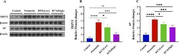
Expression of TRPV1 and SP. (A) The expression of TRPV1 in the L6-S1 DRG and the expression of SP in the L6-S1 spinal cord. Statistical results analysis of TRPV1 in the L6-S1 DRG (B) and SP in the L6-S1 spinal cord (C) between groups. Differences between groups were compared by a one-factor ANOVA. Data represents the mean ± SD, *Significant difference compared with the Control group or prostatitis group, *p < 0.05; ***p < 0.001; ****p < 0.0001; “ns” indicates p > 0.05.
Index in PubMed under a CC BY license. PMID: 40070569

Click image to see more details
Western blot analysis of TRPV1 using anti-TRPV1 antibody (A00128-4).
Electrophoresis was performed on a 5-20% SDS-PAGE gel at 70V (Stacking gel) / 90V (Resolving gel) for 2-3 hours. The sample well of each lane was loaded with 30 ug of sample under reducing conditions.
Lane 1: human 293T whole cell lysates,
Lane 2: human HepG2 whole cell lysates,
Lane 3: rat liver tissue lysates,
Lane 4: rat PC-12 whole cell lysates.
After electrophoresis, proteins were transferred to a nitrocellulose membrane at 150 mA for 50-90 minutes. Blocked the membrane with 5% non-fat milk/TBS for 1.5 hour at RT. The membrane was incubated with rabbit anti-TRPV1 antigen affinity purified polyclonal antibody (Catalog # A00128-4) at 0.5 μg/mL overnight at 4°C, then washed with TBS-0.1%Tween 3 times with 5 minutes each and probed with a goat anti-rabbit IgG-HRP secondary antibody at a dilution of 1:5000 for 1.5 hour at RT. The signal is developed using an Enhanced Chemiluminescent detection (ECL) kit (Catalog # EK1002) with Tanon 5200 system. A specific band was detected for TRPV1 at approximately 95 kDa. The expected band size for TRPV1 is at 95 kDa.

Click image to see more details
IF analysis of TRPV1 using anti-TRPV1 antibody (A00128-4).
TRPV1 was detected in an immunocytochemical section of T-47D cells. Enzyme antigen retrieval was performed using IHC enzyme antigen retrieval reagent (AR0022) for 15 mins. The cells were blocked with 10% goat serum. And then incubated with 5 μg/mL rabbit anti-TRPV1 Antibody (A00128-4) overnight at 4°C. DyLight®488 Conjugated Goat Anti-Rabbit IgG (BA1127) was used as secondary antibody at 1:100 dilution and incubated for 30 minutes at 37°C. The section was counterstained with DAPI. Visualize using a fluorescence microscope and filter sets appropriate for the label used.

Click image to see more details
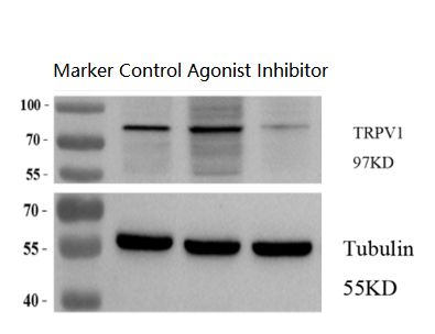
Western blot analysis of TRPV1 using anti-TRPV1 antibody (A00128-4).
Electrophoresis was performed on a 5-20% SDS-PAGE gel at 70V (Stacking gel) / 90V (Resolving gel) for 2-3 hours. The sample well of each lane was loaded with 30 ug of sample under reducing conditions.
Lane 1: Control-rat MADB106 breast cancer cells,
Lane 2: Agonist treatment group-rat MADB106 breast cancer cells,
Lane 3: Inhibitor treatment group-rat MADB106 breast cancer cells.
After electrophoresis, proteins were transferred to a nitrocellulose membrane at 150 mA for 50-90 minutes. Blocked the membrane with 5% non-fat milk/TBS for 1.5 hour at RT. The membrane was incubated with rabbit anti-TRPV1 antigen affinity purified polyclonal antibody (Catalog # A00128-4) at 1:2000 overnight at 4°C, then washed with TBS-0.1%Tween 3 times with 5 minutes each and probed with a goat anti-rabbit IgG-HRP secondary antibody at a dilution of 1:10,000 for 1 hour at RT. The signal is developed using an Enhanced Chemiluminescent detection (ECL) kit (Catalog # EK1002) with Tanon 5200 system. The expected band size for TRPV1 is at 95 kDa.

Click image to see more details
Flow Cytometry analysis of U87 cells using anti-TRPV1 antibody (A00128-4).
Overlay histogram showing U87 cells stained with A00128-4 (Blue line). To facilitate intracellular staining, cells were fixed with 4% paraformaldehyde and permeabilized with permeabilization buffer. The cells were blocked with 10% normal goat serum. And then incubated with rabbit anti-TRPV1 Antibody (A00128-4, 1 μg/1x106 cells) for 30 min at 20°C. DyLight®488 conjugated goat anti-rabbit IgG (BA1127, 5-10 μg/1x106 cells) was used as secondary antibody for 30 minutes at 20°C. Isotype control antibody (Green line) was rabbit IgG (1 μg/1x106) used under the same conditions. Unlabelled sample without incubation with primary antibody and secondary antibody (Red line) was used as a blank control.
Specific Publications For Anti-TRPV1 Antibody Picoband® (A00128-4)
Loading publications
Recommended Resources
Here are featured tools and databases that you might find useful.
- Boster's Pathways Library
- Protein Databases
- Bioscience Research Protocol Resources
- Data Processing & Analysis Software
- Photo Editing Software
- Scientific Literature Resources
- Research Paper Management Tools
- Molecular Biology Software
- Primer Design Tools
- Bioinformatics Tools
- Phylogenetic Tree Analysis
Customer Reviews
Have you used Anti-TRPV1 Antibody Picoband®?
Share your experimental results or join a short interview to earn up to $1,000 in product credits or other rewards.
1 Reviews For Anti-TRPV1 Antibody Picoband®
The Anti-TRPV1 antibody (A00128-4) showed clear and correctly positioned bands in WB of MADB106 rat breast cancer cells, with increased TRPV1 expression upon agonist treatment and decreased expression upon inhibitor treatment.
Excellent
| SKU | A00128-4 |
|---|---|
| Application | Western blot |
| Sample | Rat MADB106 breast cancer cells |
| Sample Processing Description | Normally cultured MADB106 cells, MADB106 cells treated with an agonist, MADB106 cells treated with an inhibitor |
| Other Reagents | RIPA lysis buffer (Cat. No. AR0102, Bosterbio, Wuhan) Protease inhibitor, Running buffer, Transfer buffer, Blocking buffer |
| Primary Antibody | TRPV1 Antibody Picoband® |
| Primary Incubation | 1:2000, overnight at 4 ℃ |
| Secondary Antibody | 1:10,000, HRP-conjugated Goat Anti-Rabbit IgG |
| Secondary Incubation | 1h at 37℃ |
| Detection | Substrate: ECL substrate, Imaging system:ChemiDoc MP |
| Results Summary | The role of TRPV1 in breast cancer research is highly complex, varying with cell type, tumor subtype, and tumor microenvironment, and in some cases even showing contradictory effects. The current consensus is that TRPV1 is not only a potential therapeutic target for breast cancer but also an important prognostic and regulatory factor. Experimental results show that TRPV1 protein expression markedly increases in cells treated with an agonist, while it decreases in cells treated with an inhibitor. |
Xinshuo Wang, Xi’an Jiaotong University
Verified customer
Submitted 2026-02-12
Customer Q&As
Have a question?
Find answers in Q&As, reviews.
Can't find your answer?
Submit your question
