Click image to see more details
Product Info Summary
| SKU: | A01626-1 |
|---|---|
| Size: | 100 μg/vial |
| Reactive Species: | Human, Mouse, Rat |
| Host: | Rabbit |
| Application: | ELISA, WB |
Customers Who Bought This Also Bought

Product info
Product Name
Anti-Tryptophan Hydroxylase/TPH1 Antibody Picoband®
SKU/Catalog Number
A01626-1
Size
100 μg/vial
Form
Lyophilized
Description
Boster Bio Anti-Tryptophan Hydroxylase/TPH1 Antibody Picoband® catalog # A01626-1. Tested in ELISA, WB applications. This antibody reacts with Human, Mouse, Rat. The brand Picoband indicates this is a premium antibody that guarantees superior quality, high affinity, and strong signals with minimal background in Western blot applications. Only our best-performing antibodies are designated as Picoband, ensuring unmatched performance.
Storage & Handling
Store at -20˚C for one year from date of receipt. After reconstitution, at 4˚C for one month. It can also be aliquotted and stored frozen at -20˚C for six months. Avoid repeated freeze-thaw cycles.
Cite This Product
Anti-Tryptophan Hydroxylase/TPH1 Antibody Picoband® (Boster Biological Technology, Pleasanton CA, USA, Catalog # A01626-1)
Host
Rabbit
Contents
Each vial contains 4mg Trehalose, 0.9mg NaCl, 0.2mg Na2HPO4, 0.05mg NaN3.
Clonality
Polyclonal
Isotype
Rabbit IgG
Immunogen
E. coli-derived human Tryptophan Hydroxylase recombinant protein (Position: M1-K111).
Cross-reactivity
No cross-reactivity with other proteins.
Reactive Species
A01626-1 is reactive to TPH1 in Human, Mouse, Rat
Observed Molecular Weight
51 kDa
Calculated molecular weight
51.0 kDa
Background of TPH1
Tryptophan hydroxylase 1 (TPH1) is an isoenzyme of tryptophan hydroxylase which in humans is encoded by the TPH1 gene. This gene encodes a member of the aromatic amino acid hydroxylase family. The encoded protein catalyzes the first and rate limiting step in the biosynthesis of serotonin, an important hormone and neurotransmitter. Mutations in this gene have been associated with an elevated risk for a variety of diseases and disorders, including schizophrenia, somatic anxiety, anger-related traits, bipolar disorder, suicidal behavior, addictions, and others.
Antibody Validation
Boster validates all antibodies on WB, IHC, ICC, Immunofluorescence, and ELISA with known positive control and negative samples to ensure specificity and high affinity, including thorough antibody incubations.
Application & Images
Applications
A01626-1 is guaranteed for ELISA, WB Boster Guarantee
Recommend Dilution
| Application | Dilution | Species |
|---|---|---|
| Western blot | 0.1-0.5μg/ml | |
| ELISA | 0.1-0.5μg/ml |
Tested application
Suggested blocking solution with 5% non-fat milk or BSA; (*)Recommended protein loading: 20-40 µg per lane
Validation Images & Assay Conditions
Click image to see more details
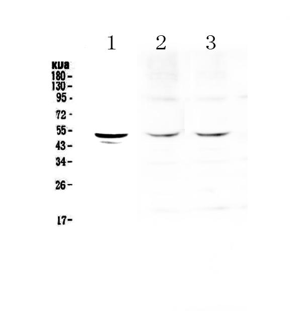
Western blot analysis of Tryptophan Hydroxylase using anti-Tryptophan Hydroxylase antibody (A01626-1).
Electrophoresis was performed on a 5-20% SDS-PAGE gel at 70V (Stacking gel) / 90V (Resolving gel) for 2-3 hours. The sample well of each lane was loaded with 50ug of sample under reducing conditions.
Lane 1: human SGC-7901 whole cell lysates,
Lane 2: rat heart tissue lysates,
Lane 3: mouse heart tissue lysates.
After Electrophoresis, proteins were transferred to a Nitrocellulose membrane at 150mA for 50-90 minutes. Blocked the membrane with 5% Non-fat Milk/ TBS for 1.5 hour at RT. The membrane was incubated with rabbit anti-Tryptophan Hydroxylase antigen affinity purified polyclonal antibody (Catalog # A01626-1) at 0.5 μg/mL overnight at 4°C, then washed with TBS-0.1%Tween 3 times with 5 minutes each and probed with a goat anti-rabbit IgG-HRP secondary antibody at a dilution of 1:10000 for 1.5 hour at RT. The signal is developed using an Enhanced Chemiluminescent detection (ECL) kit (Catalog # EK1002) with Tanon 5200 system. A specific band was detected for Tryptophan Hydroxylase at approximately 51KD. The expected band size for Tryptophan Hydroxylase is at 51KD.

Click image to see more details
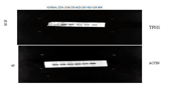
Western blot analysis of Tryptophan Hydroxylase using anti-Tryptophan Hydroxylase antibody (A01626-1).
Electrophoresis was performed on a 5-20% SDS-PAGE gel at 70V (Stacking gel) / 90V (Resolving gel) for 2-3 hours. The sample well of each lane was loaded with 50ug of sample under reducing conditions.
Lane 1: normal group-rat colon tissue lysates,
Lane 2: control group-model rat colon tissue lysates,
Lane 3: low dose Chinese medicine group-model rat colon tissue lysates,
Lane 4: medium dose Chinese medicine group-model rat colon tissue lysates,
Lane 5: high dose Chinese medicine group-model rat colon tissue lysates,
Lane 6: western medicine group-model rat colon tissue lysates.
After Electrophoresis, proteins were transferred to a Nitrocellulose membrane at 150mA for 50-90 minutes. Blocked the membrane with 5% Non-fat Milk/ TBS for 1.5 hour at RT. The membrane was incubated with rabbit anti-Tryptophan Hydroxylase antigen affinity purified polyclonal antibody (Catalog # A01626-1) at 1:1000 overnight at 4°C, then washed with TBS-0.1%Tween 3 times with 5 minutes each and probed with a goat anti-rabbit IgG-HRP secondary antibody at a dilution of 1:5000 for 1 hour at RT. The signal is developed using an Enhanced Chemiluminescent detection (ECL) kit (Catalog # EK1002) with ChemiDoc MP system. The expected band size for Tryptophan Hydroxylase is at 51KD.
Specific Publications For Anti-Tryptophan Hydroxylase/TPH1 Antibody Picoband® (A01626-1)
Loading publications
Recommended Resources
Here are featured tools and databases that you might find useful.
- Boster's Pathways Library
- Protein Databases
- Bioscience Research Protocol Resources
- Data Processing & Analysis Software
- Photo Editing Software
- Scientific Literature Resources
- Research Paper Management Tools
- Molecular Biology Software
- Primer Design Tools
- Bioinformatics Tools
- Phylogenetic Tree Analysis
Customer Reviews
Have you used Anti-Tryptophan Hydroxylase/TPH1 Antibody Picoband®?
Share your experimental results or join a short interview to earn up to $1,000 in product credits or other rewards.
1 Reviews For Anti-Tryptophan Hydroxylase/TPH1 Antibody Picoband®
WB results showed that TPH1 (A01626-1) was upregulated in the rat colon model group and reduced after Chinese medicine treatment, with the high-dose group showing the best effect, and the target bands were clear and specific.
Excellent
| SKU | A01626-1 |
|---|---|
| Application | Western Blot |
| Sample | rat colon tissue |
| Sample Processing Description | Rat colon tissues were lysed in RIPA buffer containing PMSF (100:1) for 10 min, centrifuged at 12,000 rpm for 15 min, and the supernatant was mixed with 5× loading buffer, denatured at 100°C for 10 min, and then loaded onto SDS-PAGE. |
| Other Reagents | 5% non-fat milk |
| Primary Antibody | Tryptophan Hydroxylase/TPH1 Antibody Picoband® |
| Primary Incubation | 1:1000, overnight at 4 ℃ |
| Secondary Antibody | HRP Conjugated AffiniPure Goat Anti-Rabbit IgG (H+L) (BA1054) |
| Secondary Incubation | 1:5000, 1 h in RT |
| Detection | Substrate: ECL substrate, Image system:ChemiDoc MP |
| Results Summary | The figure shows WB results of TPH1 and the internal control Actin in rat colon across different groups; expression was elevated in the model group and reduced after Chinese medicine treatment, with the high-dose group showing the best effect, and the target bands were clear and specific, indicating satisfactory results. |
Shiyu Zhang, Liaoning University of Traditional Chinese Medicine
Verified customer
Submitted 2026-03-04
Customer Q&As
Have a question?
Find answers in Q&As, reviews.
Can't find your answer?
Submit your question
1 Customer Q&As for Anti-Tryptophan Hydroxylase/TPH1 Antibody Picoband®
Question
We are currently using anti-Tryptophan Hydroxylase/TPH1 antibody A01626-1 for human tissue, and we are happy with the ELISA results. The species of reactivity given in the datasheet says human, mouse, rat. Is it likely that the antibody can work on horse tissues as well?
A. Lewis
Verified customer
Asked: 2015-12-31
Answer
The anti-Tryptophan Hydroxylase/TPH1 antibody (A01626-1) has not been tested for cross reactivity specifically with horse tissues, but there is a good chance of cross reactivity. We have an innovator award program that if you test this antibody and show it works in horse you can get your next antibody for free. Please contact me if I can help you with anything.
Boster Scientific Support
Answered: 2015-12-31
