Click image to see more details
Product Info Summary
| SKU: | RP1078 |
|---|---|
| Size: | 100 μg/vial |
| Reactive Species: | Human |
| Host: | Rabbit |
| Application: | WB |
Customers Who Bought This Also Bought

Product info
Product Name
Anti-TSG101 Antibody Picoband®
SKU/Catalog Number
RP1078
PB0550 is an alternative SKU for this antibody, used in previous lots.
Size
100 μg/vial
Form
Lyophilized
Description
Boster Bio Anti-TSG101 Antibody catalog # RP1078. Tested in WB applications. This antibody reacts with Human. The brand Picoband indicates this is a premium antibody that guarantees superior quality, high affinity, and strong signals with minimal background in Western blot applications. Only our best-performing antibodies are designated as Picoband, ensuring unmatched performance.
Storage & Handling
Store at -20˚C for one year from date of receipt. After reconstitution, at 4˚C for one month. It can also be aliquotted and stored frozen at -20˚C for six months. Avoid repeated freeze-thaw cycles.
Cite This Product
Anti-TSG101 Antibody Picoband® (Boster Biological Technology, Pleasanton CA, USA, Catalog # RP1078)
Host
Rabbit
Contents
Each vial contains 4 mg Trehalose, 0.9 mg NaCl and 0.2 mg Na2HPO4.
Clonality
Polyclonal
Isotype
Rabbit IgG
Immunogen
A synthetic peptide corresponding to a sequence at the C-terminus of human TSG101, identical to the related mouse and rat sequences.
Cross-reactivity
No cross-reactivity with other proteins
Reactive Species
RP1078 is reactive to TSG101 in Human
Observed Molecular Weight
44 kDa
Calculated molecular weight
43.9 kDa
Background of TSG101
TSG101, known as Tumor susceptibility gene 101, is mapped to 11p15. The protein encoded by this gene belongs to a group of apparently inactive homologs of ubiquitin-conjugating enzymes. The gene product contains a coiled-coil domain that interacts with stathmin, a cytosolic phosphoprotein implicated in tumorigenesis. And the protein may play a role in cell growth and differentiation and act as a negative growth regulator. In vitro steady-state expression of this tumor susceptibility gene appears to be important for maintenance of genomic stability and cell cycle regulation. Mutations and alternative splicing in this gene occur in high frequency in breast cancer and suggest that defects occur during breast cancer tumorigenesis and/or progression.
Antibody Validation
Boster validates all antibodies on WB, IHC, ICC, Immunofluorescence, and ELISA with known positive control and negative samples to ensure specificity and high affinity, including thorough antibody incubations.
Application & Images
Applications
RP1078 is guaranteed for WB Boster Guarantee
Recommend Dilution
| Application | Dilution | Species |
|---|---|---|
| Western blot | 0.1-0.5μg/ml | Human |
Tested application
Suggested blocking solution with 5% non-fat milk or BSA; (*)Recommended protein loading: 20-40 µg per lane
Validation Images & Assay Conditions
Click image to see more details
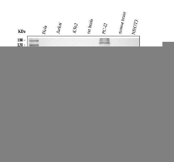
Western blot analysis of TSG101 using anti-TSG101 antibody (RP1078).
Electrophoresis was performed on a 10% SDS-PAGE gel at 80V (Stacking gel) / 120V (Resolving gel) for 2 hours. The sample well of each lane was loaded with 30 ug of sample under reducing conditions.
Lane 1: human Hela whole cell lysates,
Lane 2: human Jurkat whole cell lysates,
Lane 3: human K562 whole cell lysates,
Lane 4: rat brain tissue lysates,
Lane 5: rat PC-12 whole cell lysates,
Lane 6: mouse brain tissue lysates,
Lane 7: mouse NIH/3T3 whole cell lysates.
After electrophoresis, proteins were transferred to a nitrocellulose membrane at 150 mA for 50-90 minutes. Blocked the membrane with 5% non-fat milk/TBS for 1.5 hour at RT. The membrane was incubated with rabbit anti-TSG101 antigen affinity purified polyclonal antibody (RP1078) at 0.5 μg/mL overnight at 4°C, then washed with TBS-0.1%Tween 3 times with 5 minutes each and probed with a goat anti-rabbit IgG-HRP secondary antibody (Catalog # BA1054) at a dilution of 1:5000 for 1.5 hour at RT. The signal is developed using an ECL Plus Western Blotting Substrate (Catalog # AR1196-200) with Tanon 5200 system. A specific band was detected for TSG101 at approximately 44 kDa. The expected band size for TSG101 is at 44 kDa.
Specific Publications For Anti-TSG101 Antibody Picoband® (RP1078)
Loading publications
Recommended Resources
Here are featured tools and databases that you might find useful.
- Boster's Pathways Library
- Protein Databases
- Bioscience Research Protocol Resources
- Data Processing & Analysis Software
- Photo Editing Software
- Scientific Literature Resources
- Research Paper Management Tools
- Molecular Biology Software
- Primer Design Tools
- Bioinformatics Tools
- Phylogenetic Tree Analysis
Customer Reviews
Have you used Anti-TSG101 Antibody Picoband®?
Share your experimental results or join a short interview to earn up to $1,000 in product credits or other rewards.
0 Reviews For Anti-TSG101 Antibody Picoband®
Customer Q&As
Have a question?
Find answers in Q&As, reviews.
Can't find your answer?
Submit your question
4 Customer Q&As for Anti-TSG101 Antibody Picoband®
Question
Our team were happy with the WB result of your anti-TSG101 antibody. However we have observed positive staining in oocyte cytoplasm. using this antibody. Is that expected? Could you tell me where is TSG101 supposed to be expressed?
Verified Customer
Verified customer
Asked: 2019-12-20
Answer
From what I have seen in literature, oocyte does express TSG101. Generally TSG101 expresses in cytoplasm. Regarding which tissues have TSG101 expression, here are a few articles citing expression in various tissues:
Cervix carcinoma, and Erythroleukemia, Pubmed ID: 23186163
Eye, Pubmed ID: 15489334
Placenta, Pubmed ID: 9019400
Boster Scientific Support
Answered: 2019-12-20
Question
We are currently using anti-TSG101 antibody RP1078 for human tissue, and we are content with the WB results. The species of reactivity given in the datasheet says human, rat. Is it possible that the antibody can work on bovine tissues as well?
Verified Customer
Verified customer
Asked: 2019-05-16
Answer
The anti-TSG101 antibody (RP1078) has not been validated for cross reactivity specifically with bovine tissues, though there is a good chance of cross reactivity. We have an innovator award program that if you test this antibody and show it works in bovine you can get your next antibody for free. Please contact me if I can help you with anything.
Boster Scientific Support
Answered: 2019-05-16
Question
We have seen staining in rat eye. Do you have any suggestions? Is anti-TSG101 antibody supposed to stain eye positively?
Verified Customer
Verified customer
Asked: 2018-12-19
Answer
Based on literature eye does express TSG101. Based on Uniprot.org, TSG101 is expressed in oocyte, placenta, eye, cervix carcinoma erythroleukemia, among other tissues. Regarding which tissues have TSG101 expression, here are a few articles citing expression in various tissues:
Cervix carcinoma, and Erythroleukemia, Pubmed ID: 23186163
Eye, Pubmed ID: 15489334
Placenta, Pubmed ID: 9019400
Boster Scientific Support
Answered: 2018-12-19
Question
I was wanting to use using your anti-TSG101 antibody for viral life cycle studies. Has this antibody been tested with western blotting on hela whole cell lysate? We would like to see some validation images before ordering.
Verified Customer
Verified customer
Asked: 2017-11-28
Answer
Thank you for your inquiry. This RP1078 anti-TSG101 antibody is tested on rat brain tissue, cardiac muscle tissue, tissue lysate, hela whole cell lysate, smmc whole cell lysate. It is guaranteed to work for WB in human, rat. Our Boster guarantee will cover your intended experiment even if the sample type has not been be directly tested.
Boster Scientific Support
Answered: 2017-11-28
