Click image to see more details
-

-
-
-
-
+1
Product Info Summary
| SKU: | A03213-2 |
|---|---|
| Size: | 100 μg/vial |
| Reactive Species: | Human, Monkey, Mouse, Rat |
| Host: | Rabbit |
| Application: | ELISA, Flow Cytometry, IHC, WB |
Customers Who Bought This Also Bought

Product info
Product Name
Anti-UBA6 Antibody Picoband®
SKU/Catalog Number
A03213-2
Size
100 μg/vial
Form
Lyophilized
Description
Boster Bio Anti-UBA6 Antibody Picoband® catalog # A03213-2. Tested in ELISA, Flow Cytometry, IHC, WB applications. This antibody reacts with Human, Monkey, Mouse, Rat. The brand Picoband indicates this is a premium antibody that guarantees superior quality, high affinity, and strong signals with minimal background in Western blot applications. Only our best-performing antibodies are designated as Picoband, ensuring unmatched performance.
Storage & Handling
At -20°C for one year from date of receipt. After reconstitution, at 4°C for one month. It can also be aliquotted and stored frozen at -20°C for six months. Avoid repeated freezing and thawing.
Cite This Product
Anti-UBA6 Antibody Picoband® (Boster Biological Technology, Pleasanton CA, USA, Catalog # A03213-2)
Host
Rabbit
Contents
Each vial contains 4 mg Trehalose, 0.9 mg NaCl, 0.2 mg Na2HPO4.
Clonality
Polyclonal
Isotype
Rabbit IgG
Immunogen
E.coli-derived human UBA6 recombinant protein (Position: I85-D1033).
Cross-reactivity
No cross-reactivity with other proteins.
Reactive Species
A03213-2 is reactive to UBA6 in Human, Monkey, Mouse, Rat
Observed Molecular Weight
118 kDa
Calculated molecular weight
118.0 kDa
Background of UBA6
Modification of proteins with ubiquitin (UBB; MIM 191339) or ubiquitin-like proteins controls many signaling networks and requires a ubiquitin-activating enzyme (E1), a ubiquitin conjugating enzyme (E2), and a ubiquitin protein ligase (E3). UBE1L2 is an E1 enzyme that initiates the activation and conjugation of ubiquitin-like proteins.
Antibody Validation
Boster validates all antibodies on WB, IHC, ICC, Immunofluorescence, and ELISA with known positive control and negative samples to ensure specificity and high affinity, including thorough antibody incubations.
Application & Images
Applications
A03213-2 is guaranteed for ELISA, Flow Cytometry, IHC, WB Boster Guarantee
Recommend Dilution
| Application | Dilution | Species |
|---|---|---|
| Western blot | 0.25-0.5 μg/ml | Human, Monkey, Mouse, Rat |
| Immunohistochemistry(Paraffin-embedded Section) | 2-5 μg/ml | Human |
| Flow Cytometry (Fixed) | 1-3 μg/1x106 cells | Human |
| ELISA | 0.1-0.5 μg/ml | - |
Tested application
Suggested blocking solution with 5% non-fat milk or BSA; (*)Recommended protein loading: 20-40 µg per lane
Use TE buffer pH 9.0 for antigen retrieval; (*) citrate buffer pH 6.0 is an alternative.
Validation Images & Assay Conditions
Click image to see more details
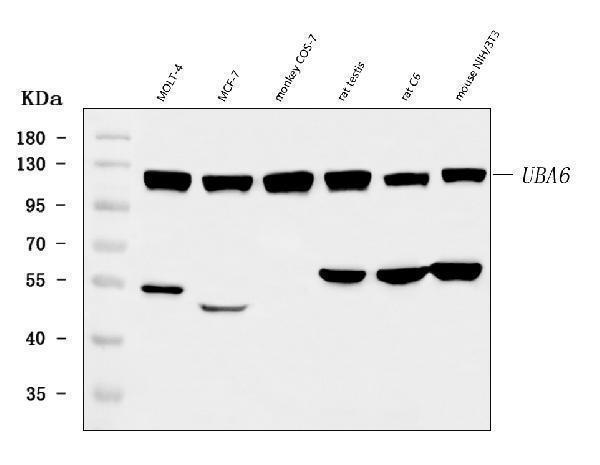
Western blot analysis of UBA6 using anti-UBA6 antibody (A03213-2).
Electrophoresis was performed on a 5-20% SDS-PAGE gel at 70V (Stacking gel) / 90V (Resolving gel) for 2-3 hours. The sample well of each lane was loaded with 30 ug of sample under reducing conditions.
Lane 1: human MOLT-4 whole cell lysates,
Lane 2: human MCF-7 whole cell lysates,
Lane 3: monkey COS-7 whole cell lysates,
Lane 4: rat testis tissue lysates,
Lane 5: rat C6 whole cell lysates,
Lane 6: mouse NIH/3T3 whole cell lysates.
After electrophoresis, proteins were transferred to a nitrocellulose membrane at 150 mA for 50-90 minutes. Blocked the membrane with 5% non-fat milk/TBS for 1.5 hour at RT. The membrane was incubated with rabbit anti-UBA6 antigen affinity purified polyclonal antibody (Catalog # A03213-2) at 0.5 μg/mL overnight at 4°C, then washed with TBS-0.1%Tween 3 times with 5 minutes each and probed with a goat anti-rabbit IgG-HRP secondary antibody at a dilution of 1:5000 for 1.5 hour at RT. The signal is developed using an Enhanced Chemiluminescent detection (ECL) kit (Catalog # EK1002) with Tanon 5200 system. A specific band was detected for UBA6 at approximately 118 kDa. The expected band size for UBA6 is at 118 kDa.

Click image to see more details
IHC analysis of UBA6 using anti-UBA6 antibody (A03213-2).
UBA6 was detected in a paraffin-embedded section of human cervical cancer tissue. Heat mediated antigen retrieval was performed in EDTA buffer (pH 8.0, epitope retrieval solution). The tissue section was blocked with 10% goat serum. The tissue section was then incubated with 2 μg/ml rabbit anti-UBA6 Antibody (A03213-2) overnight at 4°C. Peroxidase Conjugated Goat Anti-rabbit IgG was used as secondary antibody and incubated for 30 minutes at 37°C. The tissue section was developed using HRP Conjugated Rabbit IgG Super Vision Assay Kit (Catalog # SV0002) with DAB as the chromogen.

Click image to see more details
IHC analysis of UBA6 using anti-UBA6 antibody (A03213-2).
UBA6 was detected in a paraffin-embedded section of human lung squamous cell carcinoma tissue. Heat mediated antigen retrieval was performed in EDTA buffer (pH 8.0, epitope retrieval solution). The tissue section was blocked with 10% goat serum. The tissue section was then incubated with 2 μg/ml rabbit anti-UBA6 Antibody (A03213-2) overnight at 4°C. Peroxidase Conjugated Goat Anti-rabbit IgG was used as secondary antibody and incubated for 30 minutes at 37°C. The tissue section was developed using HRP Conjugated Rabbit IgG Super Vision Assay Kit (Catalog # SV0002) with DAB as the chromogen.

Click image to see more details
IHC analysis of UBA6 using anti-UBA6 antibody (A03213-2).
UBA6 was detected in a paraffin-embedded section of human ovarian cancer tissue. Heat mediated antigen retrieval was performed in EDTA buffer (pH 8.0, epitope retrieval solution). The tissue section was blocked with 10% goat serum. The tissue section was then incubated with 2 μg/ml rabbit anti-UBA6 Antibody (A03213-2) overnight at 4°C. Peroxidase Conjugated Goat Anti-rabbit IgG was used as secondary antibody and incubated for 30 minutes at 37°C. The tissue section was developed using HRP Conjugated Rabbit IgG Super Vision Assay Kit (Catalog # SV0002) with DAB as the chromogen.

Click image to see more details
Flow Cytometry analysis of PC-3 cells using anti-UBA6 antibody (A03213-2).
Overlay histogram showing PC-3 cells stained with A03213-2 (Blue line). To facilitate intracellular staining, cells were fixed with 4% paraformaldehyde and permeabilized with permeabilization buffer. The cells were blocked with 10% normal goat serum. And then incubated with rabbit anti-UBA6 Antibody (A03213-2, 1 μg/1x106 cells) for 30 min at 20°C. DyLight®488 conjugated goat anti-rabbit IgG (BA1127, 5-10 μg/1x106 cells) was used as secondary antibody for 30 minutes at 20°C. Isotype control antibody (Green line) was rabbit IgG (1 μg/1x106) used under the same conditions. Unlabelled sample without incubation with primary antibody and secondary antibody (Red line) was used as a blank control.
Specific Publications For Anti-UBA6 Antibody Picoband® (A03213-2)
Loading publications
Recommended Resources
Here are featured tools and databases that you might find useful.
- Boster's Pathways Library
- Protein Databases
- Bioscience Research Protocol Resources
- Data Processing & Analysis Software
- Photo Editing Software
- Scientific Literature Resources
- Research Paper Management Tools
- Molecular Biology Software
- Primer Design Tools
- Bioinformatics Tools
- Phylogenetic Tree Analysis
Customer Reviews
Have you used Anti-UBA6 Antibody Picoband®?
Share your experimental results or join a short interview to earn up to $1,000 in product credits or other rewards.
1 Reviews For Anti-UBA6 Antibody Picoband®
The antibody performs efficiently and specifically, with very few nonspecific bands.
Excellent
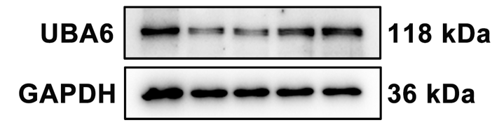
| SKU | A03213-2 |
|---|---|
| Application | Western Blot |
| Sample | MCF-7 cell |
| Sample Processing Description | Cells were directly lysed in NP-40 buffer, mixed with loading buffer at the appropriate ratio, and denatured by heating at 98 °C. Then, 20 µL of protein sample was loaded per lane onto SDS-PAGE. |
| Primary Antibody | UBA6 Antibody |
| Primary Incubation | 1:1000, overnight at 4 °C |
| Blocking Agent | 5% Non-fat milk |
| Secondary Antibody | HRP-conjugated Goat Anti-Rabbit IgG |
| Secondary Incubation | Incubate at room temperature for 1 hour |
| Detection | Signal was developed using ECL substrate on a Tanon system. |
| Results Summary | The antibody performs efficiently and specifically, with very few nonspecific bands. |
Xinyu Xiao, Peking University Health Science Center
Verified customer
Submitted 2025-09-25
Customer Q&As
Have a question?
Find answers in Q&As, reviews.
Can't find your answer?
Submit your question
