Click image to see more details
Product Info Summary
| SKU: | A00993-3 |
|---|---|
| Size: | 100 μg/vial |
| Reactive Species: | Human, Mouse, Rat |
| Host: | Rabbit |
| Application: | Flow Cytometry, WB |
Customers Who Bought This Also Bought

Product info
Product Name
Anti-uPA Receptor/PLAUR Antibody Picoband®
SKU/Catalog Number
A00993-3
Size
100 μg/vial
Form
Lyophilized
Description
Boster Bio Anti-uPA Receptor/PLAUR Antibody Picoband® catalog # A00993-3. Tested in Flow Cytometry, WB applications. This antibody reacts with Human, Mouse, Rat. The brand Picoband indicates this is a premium antibody that guarantees superior quality, high affinity, and strong signals with minimal background in Western blot applications. Only our best-performing antibodies are designated as Picoband, ensuring unmatched performance.
Storage & Handling
Store at -20˚C for one year from date of receipt. After reconstitution, at 4˚C for one month. It can also be aliquotted and stored frozen at -20˚C for six months. Avoid repeated freeze-thaw cycles.
Cite This Product
Anti-uPA Receptor/PLAUR Antibody Picoband® (Boster Biological Technology, Pleasanton CA, USA, Catalog # A00993-3)
Host
Rabbit
Contents
Each vial contains 4 mg Trehalose, 0.9 mg NaCl and 0.2 mg Na2HPO4.
Clonality
Polyclonal
Isotype
Rabbit IgG
Immunogen
A synthetic peptide corresponding to a sequence at the C-terminus of human uPA Receptor/PLAUR.
Cross-reactivity
No cross-reactivity with other proteins.
Reactive Species
A00993-3 is reactive to PLAUR in Human, Mouse, Rat
Observed Molecular Weight
39 kDa
Calculated molecular weight
37.0 kDa
Background of PLAUR
PLAUR (PLASMINOGEN ACTIVATOR RECEPTOR, UROKINASE-TYPE), also known as UPAR or CD87, is multidomain glycoprotein tethered to the cell membrane with a glycosylphosphotidylinositol (GPI) anchor. PLAUR consists of three different domains of the Ly-6/uPAR/alpha-neurotoxin family. PLAUR is originally identified as a saturable binding site for urokinase on the cell surface. And the gene plays an important role in many normal as well as pathologic processes. The PLAUR gene is localized to 19q13.31. PLAUR is a part of the plasminogen activation system, which in the healthy body is involved in tissue reorganization events such as mammary gland involution and wound healing. PLAUR binds urokinase and thus restricts plasminogen activation to the immediate vicinity of the cell membrane. Thus it seems to be an important player in the regulation of this process. In human coronary artery vascular smooth muscle cells, UPA stimulates cell migration via a UPAR signaling complex containing TYK2 and phosphatidylinositol 3-kinase.
Antibody Validation
Boster validates all antibodies on WB, IHC, ICC, Immunofluorescence, and ELISA with known positive control and negative samples to ensure specificity and high affinity, including thorough antibody incubations.
Application & Images
Applications
A00993-3 is guaranteed for Flow Cytometry, WB Boster Guarantee
Recommend Dilution
| Application | Dilution | Species |
|---|---|---|
| Western blot | 0.1-0.5μg/ml | |
| Flow Cytometry (Fixed) | 1-3μg/1x106 cells106 cells |
Tested application
Suggested blocking solution with 5% non-fat milk or BSA; (*)Recommended protein loading: 20-40 µg per lane
Validation Images & Assay Conditions
Click image to see more details
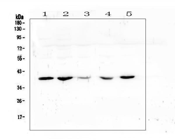
Western blot analysis of uPA Receptor using anti-uPA Receptor antibody (A00993-3).
Electrophoresis was performed on a 5-20% SDS-PAGE gel at 70V (Stacking gel) / 90V (Resolving gel) for 2-3 hours. The sample well of each lane was loaded with 50ug of sample under reducing conditions.
Lane 1: human placenta tissue lysate,
Lane 2: human U20S whole cell lysate,
Lane 3: human A431 whole cell lysate,
Lane 4: human Hela whole cell lysate,
Lane 5: human A549 whole cell lysate.
After Electrophoresis, proteins were transferred to a Nitrocellulose membrane at 150mA for 50-90 minutes. Blocked the membrane with 5% Non-fat Milk/ TBS for 1.5 hour at RT. The membrane was incubated with rabbit anti-uPA Receptor antigen affinity purified polyclonal antibody (Catalog # A00993-3) at 0.5 μg/mL overnight at 4°C, then washed with TBS-0.1%Tween 3 times with 5 minutes each and probed with a goat anti-rabbit IgG-HRP secondary antibody at a dilution of 1:10000 for 1.5 hour at RT. The signal is developed using an Enhanced Chemiluminescent detection (ECL) kit (Catalog # EK1002) with Tanon 5200 system. A specific band was detected for uPA Receptor at approximately 39KD. The expected band size for uPA Receptor is at 37KD.

Click image to see more details
Western blot analysis of uPA Receptor using anti-uPA Receptor antibody (A00993-3).
Electrophoresis was performed on a 5-20% SDS-PAGE gel at 70V (Stacking gel) / 90V (Resolving gel) for 2-3 hours. The sample well of each lane was loaded with 50ug of sample under reducing conditions.
Lane 1: rat testis tissue lysate,
Lane 2: mouse small intestine tissue lysate,
Lane 3: mouse kidney tissue lysate,
Lane 4: mouse testis tissue lysate.
After Electrophoresis, proteins were transferred to a Nitrocellulose membrane at 150mA for 50-90 minutes. Blocked the membrane with 5% Non-fat Milk/ TBS for 1.5 hour at RT. The membrane was incubated with rabbit anti-uPA Receptor antigen affinity purified polyclonal antibody (Catalog # A00993-3) at 0.5 μg/mL overnight at 4°C, then washed with TBS-0.1%Tween 3 times with 5 minutes each and probed with a goat anti-rabbit IgG-HRP secondary antibody at a dilution of 1:10000 for 1.5 hour at RT. The signal is developed using an Enhanced Chemiluminescent detection (ECL) kit (Catalog # EK1002) with Tanon 5200 system. A specific band was detected for uPA Receptor at approximately 39KD. The expected band size for uPA Receptor is at 37KD.

Click image to see more details
Flow Cytometry analysis of SiHa cells using anti-uPA Receptor antibody (A00993-3).
Overlay histogram showing SiHa cells stained with A00993-3 (Blue line). To facilitate intracellular staining, cells were fixed with 4% paraformaldehyde and permeabilized with permeabilization buffer. The cells were blocked with 10% normal goat serum. And then incubated with rabbit anti-uPA Receptor Antibody (A00993-3, 1 μg/1x106 cells) for 30 min at 20°C. DyLight®488 conjugated goat anti-rabbit IgG (BA1127, 5-10 μg/1x106 cells) was used as secondary antibody for 30 minutes at 20°C. Isotype control antibody (Green line) was rabbit IgG (1 μg/1x106) used under the same conditions. Unlabelled sample without incubation with primary antibody and secondary antibody (Red line) was used as a blank control.
Specific Publications For Anti-uPA Receptor/PLAUR Antibody Picoband® (A00993-3)
Loading publications
Recommended Resources
Here are featured tools and databases that you might find useful.
- Boster's Pathways Library
- Protein Databases
- Bioscience Research Protocol Resources
- Data Processing & Analysis Software
- Photo Editing Software
- Scientific Literature Resources
- Research Paper Management Tools
- Molecular Biology Software
- Primer Design Tools
- Bioinformatics Tools
- Phylogenetic Tree Analysis
Customer Reviews
Have you used Anti-uPA Receptor/PLAUR Antibody Picoband®?
Share your experimental results or join a short interview to earn up to $1,000 in product credits or other rewards.
0 Reviews For Anti-uPA Receptor/PLAUR Antibody Picoband®
Customer Q&As
Have a question?
Find answers in Q&As, reviews.
Can't find your answer?
Submit your question
