Click image to see more details
-

-
-
-
-
+19
Product Info Summary
| SKU: | PB9359 |
|---|---|
| Size: | 100 μg/vial |
| Reactive Species: | Human, Mouse, Rat |
| Host: | Rabbit |
| Application: | IF, IHC, WB |
Customers Who Bought This Also Bought

Product info
Product Name
Anti-Vimentin Antibody Picoband®
SKU/Catalog Number
PB9359
Size
100 μg/vial
Form
Lyophilized
Description
Boster Bio Anti-Vimentin Antibody Picoband® catalog # PB9359. Tested in IF, IHC, WB applications. This antibody reacts with Human, Mouse, Rat. The brand Picoband indicates this is a premium antibody that guarantees superior quality, high affinity, and strong signals with minimal background in Western blot applications. Only our best-performing antibodies are designated as Picoband, ensuring unmatched performance.
Storage & Handling
Store at -20˚C for one year from date of receipt. After reconstitution, at 4˚C for one month. It can also be aliquotted and stored frozen at -20˚C for six months. Avoid repeated freeze-thaw cycles.
Cite This Product
Anti-Vimentin Antibody Picoband® (Boster Biological Technology, Pleasanton CA, USA, Catalog # PB9359)
Host
Rabbit
Contents
Each vial contains 4 mg Trehalose, 0.9 mg NaCl, 0.2mg Na2HPO4, 0.05 mg NaN3.
Clonality
Polyclonal
Isotype
Rabbit IgG
Immunogen
A synthetic peptide corresponding to a sequence at the C-terminus of human Vimentin, identical to the related mouse and rat sequences.
Cross-reactivity
No cross-reactivity with other proteins.
Reactive Species
PB9359 is reactive to VIM in Human, Mouse, Rat
Observed Molecular Weight
54 kDa
Calculated molecular weight
53.7 kDa
Background of VIM
VIM (vimentin) is also known as HEL113 or CTRCT30. This gene encodes a member of the intermediate filament family. Intermediate filamentents, along with microtubules and actin microfilaments, make up the cytoskeleton. The protein encoded by this gene is responsible for maintaining cell shape, integrity of the cytoplasm, and stabilizing cytoskeletal interactions. It is also involved in the immune response, and controls the transport of low-density lipoprotein (LDL)-derived cholesterol from a lysosome to the site of esterification. It functions as an organizer of a number of critical proteins involved in attachment, migration, and cell signaling. Mutations in this gene causes a dominant, pulverulent cataract.
Antibody Validation
Boster validates all antibodies on WB, IHC, ICC, Immunofluorescence, and ELISA with known positive control and negative samples to ensure specificity and high affinity, including thorough antibody incubations.
Application & Images
Applications
PB9359 is guaranteed for IF, IHC, WB Boster Guarantee
Recommend Dilution
| Application | Dilution | Species |
|---|---|---|
| Western blot | 0.1-0.5μg/ml | Human, Mouse, Rat |
| Immunohistochemistry (Paraffin-embedded Section) | 0.5-1μg/ml | Human, Mouse, Rat |
| Immunofluorescence | 5 μg/ml | Mouse, Rat |
Tested application
Suggested blocking solution with 5% non-fat milk or BSA; (*)Recommended protein loading: 20-40 µg per lane
Use TE buffer pH 9.0 for antigen retrieval; (*) citrate buffer pH 6.0 is an alternative.
Validation Images & Assay Conditions
Click image to see more details
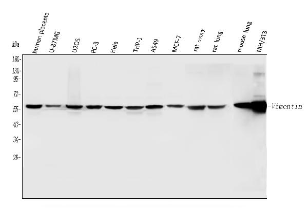
Western blot analysis of Vimentin using anti-Vimentin antibody (PB9359).
Electrophoresis was performed on a 5-20% SDS-PAGE gel at 70V (Stacking gel) / 90V (Resolving gel) for 2-3 hours. The sample well of each lane was loaded with 30 ug of sample under reducing conditions.
Lane 1: human placenta tissue lysates,
Lane 2: human U-87MG whole cell lysates,
Lane 3: human U20S whole cell lysates,
Lane 4: human PC-3 whole cell lysates,
Lane 5: human Hela whole cell lysates,
Lane 6: human THP-1 whole cell lysates,
Lane 7: human A549 whole cell lysates,
Lane 8: human MCF-7 whole cell lysates,
Lane 9: rat ovary tissue lysates,
Lane 10: rat lung tissue lysates,
Lane 11: mouse lung tissue lysates,
Lane 12: mouse NIH/3T3 whole cell lysates.
After electrophoresis, proteins were transferred to a nitrocellulose membrane at 150 mA for 50-90 minutes. Blocked the membrane with 5% non-fat milk/TBS for 1.5 hour at RT. The membrane was incubated with rabbit anti-Vimentin antigen affinity purified polyclonal antibody (Catalog # PB9359) at 0.5 μg/mL overnight at 4°C, then washed with TBS-0.1%Tween 3 times with 5 minutes each and probed with a goat anti-rabbit IgG-HRP secondary antibody at a dilution of 1:5000 for 1.5 hour at RT. The signal is developed using an Enhanced Chemiluminescent detection (ECL) kit (Catalog # EK1002) with Tanon 5200 system. A specific band was detected for Vimentin at approximately 54 kDa. The expected band size for Vimentin is at 54 kDa.

Click image to see more details
Western blot analysis of Vimentin using anti-Vimentin antibody (PB9359).
Electrophoresis was performed on a 5-20% SDS-PAGE gel at 80V (Stacking gel) / 120V (Resolving gel) for 2 hours. The sample well of each lane was loaded with 30 ug of sample under reducing conditions.
Lane 1-4: control group-human HTR8 whole cell lysates.
After electrophoresis, proteins were transferred to a nitrocellulose membrane at 150 mA for 50-90 minutes. Blocked the membrane with 5% non-fat milk/TBS for 1.5 hour at RT. The membrane was incubated with rabbit anti-Vimentin antigen affinity purified polyclonal antibody (PB9359) at 1:2500 overnight at 4°C, then washed with TBS-0.1%Tween 3 times with 5 minutes each and probed with a goat anti-rabbit IgG-HRP secondary antibody at a dilution of 1:5000 for 1 hour at RT. The signal is developed using an ECL Plus Western Blotting Substratewith ChemiDoc MP system. A specific band was detected for Vimentin at approximately 54 kDa. The expected band size for Vimentin is at 54 kDa.

Click image to see more details
IHC analysis of Vimentin using anti-Vimentin antibody (PB9359).
Vimentin was detected in a paraffin-embedded section of mouse cardiac muscle tissue. Heat mediated antigen retrieval was performed in EDTA buffer (pH 8.0, epitope retrieval solution). The tissue section was blocked with 10% goat serum. The tissue section was then incubated with 2 μg/ml rabbit anti-Vimentin Antibody (PB9359) overnight at 4°C. Biotinylated goat anti-rabbit IgG was used as secondary antibody and incubated for 30 minutes at 37°C. The tissue section was developed using Strepavidin-Biotin-Complex (SABC) (Catalog # SA1022) with DAB as the chromogen.

Click image to see more details
IHC analysis of Vimentin using anti-Vimentin antibody (PB9359).
Vimentin was detected in a paraffin-embedded section of rat cardiac muscle tissue. Heat mediated antigen retrieval was performed in EDTA buffer (pH 8.0, epitope retrieval solution). The tissue section was blocked with 10% goat serum. The tissue section was then incubated with 2 μg/ml rabbit anti-Vimentin Antibody (PB9359) overnight at 4°C. Biotinylated goat anti-rabbit IgG was used as secondary antibody and incubated for 30 minutes at 37°C. The tissue section was developed using Strepavidin-Biotin-Complex (SABC) (Catalog # SA1022) with DAB as the chromogen.

Click image to see more details
IHC analysis of Vimentin using anti-Vimentin antibody (PB9359).
Vimentin was detected in a paraffin-embedded section of human mammary cancer tissue. Heat mediated antigen retrieval was performed in EDTA buffer (pH 8.0, epitope retrieval solution). The tissue section was blocked with 10% goat serum. The tissue section was then incubated with 2 μg/ml rabbit anti-Vimentin Antibody (PB9359) overnight at 4°C. Biotinylated goat anti-rabbit IgG was used as secondary antibody and incubated for 30 minutes at 37°C. The tissue section was developed using Strepavidin-Biotin-Complex (SABC) (Catalog # SA1022) with DAB as the chromogen.

Click image to see more details
IHC analysis of Vimentin using anti-Vimentin antibody (PB9359).
Vimentin was detected in a paraffin-embedded section of human placenta tissue. Heat mediated antigen retrieval was performed in EDTA buffer (pH 8.0, epitope retrieval solution). The tissue section was blocked with 10% goat serum. The tissue section was then incubated with 2 μg/ml rabbit anti-Vimentin Antibody (PB9359) overnight at 4°C. Peroxidase Conjugated Goat Anti-rabbit IgG was used as secondary antibody and incubated for 30 minutes at 37°C. The tissue section was developed using HRP Conjugated Rabbit IgG Super Vision Assay Kit (Catalog # SV0002) with DAB as the chromogen.

Click image to see more details
IHC analysis of Vimentin using anti-Vimentin antibody (PB9359).
Vimentin was detected in a paraffin-embedded section of human lung adenocarcinoma tissue. Heat mediated antigen retrieval was performed in EDTA buffer (pH 8.0, epitope retrieval solution). The tissue section was blocked with 10% goat serum. The tissue section was then incubated with 2 μg/ml rabbit anti-Vimentin Antibody (PB9359) overnight at 4°C. Peroxidase Conjugated Goat Anti-rabbit IgG was used as secondary antibody and incubated for 30 minutes at 37°C. The tissue section was developed using HRP Conjugated Rabbit IgG Super Vision Assay Kit (Catalog # SV0002) with DAB as the chromogen.

Click image to see more details
IHC analysis of Vimentin using anti-Vimentin antibody (PB9359).
Vimentin was detected in a paraffin-embedded section of human colorectal adenocarcinoma tissue. Heat mediated antigen retrieval was performed in EDTA buffer (pH 8.0, epitope retrieval solution). The tissue section was blocked with 10% goat serum. The tissue section was then incubated with 2 μg/ml rabbit anti-Vimentin Antibody (PB9359) overnight at 4°C. Peroxidase Conjugated Goat Anti-rabbit IgG was used as secondary antibody and incubated for 30 minutes at 37°C. The tissue section was developed using HRP Conjugated Rabbit IgG Super Vision Assay Kit (Catalog # SV0002) with DAB as the chromogen.

Click image to see more details
IHC analysis of Vimentin using anti-Vimentin antibody (PB9359).
Vimentin was detected in a paraffin-embedded section of human invasive urothelial carcinoma tissue. Heat mediated antigen retrieval was performed in EDTA buffer (pH 8.0, epitope retrieval solution). The tissue section was blocked with 10% goat serum. The tissue section was then incubated with 2 μg/ml rabbit anti-Vimentin Antibody (PB9359) overnight at 4°C. Peroxidase Conjugated Goat Anti-rabbit IgG was used as secondary antibody and incubated for 30 minutes at 37°C. The tissue section was developed using HRP Conjugated Rabbit IgG Super Vision Assay Kit (Catalog # SV0002) with DAB as the chromogen.

Click image to see more details
IHC analysis of Vimentin using anti-Vimentin antibody (PB9359).
Vimentin was detected in a paraffin-embedded section of human liver cancer tissue. Heat mediated antigen retrieval was performed in EDTA buffer (pH 8.0, epitope retrieval solution). The tissue section was blocked with 10% goat serum. The tissue section was then incubated with 2 μg/ml rabbit anti-Vimentin Antibody (PB9359) overnight at 4°C. Peroxidase Conjugated Goat Anti-rabbit IgG was used as secondary antibody and incubated for 30 minutes at 37°C. The tissue section was developed using HRP Conjugated Rabbit IgG Super Vision Assay Kit (Catalog # SV0002) with DAB as the chromogen.

Click image to see more details
IHC analysis of Vimentin using anti-Vimentin antibody (PB9359).
Vimentin was detected in a paraffin-embedded section of human tonsil tissue. Heat mediated antigen retrieval was performed in EDTA buffer (pH 8.0, epitope retrieval solution). The tissue section was blocked with 10% goat serum. The tissue section was then incubated with 2 μg/ml rabbit anti-Vimentin Antibody (PB9359) overnight at 4°C. Peroxidase Conjugated Goat Anti-rabbit IgG was used as secondary antibody and incubated for 30 minutes at 37°C. The tissue section was developed using HRP Conjugated Rabbit IgG Super Vision Assay Kit (Catalog # SV0002) with DAB as the chromogen.

Click image to see more details
IF analysis of Vimentin using anti-Vimentin antibody (PB9359).
Vimentin was detected in a paraffin-embedded section of mouse brain tissue. Heat mediated antigen retrieval was performed in EDTA buffer (pH 8.0, epitope retrieval solution). The tissue section was blocked with 10% goat serum. The tissue section was then incubated with 5 μg/mL rabbit anti-Vimentin Antibody (PB9359) overnight at 4°C. DyLight488 Conjugated Goat Anti-Rabbit IgG (BA1127) was used as secondary antibody at 1:500 dilution and incubated for 30 minutes at 37°C. The section was counterstained with DAPI. Visualize using a fluorescence microscope and filter sets appropriate for the label used.

Click image to see more details
IF analysis of Vimentin using anti-Vimentin antibody (PB9359).
Vimentin was detected in a paraffin-embedded section of rat brain tissue. Heat mediated antigen retrieval was performed in EDTA buffer (pH 8.0, epitope retrieval solution). The tissue section was blocked with 10% goat serum. The tissue section was then incubated with 5 μg/mL rabbit anti-Vimentin Antibody (PB9359) overnight at 4°C. DyLight488 Conjugated Goat Anti-Rabbit IgG (BA1127) was used as secondary antibody at 1:500 dilution and incubated for 30 minutes at 37°C. The section was counterstained with DAPI. Visualize using a fluorescence microscope and filter sets appropriate for the label used.

Click image to see more details
MiR-3619 induces p21, downregulates β-catenin and CDK2 expression and inhibits the EMT process. a p21, E-cadherin, and Cyclin D1 mRNA expression were detected by qRT-PCR. b , c The expression of proteins from potential miR-3619 targets is shown in a western blot of BCa cells from 3 days after transfection of miR-3619 or controls. d , e Immunofluorescence staining of β-catenin in 5637 and T24 cells. f N-cadherin, Vimentin, and Snail mRNA expression in 5637 and T24 cells were measured using qRT-PCR. g The N-cadherin, Vimentin, and Snail protein expression in 5637 and T24 cells were measured using western blot. * P < 0.05, ** P < 0.01 compared with the dsControl group
Index in PubMed under a CC BY license. PMID: 30237499

Click image to see more details
Generation of 3D-spheroid GMSCs. ( A ) 4 × 10 4 of GMSCs/well in 200 µl of complete stemgro culture medium was seeded into each well of ultra-low attachment round-shaped 96-U well plates and cultured for 48 h. H & E staining of cryosections of GMSC spheroids. Scale bar: 50 µm. ( B ) Immunocytochemistry showed the expression of a panel of MSC-associated markers CD29, CD73 and CD90 and extracellular components such as type I collagen (col-I), vimentin, fibronectin, and laminin in GMSC-derived spheroids. ( C ) GMSC spheroids were dissociated into single cells and stained with Annexin V-FITC and 7-AAD to detect early apoptosis and late apoptotic/necrotic cells by flow cytometric analysis. ( D ) Immunocytochemistry showed that less than 10% of cells inside GMSC spheroids were positive for the cleaved caspase-3. Cell nuclei were counter-stained by DAPI (blue). Scale bar: 50 µm. Data are representative of 3 independent experiments.
Index in PubMed under a CC BY license. PMID: 29700345

Click image to see more details
MiR-139 suppresses HCC migration and invasion. a - e Hep3B and SMMC7721 cells were transfected with 100 nM miR-139 mimic oligos or control oligos. The migration capacity of ( a ) Hep3B and ( b ) SMMC7721 cells were analyzed with the transwell migration assay. The invasion ability of ( c ) Hep3B and ( d ) SMMC7721 cells were measured with the matrigel transwell invasion assay. e The protein expression levels of E-cadherin, Vimentin and SNAIL1 were assayed with western blot in both Hep3B and SMMC7721 cells. f Hep3B and SMMC7721 cells were transfected with 100 nM miR-139 inhibitory or control oligos. The protein levels of E-cadherin, Vimentin and SNAIL1 were measured by western blot in both cell lines. All experiments were repeated 3 times. ** P < 0.01, *** P < 0.001
Index in PubMed under a CC BY license. PMID: 31046781

Click image to see more details
Down-regulating KPNA2 leads to decreased growth of HCC cells. Hep3B cells were transfected with non-targeting control oligo or with siRNA specifically targeting KPNA2 using lipofectamine 2000. a KPNA2 mRNA level was measured with qRT-PCR. b Western blot was used to assay the protein level of KPNA2. c Cell viability was measured using the MTT assay. d Cell apoptosis was measured with the Annxin V-FITC/Hoechst 33258 double staining flow cytometry. The cells were incubated for 72 h after transfection of the oligos. e colonogenic, ( f ) migratory and ( g ) invading capacities of cells were assayed as described above. h The protein levels of E-cadherin and Vimentin were measured using western blot. i The OS of HCC patients with low or high KPNA2 level was analyzed with Kaplan-Meier survival analysis and the curves were compared with the Log-rank test. The Q1–4 represent the 25% quantile of KPNA2 expression. Q1 represents the lowest 25%, Q2 represents 25–50%, Q3 is the 50–75% and Q4 is the 75–100%. All experiments were repeated 3 times. * P < 0.05, ** P < 0.01
Index in PubMed under a CC BY license. PMID: 31046781

Click image to see more details
LRRC4 inhibits collective cells invasion by down-regulating E-cadherin without EMT induction. (A) Phalloidin staining was performed in SKOV3 cells stably expressing LRRC4 through lentivirus infection. F-actin aggregates along control cell peripherals, while LRRC4 inhibites the accumulation and aggregation of F-actin (indicated with arrowheads). (B) The mRNA levels of EMT associated-genes, including E-cadherin, N-cadherin, slug, twist, ZEB1 and ZEB2, as assessed with RT-qPCR. (C) Western blotting analysis of E-cadherin and Vimentin protein in SKOV3 cells transiently transfected with different doses of LRRC4 expression vector (0.5, 1, and 2 μg). (D,E) E-cadherin and LRRC4 protein expression and localization as detected with immunohistochemistry (IHC) in human normal ovarian epithelial cells ( D , n = 5), human HGSC ( E , n = 17) and ascites of ovarian cancer patients. Scale bars: the left is 50 μm while the right is 20 μm. The area in the red boxes to the right is magnified. (F) E-cadherin and LRRC4 in EOC ascitic tissue as stained with IHC. Scale bars: 20 μm. (G) Expression of E-cadherin, Vimentin and Pan-cadherin in intraperitoneal xenografted primary tumor tissues from the mouse model was examined by the use of IHC in. Scale bars: 20 μm and 50 μm. * p < 0.05, ** p < 0.01.
Index in PubMed under a CC BY license. PMID: 32117780

Click image to see more details
Key characteristics of the immortalized HP-1 human PSC cell line. Phase contrast microscopy in HP-1 cells after seeding for 12 h ( A ); Double-immunofluorescence labelling using antibodies for SV40 antigen and GFAP, green, localization of SV40 antigen, red, localization of GFAP (B); Immunofluorescence showing the presence of α-SMA ( C ); Double-immunofluorescence labelling for vimentin and desmin. Green, localization of vimentin (D), red, localization of desmin (E), yellow, colocalization of vimentin and desmin (F). (Original magnification × 200 in A ; × 400 in B-F ).
Index in PubMed under a CC BY license. PMID: 31929747

Click image to see more details
ZFPM2-AS1 promoted cell metastatic properties by affecting EMT in HCC cells. A , B The migration abilities were evaluated in HCC cells infected with sh-ZFPM2-AS1-1 or sh-ZFPM2-AS1-2 lentivirus, by wound-healing assays. Scale bar = 50 μm. C Transwell assays detected the invasion capacities of HCC cells after ZFPM2-AS1-2 depletion. Scale bar = 50 μm. D Western blot examined the protein levels of N-cadherin and vimentin in HCC cells. Data are presented as the mean ± SD from three independent experiments. * P < 0.05, ** P < 0.01.
Index in PubMed under a CC BY license. PMID: 33414427

Click image to see more details
The characterization of PDLCs. Primary culture of PDLCs at the Day 7 (×100) ( a ); Morphology of PDLCs at Passage 1 (× 100) ( b ); Immunohistochemistry staining results of PDLCs, positive for vimentin (× 100) ( c ) and negative for cytokeratin (× 100) ( d )
Index in PubMed under a CC BY license. PMID: 33485352

Click image to see more details
LPC inhibits pulmonary metastasis of CT26 colon cancer. a Schematic view of the experimental procedures of CT26 pulmonary metastatic mouse model. b , c Image and corresponding H&E staining of lung tissue. d Percentage change of body weight. e – g Lung weight, the number of lung tumor nodule, and metastasis rate ( n = 6 mice). h Protein expression of PCNA, vimentin and E-cadherin in lungs ( n = 3 mice). Data were presented as mean ± SEM, * p < 0.05, ** p < 0.01.
Index in PubMed under a CC BY license. PMID: 36774343

Click image to see more details
Western blot results showing the expression levels of A549 cells treated with different concentrations of GLXB-D drug serum and TGF-β1 for 24 h . (A): The protein expression straps of E-cadherin and Vimentin; (B): The grey value ratio of expressions of E-cadherin and Vimentin. (1) control group; (2) 5ng/mL TGF-β1 induced group; (3) 5% GLXB-D drug serum group; (4) 10% GLXB-D drug serum group; (5) 15% GLXB-D drug serum group.
Index in De Gruyter Brill under a CC BY license. DOI: 10.1515/chem-2018-0042
Specific Publications For Anti-Vimentin Antibody Picoband® (PB9359)
Loading publications
Recommended Resources
Here are featured tools and databases that you might find useful.
- Boster's Pathways Library
- Protein Databases
- Bioscience Research Protocol Resources
- Data Processing & Analysis Software
- Photo Editing Software
- Scientific Literature Resources
- Research Paper Management Tools
- Molecular Biology Software
- Primer Design Tools
- Bioinformatics Tools
- Phylogenetic Tree Analysis
Customer Reviews
Have you used Anti-Vimentin Antibody Picoband®?
Share your experimental results or join a short interview to earn up to $1,000 in product credits or other rewards.
1 Reviews For Anti-Vimentin Antibody Picoband®
This antibody is highly specific and efficient, suitable for detecting Vimentin protein in HTR8 cells by Western blot. Only minor nonspecific bands are observed.
Excellent

| SKU | PB9359 |
|---|---|
| Application | Western Blot |
| Sample | human HTR8 cell |
| Sample Processing Description | The samples were lysed in RIPA buffer, mixed with β-mercaptoethanol, and denatured at 100°C for 10 minutes before loading onto an SDS-PAGE gel. |
| Other Reagents | 5% Non-fat milk |
| Primary Antibody | Vimentin Antibody Picoband® |
| Primary Incubation | 1:2500, overnight at 4 ℃ |
| Secondary Antibody | HRP-conjugated Anti-Rabbit IgG Secondary Antibody |
| Secondary Incubation | 1 hour in room temperature |
| Detection | Substrate: ECL reagent, Imaging system:ChemiDoc MP |
| Results Summary | This antibody is highly specific and efficient, suitable for detecting Vimentin protein in HTR8 cells by Western blot, with only minor nonspecific bands observed. |
Xiaoqin Yu, NIHWR
Verified customer
Submitted 2025-11-11
Customer Q&As
Have a question?
Find answers in Q&As, reviews.
Can't find your answer?
Submit your question
5 Customer Q&As for Anti-Vimentin Antibody Picoband®
Question
We purchased anti-Vimentin antibody for IHC on leukemic t-cell in a previous project. I am using human, and We intend to use the antibody for WB next. We are interested in examining leukemic t-cell as well as cervix in our next experiment. Could you please give me some suggestion on which antibody would work the best for WB?
Verified Customer
Verified customer
Asked: 2020-04-03
Answer
I looked at the website and datasheets of our anti-Vimentin antibody and I see that PB9359 has been validated on human in both IHC and WB. Thus PB9359 should work for your application. Our Boster satisfaction guarantee will cover this product for WB in human even if the specific tissue type has not been validated. We do have a comprehensive range of products for WB detection and you can check out our website bosterbio.com to find out more information about them.
Boster Scientific Support
Answered: 2020-04-03
Question
My team were well pleased with the WB result of your anti-Vimentin antibody. However we have seen positive staining in lymphoblast cytoplasm using this antibody. Is that expected? Could you tell me where is VIM supposed to be expressed?
Verified Customer
Verified customer
Asked: 2020-02-11
Answer
Based on literature, lymphoblast does express VIM. Generally VIM expresses in cytoplasm. Regarding which tissues have VIM expression, here are a few articles citing expression in various tissues:
Cervix carcinoma, Pubmed ID: 16964243, 17081983, 17924679, 18220336, 18669648, 18691976, 20068231
Cervix carcinoma, and Erythroleukemia, Pubmed ID: 23186163
Embryo, Placenta, and Stomach, Pubmed ID: 14702039
Fibroblast, Pubmed ID: 3371665
Leukemic T-cell, Pubmed ID: 19690332
Liver, Pubmed ID: 24275569
Lymphoblast, Pubmed ID: 14654843
Lymphoma, Pubmed ID: 14996095
Mammary carcinoma, Pubmed ID: 9150946
Osteosarcoma, Pubmed ID: 2323579
Boster Scientific Support
Answered: 2020-02-11
Question
We have observed staining in human cervix carcinoma. Are there any suggestions? Is anti-Vimentin antibody supposed to stain cervix carcinoma positively?
Verified Customer
Verified customer
Asked: 2019-12-20
Answer
From literature cervix carcinoma does express VIM. From Uniprot.org, VIM is expressed in dorsal root ganglion, lymphoma, testis, embryo, placenta stomach, adipose tissue coronary artery, cervix, placenta testis, mammary carcinoma, t-cell, hepatoma, brain, cajal-retzius cell fetal brain cortex, fibroblast, osteosarcoma, lymphoblast, cervix carcinoma, leukemic t-cell, cervix carcinoma erythroleukemia, liver, among other tissues. Regarding which tissues have VIM expression, here are a few articles citing expression in various tissues:
Cervix carcinoma, Pubmed ID: 16964243, 17081983, 17924679, 18220336, 18669648, 18691976, 20068231
Cervix carcinoma, and Erythroleukemia, Pubmed ID: 23186163
Embryo, Placenta, and Stomach, Pubmed ID: 14702039
Fibroblast, Pubmed ID: 3371665
Leukemic T-cell, Pubmed ID: 19690332
Liver, Pubmed ID: 24275569
Lymphoblast, Pubmed ID: 14654843
Lymphoma, Pubmed ID: 14996095
Mammary carcinoma, Pubmed ID: 9150946
Osteosarcoma, Pubmed ID: 2323579
Boster Scientific Support
Answered: 2019-12-20
Question
We are currently using anti-Vimentin antibody PB9359 for mouse tissue, and we are happy with the IHC results. The species of reactivity given in the datasheet says human, mouse, rat. Is it possible that the antibody can work on monkey tissues as well?
A. Roberts
Verified customer
Asked: 2017-10-12
Answer
The anti-Vimentin antibody (PB9359) has not been tested for cross reactivity specifically with monkey tissues, though there is a good chance of cross reactivity. We have an innovator award program that if you test this antibody and show it works in monkey you can get your next antibody for free. Please contact me if I can help you with anything.
Boster Scientific Support
Answered: 2017-10-12
Question
I am interested in using your anti-Vimentin antibody for cellular response to interferon-gamma studies. Has this antibody been tested with western blotting on rat kidney tissue? We would like to see some validation images before ordering.
G. Rodriguez
Verified customer
Asked: 2015-02-16
Answer
We appreciate your inquiry. This PB9359 anti-Vimentin antibody is validated on ht1080 whole cell lysate, nih whole cell lysate, jurkat whole cell lysate, hut whole cell lysate, hela whole cell lysate, human placenta tissue, tissue lysate, rat kidney tissue, testis tissue, mouse kidney tissue, cardiac muscle tissue, mammary cancer tissue. It is guaranteed to work for IHC, WB in human, mouse, rat. Our Boster guarantee will cover your intended experiment even if the sample type has not been be directly tested.
Boster Scientific Support
Answered: 2015-02-16
