Click image to see more details
-

-
-
-
-
+24
Product Info Summary
| SKU: | M00235-1 |
|---|---|
| Size: | 100 μl |
| Reactive Species: | Human, Mouse, Rat |
| Host: | Rabbit |
| Application: | Flow Cytometry, IF, IHC, ICC, WB |
Customers Who Bought This Also Bought

Product info
Product Name
Anti-Vimentin Rabbit Monoclonal Antibody
SKU/Catalog Number
M00235-1
BM4029 is an alternative SKU for this antibody, used in previous lots.
Size
100 μl
Form
Liquid
Description
Boster Bio Anti-Vimentin Rabbit Monoclonal Antibody catalog # M00235-1. Tested in WB, IHC, ICC/IF, Flow Cytometry applications. This antibody reacts with Human, Mouse, Rat.
Storage & Handling
Store at -20°C for one year. For short term storage and frequent use, store at 4°C for up to one month. Avoid repeated freeze-thaw cycles.
Cite This Product
Anti-Vimentin Rabbit Monoclonal Antibody (Boster Biological Technology, Pleasanton CA, USA, Catalog # M00235-1)
Host
Rabbit
Contents
Rabbit IgG in stabilizing components, phosphate buffered saline, pH 7.4, 150mM NaCl, 0.02% sodium azide and 50% glycerol.
*This antibody is supplied in a stabilized formulation.
Compatibility with conjugation reactions depends on the chemistry of the conjugation method used.
For conjugation methods that are not compatible with the stabilizing components present in this formulation, a carrier-free antibody format is required.
Clonality
Monoclonal
Clone Number
AGF-22
Isotype
Rabbit IgG
Immunogen
A synthesized peptide derived from human Vimentin
Reactive Species
M00235-1 is reactive to VIM in Human, Mouse, Rat
Observed Molecular Weight
54 kDa
Calculated molecular weight
53.7 kDa
Antibody Validation
Boster validates all antibodies on WB, IHC, ICC, Immunofluorescence, and ELISA with known positive control and negative samples to ensure specificity and high affinity, including thorough antibody incubations.
Application & Images
Applications
M00235-1 is guaranteed for Flow Cytometry, IF, IHC, ICC, WB Boster Guarantee
Recommend Dilution
WB 1:500-2000
IHC 1:50-200
ICC/IF 1:50-200
FC 1:50
Tested application
Suggested blocking solution with 5% non-fat milk or BSA; (*)Recommended protein loading: 20-40 µg per lane
Use TE buffer pH 9.0 for antigen retrieval; (*) citrate buffer pH 6.0 is an alternative.
Validation Images & Assay Conditions
Click image to see more details
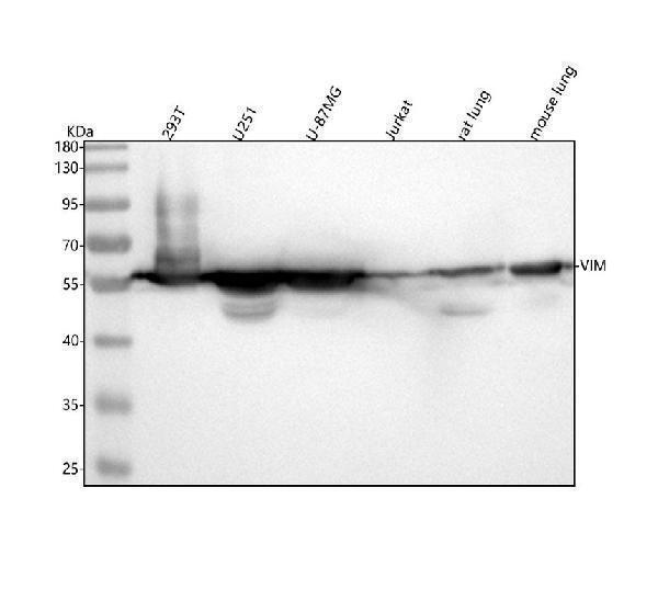
Western blot analysis of Vimentin using anti-Vimentin antibody (M00235-1).
Electrophoresis was performed on a 5-20% SDS-PAGE gel at 70V (Stacking gel) / 90V (Resolving gel) for 2-3 hours. The sample well of each lane was loaded with 30 ug of sample under reducing conditions.
Lane 1: human 293T whole cell lysates,
Lane 2: human U251 whole cell lysates,
Lane 3: human U-87MG whole cell lysates,
Lane 4: human Jurkat whole cell lysates,
Lane 5: rat lung tissue lysates,
Lane 6: mouse lung tissue lysates.
After electrophoresis, proteins were transferred to a nitrocellulose membrane at 150 mA for 50-90 minutes. Blocked the membrane with 5% non-fat milk/TBS for 1.5 hour at RT. The membrane was incubated with rabbit anti-Vimentin antigen affinity purified monoclonal antibody (Catalog # M00235-1) at 1:1000 overnight at 4°C, then washed with TBS-0.1%Tween 3 times with 5 minutes each and probed with a goat anti-rabbit IgG-HRP secondary antibody at a dilution of 1:5000 for 1.5 hour at RT. The signal is developed using an Enhanced Chemiluminescent detection (ECL) kit (Catalog # EK1002) with Tanon 5200 system. A specific band was detected for Vimentin at approximately 54 kDa. The expected band size for Vimentin is at 54 kDa.

Click image to see more details
Therapeutic potentials of Pg-Fe-hMSC in both monocellular and multicellular humanized fibrotic models. a Schematic illustration and representative image of EpCAM immunostaining in primary human lung epithelial cells (hLECs). Scale bar, 50 μm. b Mitochondrial transfer rates from the indicated hMSC to the primary hLEC ( n = 3 biologically independent cells). c Relative intracellular ROS levels ( n = 3 biologically independent cells), d TGF- β expression levels ( n = 4 biologically independent cells), and e Viability of BLM-treated hLEC after the indicated treatment using different engineered hMSCs ( n = 3 biologically independent cells). f Schematic illustration showing the preparation of the 3D multicellular human fibrotic model. g Representative immunostaining images showing the expression of α -smooth muscle actin ( α -SMA), vimentin, collagen-I, and Ki-67 in the healthy and fibrotic multicellular human spheroid models. Blue fluorescent signals indicate the cell nuclei and green signals indicate the biomarkers. Scale bar, 20 μm. h Mitochondrial transfer rates of different engineered hMSC in fibrotic human lung spheroids ( n = 3 biologically independent experiments). i Relative intracellular ROS levels ( n = 3 biologically independent experiments) and j TGF- β expression levels of fibrotic human lung spheroids after the indicated treatment using different engineered hMSCs ( n = 4 biologically independent experiments). k Viability of fibrotic human lung spheroids after the indicated treatment using different engineered hMSCs ( n = 3 biologically independent experiments). Data are presented as means ± SD. Statistical significance was analyzed using ordinary one-way ANOVA. ECs epithelial cells.
Index in PubMed under a CC BY license. PMID: 37723135

Click image to see more details
IHC analysis of Vimentin using anti-Vimentin antibody (M00235-1).
Vimentin was detected in a paraffin-embedded section of human colon cancer tissue. Heat mediated antigen retrieval was performed in EDTA buffer (pH 8.0, epitope retrieval solution). The tissue section was blocked with 10% goat serum. The tissue section was then incubated with 1:200 rabbit anti-Vimentin Antibody (M00235-1) overnight at 4°C. Peroxidase Conjugated Goat Anti-rabbit IgG was used as secondary antibody and incubated for 30 minutes at 37°C. The tissue section was developed using HRP Conjugated Rabbit IgG Super Vision Assay Kit (Catalog # SV0002) with DAB as the chromogen.

Click image to see more details
IHC analysis of Vimentin using anti-Vimentin antibody (M00235-1).
Vimentin was detected in a paraffin-embedded section of human glioma tissue. Heat mediated antigen retrieval was performed in EDTA buffer (pH 8.0, epitope retrieval solution). The tissue section was blocked with 10% goat serum. The tissue section was then incubated with 2 μg/ml rabbit anti-Vimentin Antibody (M00235-1) overnight at 4°C. Peroxidase Conjugated Goat Anti-rabbit IgG was used as secondary antibody and incubated for 30 minutes at 37°C. The tissue section was developed using HRP Conjugated Rabbit IgG Super Vision Assay Kit (Catalog # SV0002) with DAB as the chromogen.

Click image to see more details
IHC analysis of Vimentin using anti-Vimentin antibody (M00235-1).
Vimentin was detected in a paraffin-embedded section of human liver cancer tissue. Heat mediated antigen retrieval was performed in EDTA buffer (pH 8.0, epitope retrieval solution). The tissue section was blocked with 10% goat serum. The tissue section was then incubated with 1:200 rabbit anti-Vimentin Antibody (M00235-1) overnight at 4°C. Peroxidase Conjugated Goat Anti-rabbit IgG was used as secondary antibody and incubated for 30 minutes at 37°C. The tissue section was developed using HRP Conjugated Rabbit IgG Super Vision Assay Kit (Catalog # SV0002) with DAB as the chromogen.

Click image to see more details
IHC analysis of Vimentin using anti-Vimentin antibody (M00235-1).
Vimentin was detected in a paraffin-embedded section of human non small cell lung cancer tissue. Heat mediated antigen retrieval was performed in EDTA buffer (pH 8.0, epitope retrieval solution). The tissue section was blocked with 10% goat serum. The tissue section was then incubated with 1:200 rabbit anti-Vimentin Antibody (M00235-1) overnight at 4°C. Peroxidase Conjugated Goat Anti-rabbit IgG was used as secondary antibody and incubated for 30 minutes at 37°C. The tissue section was developed using HRP Conjugated Rabbit IgG Super Vision Assay Kit (Catalog # SV0002) with DAB as the chromogen.

Click image to see more details
IHC analysis of Vimentin using anti-Vimentin antibody (M00235-1).
Vimentin was detected in a paraffin-embedded section of human ovarian cancer tissue. Heat mediated antigen retrieval was performed in EDTA buffer (pH 8.0, epitope retrieval solution). The tissue section was blocked with 10% goat serum. The tissue section was then incubated with 1:200 rabbit anti-Vimentin Antibody (M00235-1) overnight at 4°C. Peroxidase Conjugated Goat Anti-rabbit IgG was used as secondary antibody and incubated for 30 minutes at 37°C. The tissue section was developed using HRP Conjugated Rabbit IgG Super Vision Assay Kit (Catalog # SV0002) with DAB as the chromogen.

Click image to see more details
IHC analysis of Vimentin using anti-Vimentin antibody (M00235-1).
Vimentin was detected in a paraffin-embedded section of human placenta tissue. Heat mediated antigen retrieval was performed in EDTA buffer (pH 8.0, epitope retrieval solution). The tissue section was blocked with 10% goat serum. The tissue section was then incubated with 1:200 rabbit anti-Vimentin Antibody (M00235-1) overnight at 4°C. Peroxidase Conjugated Goat Anti-rabbit IgG was used as secondary antibody and incubated for 30 minutes at 37°C. The tissue section was developed using HRP Conjugated Rabbit IgG Super Vision Assay Kit (Catalog # SV0002) with DAB as the chromogen.

Click image to see more details
IHC analysis of Vimentin using anti-Vimentin antibody (M00235-1).
Vimentin was detected in a paraffin-embedded section of human tonsil tissue. Heat mediated antigen retrieval was performed in EDTA buffer (pH 8.0, epitope retrieval solution). The tissue section was blocked with 10% goat serum. The tissue section was then incubated with 1:200 rabbit anti-Vimentin Antibody (M00235-1) overnight at 4°C. Peroxidase Conjugated Goat Anti-rabbit IgG was used as secondary antibody and incubated for 30 minutes at 37°C. The tissue section was developed using HRP Conjugated Rabbit IgG Super Vision Assay Kit (Catalog # SV0002) with DAB as the chromogen.

Click image to see more details
IHC analysis of Vimentin/VIM using anti-Vimentin/VIM antibody (M00235-1).
Vimentin/VIM was detected in a paraffin-embedded section of human tonsil tissue. Heat mediated antigen retrieval was performed in EDTA buffer (pH 8.0, epitope retrieval solution). The tissue section was blocked with 10% goat serum. The tissue section was then incubated with 5 μg/ml rabbit anti-Vimentin/VIM Antibody (M00235-1) overnight at 4°C. HRP-AffiniPure Goat Anti-Rabbit IgG was used as secondary antibody and incubated for 30 minutes at 37°C. The tissue section was developed using HRP Conjugated Rabbit IgG Super Vision Assay Kit (Catalog # SV0002) with DAB as the chromogen.

Click image to see more details
IHC analysis of Vimentin/VIM using anti-Vimentin/VIM antibody (M00235-1).
Vimentin/VIM was detected in a paraffin-embedded section of rat brain tissue. Heat mediated antigen retrieval was performed in EDTA buffer (pH 8.0, epitope retrieval solution). The tissue section was blocked with 10% goat serum. The tissue section was then incubated with 5 μg/ml rabbit anti-Vimentin/VIM Antibody (M00235-1) overnight at 4°C. HRP-AffiniPure Goat Anti-Rabbit IgG was used as secondary antibody and incubated for 30 minutes at 37°C. The tissue section was developed using HRP Conjugated Rabbit IgG Super Vision Assay Kit (Catalog # SV0002) with DAB as the chromogen.

Click image to see more details
IHC analysis of Vimentin/VIM using anti-Vimentin/VIM antibody (M00235-1).
Vimentin/VIM was detected in a paraffin-embedded section of rat lung tissue. Heat mediated antigen retrieval was performed in EDTA buffer (pH 8.0, epitope retrieval solution). The tissue section was blocked with 10% goat serum. The tissue section was then incubated with 5 μg/ml rabbit anti-Vimentin/VIM Antibody (M00235-1) overnight at 4°C. HRP-AffiniPure Goat Anti-Rabbit IgG was used as secondary antibody and incubated for 30 minutes at 37°C. The tissue section was developed using HRP Conjugated Rabbit IgG Super Vision Assay Kit (Catalog # SV0002) with DAB as the chromogen.

Click image to see more details
IHC analysis of Vimentin/VIM using anti-Vimentin/VIM antibody (M00235-1).
Vimentin/VIM was detected in a paraffin-embedded section of human liver cancer tissue. Heat mediated antigen retrieval was performed in EDTA buffer (pH 8.0, epitope retrieval solution). The tissue section was blocked with 10% goat serum. The tissue section was then incubated with 5 μg/ml rabbit anti-Vimentin/VIM Antibody (M00235-1) overnight at 4°C. HRP-AffiniPure Goat Anti-Rabbit IgG was used as secondary antibody and incubated for 30 minutes at 37°C. The tissue section was developed using HRP Conjugated Rabbit IgG Super Vision Assay Kit (Catalog # SV0002) with DAB as the chromogen.

Click image to see more details
IHC analysis of Vimentin/VIM using anti-Vimentin/VIM antibody (M00235-1).
Vimentin/VIM was detected in a paraffin-embedded section of human lung cancer tissue. Heat mediated antigen retrieval was performed in EDTA buffer (pH 8.0, epitope retrieval solution). The tissue section was blocked with 10% goat serum. The tissue section was then incubated with 5 μg/ml rabbit anti-Vimentin/VIM Antibody (M00235-1) overnight at 4°C. HRP-AffiniPure Goat Anti-Rabbit IgG was used as secondary antibody and incubated for 30 minutes at 37°C. The tissue section was developed using HRP Conjugated Rabbit IgG Super Vision Assay Kit (Catalog # SV0002) with DAB as the chromogen.

Click image to see more details
IHC analysis of Vimentin/VIM using anti-Vimentin/VIM antibody (M00235-1).
Vimentin/VIM was detected in a paraffin-embedded section of human stomach cancer tissue. Heat mediated antigen retrieval was performed in EDTA buffer (pH 8.0, epitope retrieval solution). The tissue section was blocked with 10% goat serum. The tissue section was then incubated with 5 μg/ml rabbit anti-Vimentin/VIM Antibody (M00235-1) overnight at 4°C. HRP-AffiniPure Goat Anti-Rabbit IgG was used as secondary antibody and incubated for 30 minutes at 37°C. The tissue section was developed using HRP Conjugated Rabbit IgG Super Vision Assay Kit (Catalog # SV0002) with DAB as the chromogen.

Click image to see more details
IHC analysis of Vimentin/VIM using anti-Vimentin/VIM antibody (M00235-1).
Vimentin/VIM was detected in a paraffin-embedded section of human thyroid cancer tissue. Heat mediated antigen retrieval was performed in EDTA buffer (pH 8.0, epitope retrieval solution). The tissue section was blocked with 10% goat serum. The tissue section was then incubated with 5 μg/ml rabbit anti-Vimentin/VIM Antibody (M00235-1) overnight at 4°C. HRP-AffiniPure Goat Anti-Rabbit IgG was used as secondary antibody and incubated for 30 minutes at 37°C. The tissue section was developed using HRP Conjugated Rabbit IgG Super Vision Assay Kit (Catalog # SV0002) with DAB as the chromogen.

Click image to see more details
IHC analysis of Vimentin/VIM using anti-Vimentin/VIM antibody (M00235-1).
Vimentin/VIM was detected in a paraffin-embedded section of human cervical cancer tissue. Heat mediated antigen retrieval was performed in EDTA buffer (pH 8.0, epitope retrieval solution). The tissue section was blocked with 10% goat serum. The tissue section was then incubated with 5 μg/ml rabbit anti-Vimentin/VIM Antibody (M00235-1) overnight at 4°C. HRP-AffiniPure Goat Anti-Rabbit IgG was used as secondary antibody and incubated for 30 minutes at 37°C. The tissue section was developed using HRP Conjugated Rabbit IgG Super Vision Assay Kit (Catalog # SV0002) with DAB as the chromogen.

Click image to see more details
IHC analysis of Vimentin/VIM using anti-Vimentin/VIM antibody (M00235-1).
Vimentin/VIM was detected in a paraffin-embedded section of human pancreatic cancer tissue. Heat mediated antigen retrieval was performed in EDTA buffer (pH 8.0, epitope retrieval solution). The tissue section was blocked with 10% goat serum. The tissue section was then incubated with 5 μg/ml rabbit anti-Vimentin/VIM Antibody (M00235-1) overnight at 4°C. HRP-AffiniPure Goat Anti-Rabbit IgG was used as secondary antibody and incubated for 30 minutes at 37°C. The tissue section was developed using HRP Conjugated Rabbit IgG Super Vision Assay Kit (Catalog # SV0002) with DAB as the chromogen.

Click image to see more details
Immunofluorescent analysis of Hela cells, using Vimentin Antibody.

Click image to see more details
IF analysis of Vimentin/VIM using anti-Vimentin/VIM antibody (M00235-1).
Vimentin/VIM was detected in a paraffin-embedded section of human tonsil tissue. Heat mediated antigen retrieval was performed in EDTA buffer (pH 8.0, epitope retrieval solution). The tissue section was blocked with 10% goat serum. The tissue section was then incubated with 25 μg/mL rabbit anti-Vimentin/VIM Antibody (M00235-1) overnight at 4°C. DyLight 594 Conjugated AffiniPure Goat Anti-mouse IgG (H+L) (BA1142) was used as secondary antibody at 1:100 dilution and incubated for 30 minutes at 37°C. The section was counterstained with DAPI. Visualize using a fluorescence microscope and filter sets appropriate for the label used.

Click image to see more details
IF analysis of Vimentin/VIM using anti-Vimentin/VIM antibody (M00235-1).
Vimentin/VIM was detected in a paraffin-embedded section of human placenta tissue. Heat mediated antigen retrieval was performed in EDTA buffer (pH 8.0, epitope retrieval solution). The tissue section was blocked with 10% goat serum. The tissue section was then incubated with 25 μg/mL rabbit anti-Vimentin/VIM Antibody (M00235-1) overnight at 4°C. DyLight 594 Conjugated AffiniPure Goat Anti-mouse IgG (H+L) (BA1142) was used as secondary antibody at 1:100 dilution and incubated for 30 minutes at 37°C. The section was counterstained with DAPI. Visualize using a fluorescence microscope and filter sets appropriate for the label used.

Click image to see more details
IF analysis of Vimentin/VIM using anti-Vimentin/VIM antibody (M00235-1).
Vimentin/VIM was detected in a paraffin-embedded section of mouse brain tissue. Heat mediated antigen retrieval was performed in EDTA buffer (pH 8.0, epitope retrieval solution). The tissue section was blocked with 10% goat serum. The tissue section was then incubated with 25 μg/mL rabbit anti-Vimentin/VIM Antibody (M00235-1) overnight at 4°C. DyLight 594 Conjugated AffiniPure Goat Anti-mouse IgG (H+L) (BA1142) was used as secondary antibody at 1:100 dilution and incubated for 30 minutes at 37°C. The section was counterstained with DAPI. Visualize using a fluorescence microscope and filter sets appropriate for the label used.

Click image to see more details
IF analysis of Vimentin/VIM using anti-Vimentin/VIM antibody (M00235-1).
Vimentin/VIM was detected in a paraffin-embedded section of human liver caner tissue. Heat mediated antigen retrieval was performed in EDTA buffer (pH 8.0, epitope retrieval solution). The tissue section was blocked with 10% goat serum. The tissue section was then incubated with 25 μg/mL rabbit anti-Vimentin/VIM Antibody (M00235-1) overnight at 4°C. DyLight 594 Conjugated AffiniPure Goat Anti-mouse IgG (H+L) (BA1142) was used as secondary antibody at 1:100 dilution and incubated for 30 minutes at 37°C. The section was counterstained with DAPI. Visualize using a fluorescence microscope and filter sets appropriate for the label used.

Click image to see more details
IF analysis of Vimentin/VIM using anti-Vimentin/VIM antibody (M00235-1).
Vimentin/VIM was detected in a paraffin-embedded section of human lung caner tissue. Heat mediated antigen retrieval was performed in EDTA buffer (pH 8.0, epitope retrieval solution). The tissue section was blocked with 10% goat serum. The tissue section was then incubated with 25 μg/mL rabbit anti-Vimentin/VIM Antibody (M00235-1) overnight at 4°C. DyLight 594 Conjugated AffiniPure Goat Anti-mouse IgG (H+L) (BA1142) was used as secondary antibody at 1:100 dilution and incubated for 30 minutes at 37°C. The section was counterstained with DAPI. Visualize using a fluorescence microscope and filter sets appropriate for the label used.

Click image to see more details
IF analysis of Vimentin/VIM using anti-Vimentin/VIM antibody (M00235-1).
Vimentin/VIM was detected in a paraffin-embedded section of human thyroid caner tissue. Heat mediated antigen retrieval was performed in EDTA buffer (pH 8.0, epitope retrieval solution). The tissue section was blocked with 10% goat serum. The tissue section was then incubated with 25 μg/mL rabbit anti-Vimentin/VIM Antibody (M00235-1) overnight at 4°C. DyLight 594 Conjugated AffiniPure Goat Anti-mouse IgG (H+L) (BA1142) was used as secondary antibody at 1:100 dilution and incubated for 30 minutes at 37°C. The section was counterstained with DAPI. Visualize using a fluorescence microscope and filter sets appropriate for the label used.

Click image to see more details
IF analysis of Vimentin/VIM using anti-Vimentin/VIM antibody (M00235-1).
Vimentin/VIM was detected in a paraffin-embedded section of human cervical caner tissue. Heat mediated antigen retrieval was performed in EDTA buffer (pH 8.0, epitope retrieval solution). The tissue section was blocked with 10% goat serum. The tissue section was then incubated with 25 μg/mL rabbit anti-Vimentin/VIM Antibody (M00235-1) overnight at 4°C. DyLight 594 Conjugated AffiniPure Goat Anti-mouse IgG (H+L) (BA1142) was used as secondary antibody at 1:100 dilution and incubated for 30 minutes at 37°C. The section was counterstained with DAPI. Visualize using a fluorescence microscope and filter sets appropriate for the label used.

Click image to see more details
IF analysis of Vimentin/VIM using anti-Vimentin/VIM antibody (M00235-1).
Vimentin/VIM was detected in a paraffin-embedded section of human pancreatic caner tissue. Heat mediated antigen retrieval was performed in EDTA buffer (pH 8.0, epitope retrieval solution). The tissue section was blocked with 10% goat serum. The tissue section was then incubated with 25 μg/mL rabbit anti-Vimentin/VIM Antibody (M00235-1) overnight at 4°C. DyLight 594 Conjugated AffiniPure Goat Anti-mouse IgG (H+L) (BA1142) was used as secondary antibody at 1:100 dilution and incubated for 30 minutes at 37°C. The section was counterstained with DAPI. Visualize using a fluorescence microscope and filter sets appropriate for the label used.

Click image to see more details
IF analysis of Vimentin/VIM using anti-Vimentin/VIM antibody (M00235-1).
Vimentin/VIM was detected in a paraffin-embedded section of human stomach caner tissue. Heat mediated antigen retrieval was performed in EDTA buffer (pH 8.0, epitope retrieval solution). The tissue section was blocked with 10% goat serum. The tissue section was then incubated with 25 μg/mL rabbit anti-Vimentin/VIM Antibody (M00235-1) overnight at 4°C. DyLight 594 Conjugated AffiniPure Goat Anti-mouse IgG (H+L) (BA1142) was used as secondary antibody at 1:100 dilution and incubated for 30 minutes at 37°C. The section was counterstained with DAPI. Visualize using a fluorescence microscope and filter sets appropriate for the label used.
Specific Publications For Anti-Vimentin Rabbit Monoclonal Antibody (M00235-1)
Loading publications
Recommended Resources
Here are featured tools and databases that you might find useful.
- Boster's Pathways Library
- Protein Databases
- Bioscience Research Protocol Resources
- Data Processing & Analysis Software
- Photo Editing Software
- Scientific Literature Resources
- Research Paper Management Tools
- Molecular Biology Software
- Primer Design Tools
- Bioinformatics Tools
- Phylogenetic Tree Analysis
Customer Reviews
Have you used Anti-Vimentin Rabbit Monoclonal Antibody?
Share your experimental results or join a short interview to earn up to $1,000 in product credits or other rewards.
0 Reviews For Anti-Vimentin Rabbit Monoclonal Antibody
Customer Q&As
Have a question?
Find answers in Q&As, reviews.
Can't find your answer?
Submit your question
16 Customer Q&As for Anti-Vimentin Rabbit Monoclonal Antibody
Question
Could you let me know whether the following antibodies bind to the extracellular domain of the target proteins or not?
Verified customer
Asked: 2022-09-21
Answer
The immunogen sequence of M00235-1 is TRDGQVINETSQHHDDLE
Boster Scientific Support
Answered: 2022-09-21
Question
We ordered your anti-Vimentin Rabbit Monoclonal antibody for IF on lymphoma in the past. I am using mouse, and We intend to use the antibody for IHC next. We are interested in examining lymphoma as well as brain in our next experiment. Could give a recommendation on which antibody would work the best for IHC?
Verified Customer
Verified customer
Asked: 2020-05-04
Answer
I looked at the website and datasheets of our anti-Vimentin Rabbit Monoclonal antibody and it seems that M00235-1 has been tested on mouse in both IF and IHC. Thus M00235-1 should work for your application. Our Boster satisfaction guarantee will cover this product for IHC in mouse even if the specific tissue type has not been validated. We do have a comprehensive range of products for IHC detection and you can check out our website bosterbio.com to find out more information about them.
Boster Scientific Support
Answered: 2020-05-04
Question
you antibody to test anti-Vimentin Rabbit Monoclonal antibody M00235-1 on rat embryo for research purposes, then I may be interested in using anti-Vimentin Rabbit Monoclonal antibody M00235-1 for diagnostic purposes as well. Is the antibody suitable for diagnostic purposes?
Verified Customer
Verified customer
Asked: 2020-05-01
Answer
The products we sell, including anti-Vimentin Rabbit Monoclonal antibody M00235-1, are only intended for research use. They would not be suitable for use in diagnostic work. If you have the means to develop a product into diagnostic use, and are interested in collaborating with us and develop our product into an IVD product, please contact us for more discussions.
Boster Scientific Support
Answered: 2020-05-01
Question
I appreciate helping with my inquiry over the phone. Here are the WB image, lot number and protocol we used for embryo using anti-Vimentin Rabbit Monoclonal antibody M00235-1. Let me know if you need anything else.
Verified Customer
Verified customer
Asked: 2019-12-30
Answer
We appreciate the data. You have provided everything we needed. Our lab team are working to resolve your inquiry as quickly as possible, and we appreciate your patience and understanding! Please let me know if there is anything you need in the meantime.
Boster Scientific Support
Answered: 2019-12-30
Question
Is there a BSA free version of anti-Vimentin Rabbit Monoclonal antibody M00235-1 available?
Verified Customer
Verified customer
Asked: 2019-12-30
Answer
We appreciate your recent telephone inquiry. I can confirm that some lots of this anti-Vimentin Rabbit Monoclonal antibody M00235-1 are BSA free. For now, these lots are available and we can make a BSA free formula for you free of charge. It will take 3 extra days to prepare. If you require this antibody BSA free again in future, please do not hesitate to contact me and I will be pleased to check which lots we have in stock that are BSA free.
Boster Scientific Support
Answered: 2019-12-30
Question
We are currently using anti-Vimentin Rabbit Monoclonal antibody M00235-1 for human tissue, and we are content with the IHC results. The species of reactivity given in the datasheet says human, mouse, rat. Is it true that the antibody can work on pig tissues as well?
E. Carter
Verified customer
Asked: 2019-12-02
Answer
The anti-Vimentin Rabbit Monoclonal antibody (M00235-1) has not been tested for cross reactivity specifically with pig tissues, though there is a good chance of cross reactivity. We have an innovator award program that if you test this antibody and show it works in pig you can get your next antibody for free. Please contact me if I can help you with anything.
Boster Scientific Support
Answered: 2019-12-02
Question
I see that the anti-Vimentin Rabbit Monoclonal antibody M00235-1 works with IHC, what is the protocol used to produce the result images on the product page?
Verified Customer
Verified customer
Asked: 2019-10-23
Answer
You can find protocols for IHC on the "support/technical resources" section of our navigation menu. If you have any further questions, please send an email to support@bosterbio.com
Boster Scientific Support
Answered: 2019-10-23
Question
See below the WB image, lot number and protocol we used for embryo using anti-Vimentin Rabbit Monoclonal antibody M00235-1. Please let me know if you require anything else.
Verified Customer
Verified customer
Asked: 2019-10-09
Answer
Thank you very much for the data. Our lab team are working to resolve this as quickly as possible, and we appreciate your patience and understanding! You have provided everything we needed. Please let me know if there is anything you need in the meantime.
Boster Scientific Support
Answered: 2019-10-09
Question
Is a blocking peptide available for product anti-Vimentin Rabbit Monoclonal antibody (M00235-1)?
Verified Customer
Verified customer
Asked: 2019-08-29
Answer
We do provide the blocking peptide for product anti-Vimentin Rabbit Monoclonal antibody (M00235-1). If you would like to place an order for it please contact support@bosterbio.com and make a special request.
Boster Scientific Support
Answered: 2019-08-29
Question
Does anti-Vimentin Rabbit Monoclonal antibody M00235-1 work for IHC with embryo?
Verified Customer
Verified customer
Asked: 2019-07-26
Answer
According to the expression profile of embryo, VIM is highly expressed in embryo. So, it is likely that anti-Vimentin Rabbit Monoclonal antibody M00235-1 will work for IHC with embryo.
Boster Scientific Support
Answered: 2019-07-26
Question
I was wanting to use your anti-Vimentin Rabbit Monoclonal antibody for IHC for rat embryo on frozen tissues, but I want to know if it has been tested for this particular application. Has this antibody been tested and is this antibody a good choice for rat embryo identification?
Verified Customer
Verified customer
Asked: 2019-04-19
Answer
You can see on the product datasheet, M00235-1 anti-Vimentin Rabbit Monoclonal antibody has been validated for IF, IHC, WB on human, mouse, rat tissues. We have an innovator award program that if you test this antibody and show it works in rat embryo in IHC-frozen, you can get your next antibody for free.
Boster Scientific Support
Answered: 2019-04-19
Question
Would M00235-1 anti-Vimentin Rabbit Monoclonal antibody work on parafin embedded sections? If so, which fixation method do you recommend we use (PFA, paraformaldehyde, other)?
Verified Customer
Verified customer
Asked: 2018-02-22
Answer
It shows on the product datasheet, M00235-1 anti-Vimentin Rabbit Monoclonal antibody as been validated on IHC. It is best to use PFA for fixation because it has better tissue penetration ability. PFA needs to be prepared fresh before use. Long term stored PFA turns into formalin, as the PFA molecules congregate and become formalin.
Boster Scientific Support
Answered: 2018-02-22
Question
I have a question about product M00235-1, anti-Vimentin Rabbit Monoclonal antibody. I was wondering if it would be possible to conjugate this antibody with biotin. I would need it to be without BSA or sodium azide. I am planning on using a buffer exchange of sodium azide with PBS only. Would there be problems for me to conjugate the antibody and store it in -20 degrees in small aliquots?
Verified Customer
Verified customer
Asked: 2017-10-25
Answer
It is not recommended storing this antibody with PBS buffer only in -20 degrees. If you want to store it in -20 degrees it is best to add some cryoprotectant like glycerol. If you want carrier free M00235-1 anti-Vimentin Rabbit Monoclonal antibody, we can provide it to you in a special formula with trehalose and/or glycerol. These molecules will not interfere with conjugation chemistry and provide a good level of protection for the antibody from degradation. Please be sure to specify this in your purchase order.
Boster Scientific Support
Answered: 2017-10-25
Question
Is this M00235-1 anti-Vimentin Rabbit Monoclonal antibody reactive to the isotypes of VIM?
H. Baker
Verified customer
Asked: 2015-05-27
Answer
The immunogen of M00235-1 anti-Vimentin Rabbit Monoclonal antibody is A synthesized peptide derived from human Vimentin. Could you tell me which isotype you are interested in so I can help see if the immunogen is part of this isotype?
Boster Scientific Support
Answered: 2015-05-27
Question
Our lab were satisfied with the WB result of your anti-Vimentin Rabbit Monoclonal antibody. However we have seen positive staining in hepatoma cytoplasm using this antibody. Is that expected? Could you tell me where is VIM supposed to be expressed?
L. Edwards
Verified customer
Asked: 2013-07-31
Answer
From what I have seen in literature, hepatoma does express VIM. Generally VIM expresses in cytoplasm. Regarding which tissues have VIM expression, here are a few articles citing expression in various tissues:
Cervix carcinoma, Pubmed ID: 16964243, 17081983, 17924679, 18220336, 18669648, 18691976, 20068231
Cervix carcinoma, and Erythroleukemia, Pubmed ID: 23186163
Embryo, Placenta, and Stomach, Pubmed ID: 14702039
Fibroblast, Pubmed ID: 3371665
Leukemic T-cell, Pubmed ID: 19690332
Liver, Pubmed ID: 24275569
Lymphoblast, Pubmed ID: 14654843
Lymphoma, Pubmed ID: 14996095
Mammary carcinoma, Pubmed ID: 9150946
Osteosarcoma, Pubmed ID: 2323579
Boster Scientific Support
Answered: 2013-07-31
Question
We have been able to see staining in human testis. Do you have any suggestions? Is anti-Vimentin Rabbit Monoclonal antibody supposed to stain testis positively?
M. Moore
Verified customer
Asked: 2013-03-12
Answer
From literature testis does express VIM. From Uniprot.org, VIM is expressed in dorsal root ganglion, lymphoma, testis, embryo, placenta stomach, adipose tissue coronary artery, cervix, placenta testis, mammary carcinoma, t-cell, hepatoma, brain, cajal-retzius cell fetal brain cortex, fibroblast, osteosarcoma, lymphoblast, cervix carcinoma, leukemic t-cell, cervix carcinoma erythroleukemia, liver, among other tissues. Regarding which tissues have VIM expression, here are a few articles citing expression in various tissues:
Cervix carcinoma, Pubmed ID: 16964243, 17081983, 17924679, 18220336, 18669648, 18691976, 20068231
Cervix carcinoma, and Erythroleukemia, Pubmed ID: 23186163
Embryo, Placenta, and Stomach, Pubmed ID: 14702039
Fibroblast, Pubmed ID: 3371665
Leukemic T-cell, Pubmed ID: 19690332
Liver, Pubmed ID: 24275569
Lymphoblast, Pubmed ID: 14654843
Lymphoma, Pubmed ID: 14996095
Mammary carcinoma, Pubmed ID: 9150946
Osteosarcoma, Pubmed ID: 2323579
Boster Scientific Support
Answered: 2013-03-12
