Click image to see more details
Product Info Summary
| SKU: | PA1218 |
|---|---|
| Size: | 100 μg/vial |
| Reactive Species: | Human |
| Host: | Rabbit |
| Application: | Flow Cytometry, IF, ICC, WB |
Customers Who Bought This Also Bought

Product info
Product Name
Anti-XIAP-associated factor 1 XAF1 Antibody Picoband®
SKU/Catalog Number
PA1218
Size
100 μg/vial
Form
Lyophilized
Description
Boster Bio Anti-XIAP-associated factor 1 XAF1 Antibody catalog # PA1218. Tested in Flow Cytometry, IF, ICC, WB applications. This antibody reacts with Human. The brand Picoband indicates this is a premium antibody that guarantees superior quality, high affinity, and strong signals with minimal background in Western blot applications. Only our best-performing antibodies are designated as Picoband, ensuring unmatched performance.
Storage & Handling
Store at -20˚C for one year from date of receipt. After reconstitution, at 4˚C for one month. It can also be aliquotted and stored frozen at -20˚C for six months. Avoid repeated freeze-thaw cycles.
Cite This Product
Anti-XIAP-associated factor 1 XAF1 Antibody Picoband® (Boster Biological Technology, Pleasanton CA, USA, Catalog # PA1218)
Host
Rabbit
Contents
Each vial contains 4 mg Trehalose, 0.9 mg NaCl and 0.2 mg Na2HPO4.
Clonality
Polyclonal
Isotype
Rabbit IgG
Immunogen
A synthetic peptide corresponding to a sequence at the C-terminus of human XAF1.
Cross-reactivity
No cross-reactivity with other proteins
Reactive Species
PA1218 is reactive to XAF1 in Human
Observed Molecular Weight
35 kDa
Calculated molecular weight
34.6 kDa
Background of XAF1
XIAP associated factor-1, also known as XAF1, is a human gene. X-linked inhibitor of apoptosis (XIAP; MIM 300079) is a potent member of the IAP family. All members of this family possess baculoviral IAP (BIR) repeats, cysteine-rich domains of approximately 80 amino acids that bind and inhibit caspases. XAF1 antagonizes the anticaspase activity of XIAP and may be important in mediating apoptosis resistance in cancer cells. And alteration in XAF1 and XIAP RNA expression levels may lead to increased apoptotic resistance and proliferation due to unregulated XIAP function in cancer cells.
Antibody Validation
Boster validates all antibodies on WB, IHC, ICC, Immunofluorescence, and ELISA with known positive control and negative samples to ensure specificity and high affinity, including thorough antibody incubations.
Application & Images
Applications
PA1218 is guaranteed for Flow Cytometry, IF, ICC, WB Boster Guarantee
Recommend Dilution
| Application | Dilution | Species |
|---|---|---|
| Western blot | 0.1-0.5μg/ml | Human |
| Immunocytochemistry/Immunofluorescence | 5 μg/ml | Human |
| Flow Cytometry(Fixed) | 1-3 μg/1x106 cells | Human |
Tested application
Suggested blocking solution with 5% non-fat milk or BSA; (*)Recommended protein loading: 20-40 µg per lane
Validation Images & Assay Conditions
Click image to see more details
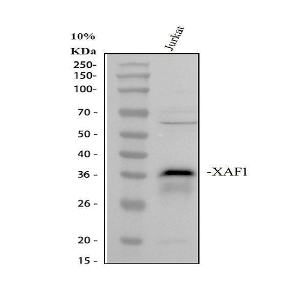
Western blot analysis of XAF1 using anti-XAF1 antibody (PA1218).
Electrophoresis was performed on a 10% SDS-PAGE gel at 80V (Stacking gel) / 120V (Resolving gel) for 2 hours. The sample well of each lane was loaded with 30 ug of sample under reducing conditions.
Lane 1: human Jurkat whole cell lysates.
After electrophoresis, proteins were transferred to a nitrocellulose membrane at 150 mA for 50-90 minutes. Blocked the membrane with 5% non-fat milk/TBS for 1.5 hour at RT. The membrane was incubated with rabbit anti-XAF1 antigen affinity purified polyclonal antibody (PA1218) at 0.5 μg/mL overnight at 4°C, then washed with TBS-0.1%Tween 3 times with 5 minutes each and probed with a goat anti-rabbit IgG-HRP secondary antibody (Catalog # BA1054) at a dilution of 1:5000 for 1.5 hour at RT. The signal is developed using an ECL Plus Western Blotting Substrate (Catalog # AR1196-200) with Tanon 5200 system. A specific band was detected for XAF1 at approximately 35 kDa. The expected band size for XAF1 is at 35 kDa.

Click image to see more details
IF analysis of XAF1 using anti-XAF1 antibody (PA1218).
XAF1 was detected in an immunocytochemical section of U2OS cells. Enzyme antigen retrieval was performed using IHC enzyme antigen retrieval reagent (AR0022) for 15 mins. The cells were blocked with 10% goat serum. And then incubated with 5 μg/mL rabbit anti-XAF1 Antibody (PA1218) overnight at 4°C. DyLight®488 Conjugated Goat Anti-Rabbit IgG (BA1127) was used as secondary antibody at 1:500 dilution and incubated for 30 minutes at 37°C. Visualize using a fluorescence microscope and filter sets appropriate for the label used.

Click image to see more details
Flow Cytometry analysis of SiHa cells using anti-XAF1 antibody (PA1218).
Overlay histogram showing SiHa cells stained with PA1218 (Blue line). To facilitate intracellular staining, cells were fixed with 4% paraformaldehyde and permeabilized with permeabilization buffer. The cells were blocked with 10% normal goat serum. And then incubated with rabbit anti-XAF1 Antibody (PA1218, 1 μg/1x106 cells) for 30 min at 20°C. DyLight®488 conjugated goat anti-rabbit IgG (BA1127, 5-10 μg/1x106 cells) was used as secondary antibody for 30 minutes at 20°C. Isotype control antibody (Green line) was rabbit IgG (1 μg/1x106) used under the same conditions. Unlabelled sample without incubation with primary antibody and secondary antibody (Red line) was used as a blank control.
Specific Publications For Anti-XIAP-associated factor 1 XAF1 Antibody Picoband® (PA1218)
Loading publications
Recommended Resources
Here are featured tools and databases that you might find useful.
- Boster's Pathways Library
- Protein Databases
- Bioscience Research Protocol Resources
- Data Processing & Analysis Software
- Photo Editing Software
- Scientific Literature Resources
- Research Paper Management Tools
- Molecular Biology Software
- Primer Design Tools
- Bioinformatics Tools
- Phylogenetic Tree Analysis
Customer Reviews
Have you used Anti-XIAP-associated factor 1 XAF1 Antibody Picoband®?
Share your experimental results or join a short interview to earn up to $1,000 in product credits or other rewards.
0 Reviews For Anti-XIAP-associated factor 1 XAF1 Antibody Picoband®
Customer Q&As
Have a question?
Find answers in Q&As, reviews.
Can't find your answer?
Submit your question
5 Customer Q&As for Anti-XIAP-associated factor 1 XAF1 Antibody Picoband®
Question
you antibody to test anti-XAF1 antibody PA1218 on human thymus for research purposes, then I may be interested in using anti-XAF1 antibody PA1218 for diagnostic purposes as well. Is the antibody suitable for diagnostic purposes?
Verified Customer
Verified customer
Asked: 2020-05-01
Answer
The products we sell, including anti-XAF1 antibody PA1218, are only intended for research use. They would not be suitable for use in diagnostic work. If you have the means to develop a product into diagnostic use, and are interested in collaborating with us and develop our product into an IVD product, please contact us for more discussions.
Boster Scientific Support
Answered: 2020-05-01
Question
I was wanting to use your anti-XAF1 antibody for IHC for human thymus on frozen tissues, but I want to know if it has been validated for this particular application. Has this antibody been validated and is this antibody a good choice for human thymus identification?
Verified Customer
Verified customer
Asked: 2020-01-16
Answer
It shows on the product datasheet, PA1218 anti-XAF1 antibody has been tested for IHC, WB on human tissues. We have an innovator award program that if you test this antibody and show it works in human thymus in IHC-frozen, you can get your next antibody for free.
Boster Scientific Support
Answered: 2020-01-16
Question
We are currently using anti-XAF1 antibody PA1218 for human tissue, and we are content with the WB results. The species of reactivity given in the datasheet says human. Is it true that the antibody can work on primate tissues as well?
Verified Customer
Verified customer
Asked: 2019-05-15
Answer
The anti-XAF1 antibody (PA1218) has not been tested for cross reactivity specifically with primate tissues, though there is a good chance of cross reactivity. We have an innovator award program that if you test this antibody and show it works in primate you can get your next antibody for free. Please contact me if I can help you with anything.
Boster Scientific Support
Answered: 2019-05-15
Question
Can you help my question with product PA1218, anti-XAF1 antibody. I was wondering if it would be possible to conjugate this antibody with biotin. I would need it to be without BSA or sodium azide. I am planning on using a buffer exchange of sodium azide with PBS only. Would there be problems for me to conjugate the antibody and store it in -20 degrees in small aliquots?
E. Wu
Verified customer
Asked: 2014-10-29
Answer
We do not recommend storing this antibody with PBS buffer only in -20 degrees. If you want to store it in -20 degrees it is best to add some cryoprotectant like glycerol. If you want carrier free PA1218 anti-XAF1 antibody, we can provide it to you in a special formula with trehalose and/or glycerol. These molecules will not interfere with conjugation chemistry and provide a good level of protection for the antibody from degradation. Please be sure to specify this in your purchase order.
Boster Scientific Support
Answered: 2014-10-29
Question
I have attached the WB image, lot number and protocol we used for thymus using anti-XAF1 antibody PA1218. Please let me know if you require anything else.
L. Gonzalez
Verified customer
Asked: 2013-02-13
Answer
Thank you very much for the data. Our lab team are working to resolve this as quickly as possible, and we appreciate your patience and understanding! You have provided everything we needed. Please let me know if there is anything you need in the meantime.
Boster Scientific Support
Answered: 2013-02-13
