This website uses cookies to ensure you get the best experience on our website.
- Table of Contents
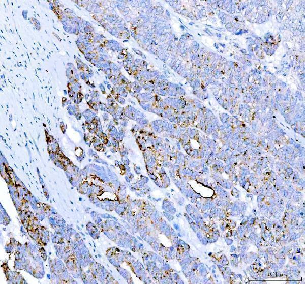
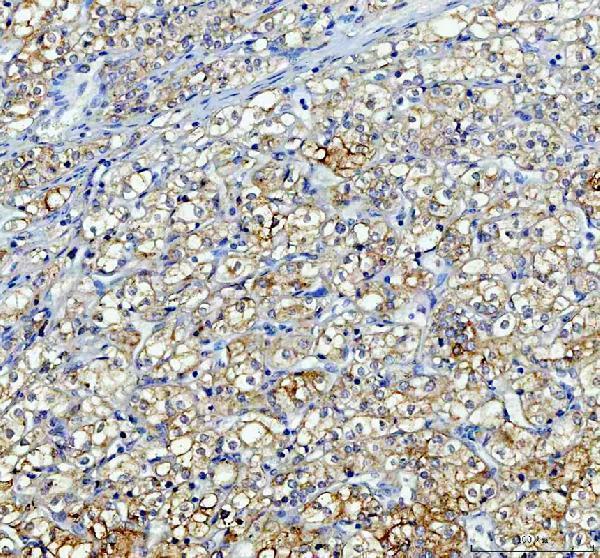
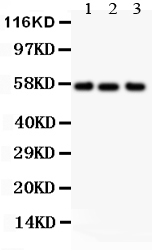
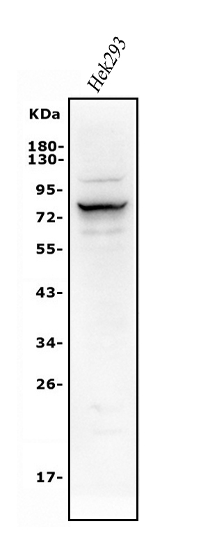
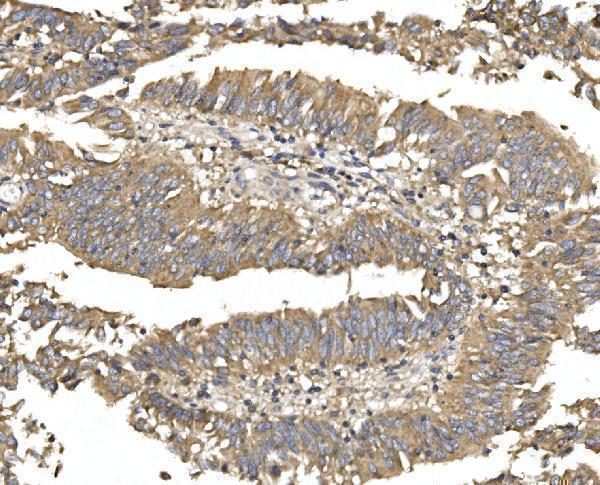
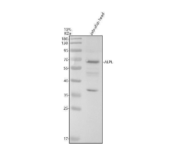
Facts about Alkaline phosphatase, tissue-nonspecific isozyme.
Human | |
---|---|
Gene Name: | ALPL |
Uniprot: | P05186 |
Entrez: | 249 |
Belongs to: |
---|
alkaline phosphatase family |
Akp2; Alkaline phosphatase liver/bone/kidney isozyme; alkaline phosphatase, liver/bone/kidney; alkaline phosphatase, tissue-nonspecific isozyme; alkaline phosphomonoesterase; ALPL; APTNAP; AP-TNAP; EC 3.1.3.1; FLJ40094; FLJ93059; glycerophosphatase; HOPS; liver/bone/kidney-type alkaline phosphatase; MGC161443; tissue-nonspecific ALP; TNAP; TNSALP; TNSALPMGC167935
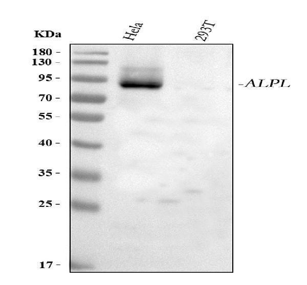
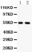
Mass (kDA):
57.305 kDA
Human | |
---|---|
Location: | 1p36.12 |
Sequence: | 1; NC_000001.11 (21508984..21578412) |
Cell membrane; Lipid-anchor, GPI-anchor.
This article will discuss some of the most effective ways to utilize Boster Bio's ALPL Marker. This antibody will detect the apoptosis-associated protein which is also known as ALPL. This antibody is vital in cancer research , but it is also accessible for other uses. If you're looking to test your brand new antibody in cell culture , or any other application, Boster has you covered.
Selecting the right IHC staining protocol is essential. There are numerous options to choose from that include a stepwise procedure illustrated by a flowchart, recommended reagents, as well as an IHC workflow with the ALPL marker. You can also go for more detailed guides that includes 101 Steps to Better Histology, which provides useful tips on IHC sample preparation. To start your IHC workflow, you'll require an IHC tissue sample. You can use biopsy tissue surgical specimens, surgical specimens, or animal models. The autopsy specimen is more of a postmortem autolysis. This is due to the fact that after two hours the antigens could have lost their luster and disappear from the tissue.
To prepare the ALPL marker-based IHC stain make the paraffin-embedded section using a rotating microtome. slice it to a thickness of 2-7 millimeters. After that, the section is fixed at 4°C for long-term storage. The ALPL-containing antibodies are detected using a variety of IHC methods for detection, which range from a single-molecule probe to the phosphorylated chromophore.
Multiplexed IHC has several benefits that include the ability to label multiple markers within one tissue. Since it provides comprehensive information on cell composition and spatial arrangement it is an important tool in the IHC workflow. As a result, multiplexed IHC is an essential tool in cancer immunotherapy. Multiplexed IHC for instance, can be used to detect multiple tumor markers in one specimen. This permits multicolored research in cancer immunotherapy.
Cytokeratin 18 is a different IHC marker. Cytokeratin 18 is found on human colon cancer sections by indirect detection. Through the use of poly-HRP conjugate Cytokeratin 18 can be detected. The signal is then visualized by using the DAB substrate that has a metal enhancement. However, cytokeratin 18 antibodies are not commonly used for IHC. Commercially available a limited number of GO/BGAL conjugates
When using immunoenzymatic staining of tissue the enzyme reacts an unlabeled primary antibody , resulting in an insoluble colored product. The intensity of color produced is correlated with the concentration of the tissue antigen and the primary antibody. The enzymes used are calf intestinal alkaline HRP and phosphatase. The activity of the enzyme will depend on its concentration in the substrate and the buffer. The activity of the enzyme can also be affected by light levels.
HALO is an IHC tool that allows you to analyze the tissue in sections that are serial. It lets you find cells that express the selected biomarker at different locations as well as segment the tumor and then analyze its spatial distribution. In addition, HALO comes with add-ons, including Tissue Classifier FISH-IF Quantification and Spatial Analysis. The software can analyze stained sections of serial sections using additional IHC markers. HALO can be used with many image formats, including ones with multiple biomarkers.
An established method of testing antibodies is required to confirm an IHC validation protocol for the ALPL mark. It is simple to validate an antibody if it is tested on a reputable tissue sample. Furthermore, the validation of an antibody must yield the desired results for both positive and negative tissues and have a good signal-to-noise ratio. If the antibody produces inconsistent or insufficient results the Level 3 validation is advised.
IHC is a specialised stain in histology with significant diagnostic value and is increasingly used in research and development of drugs to determine patients with specific pathologies. It is also a powerful method to select patients in the clinic. The development of an IHC biomarker is dependent on confirmation of the antibody and approval from regulatory organizations. Although thousands of biomarker-related antibodies are sold, only a few have been tested.
The criteria for evaluating the IHC test are usually determined by the number cells stained and the intensity of staining. However, there are a few important distinctions in the scoring systems employed. Megumi et al. scoring method used in the Megumi al. study was based on the percentage BMP-7-positive cells as well as the intensity of staining. These differences in quality should be assessed in relation to the overall quality of test.
The procedure for staining with immunoreactive dye is based on an IHC validation protocol. This method requires that you make use of a positive and negative cultured cell as a control. The buffers used to fix the positive cells must be the same as those used for the controls. The FFPE cell blocks must be processed in a way that is similar to tissue. This validation requires standardizing the processing of FFPE cells blocks and using the microarray of cells to determine the interaction between multiple cells.
To further enhance the quality of IHC data To further improve the quality of IHC data, a standardised validation protocol is highly advised. It lists the minimum requirements for the validation of an antibody. These requirements are similar to those that are used for tissue microarrays or the Clinical Laboratory Standards Institute standardisation efforts. This validation protocol can be applied to frozen and wholemount IHC. The protocol should be able to assist IHC researchers to make reliable and accurate biomarker comparisons.
An IHC validation protocol for the ALPL marker may have several advantages over other scoring methods. It makes it easier to interpret IHC results, increase the quality of data that scientists can use, and save you time and money. A scoring system can increase the productivity of scientists. It clarifies the reporting of data. Here are some advantages of using an established IHC validation procedure for the ALPL marker
The ALPL marker is an epithelial protein with numerous applications. Boster Bio offers the anti-Cytokeratin, Basic (Type II) Monoclonal Antibody. It is a tiny protein, yet highly immunogenic, making it an excellent choice for many different applications. Additionally, Boster offers antibodies with high affinity that have been widely cited in the scientific literature in the past 25 years. Additionally, these antibodies have been validated on Western Blotting, Immunohistochemistry, and ELISA.
Cells are then incubated with fluorescently conjugated antibodies , or an unlabeled antibody in order to utilize the ALPL marker. Then, they are analyzed using an instrument called a flow-cytometer. Make sure that fluorescently conjugated antibodies are biotin-conjugated. Alternately indirect staining can be done using the avidin-biotin method.
The PAP method consists of immunization of an antigen with the HRP moiety. The antibody is able to bind to the HRP moiety, forming a stable polygon. The PAP method is superior because it has a low background for staining tissues. It should be observed shortly after staining and mounted using buffered glucose medium. The anti-fluorescence degrading medium should not be stored at 4° Celsius for more than a week. It may not degrade the signal for less than a month.
Indirect detection is a more effective method than direct detection. The indirect method requires relatively only a few standard conjugated secondary antibodies and is extremely sensitive and specific. This method is widely employed to screen monoclonal antibody prior to scaling up to manufacturing at a large scale. There are disadvantages for both direct and indirect detection methods. Direct detection is usually less sensitive than indirect detection. However, the latter has higher sensitivity. Indirect detection allows you to apply various types of controls to the results.
PMID: 3532105 by Weiss M.J., et al. Isolation and characterization of a cDNA encoding a human liver/bone/kidney-type alkaline phosphatase.
PMID: 3165380 by Weiss M.J., et al. Structure of the human liver/bone/kidney alkaline phosphatase gene.
*Showing only the more recent 20. More publications can be found for each product on its corresponding product page