This website uses cookies to ensure you get the best experience on our website.
- Table of Contents
1 Citations 9 Q&As

2 Citations 17 Q&As
2 Citations 15 Q&As


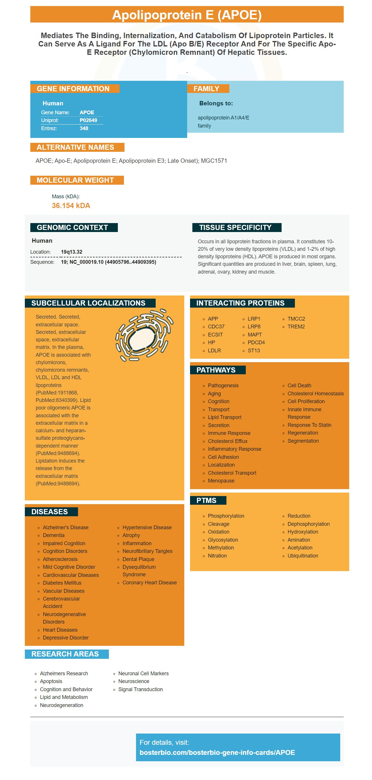
Facts about Apolipoprotein E.

.
| Human | |
|---|---|
| Gene Name: | APOE |
| Uniprot: | P02649 |
| Entrez: | 348 |

| Belongs to: |
|---|
| apolipoprotein A1/A4/E family |

APOE; Apo-E; Apolipoprotein E; apolipoprotein E3; late onset); MGC1571
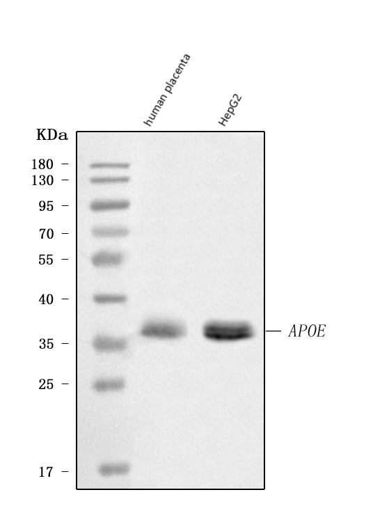
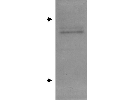
Mass (kDA):
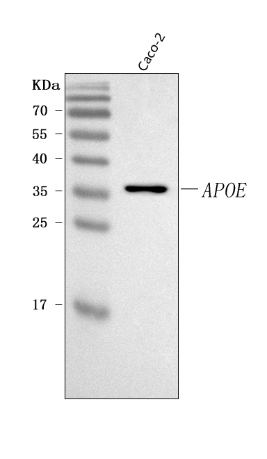
36.154 kDA

| Human | |
|---|---|
| Location: | 19q13.32 |
| Sequence: | 19; NC_000019.10 (44905796..44909395) |
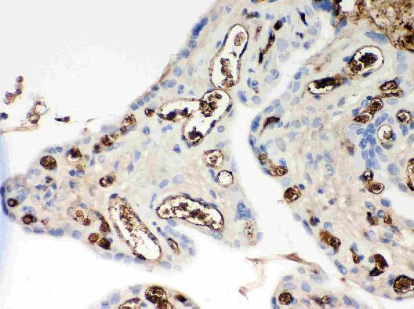
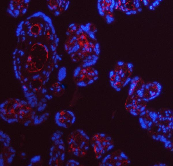
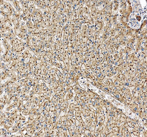
Occurs in all lipoprotein fractions in plasma. It constitutes 10-20% of very low density lipoproteins (VLDL) and 1-2% of high density lipoproteins (HDL). APOE is produced in most organs. Significant quantities are produced in liver, brain, spleen, lung, adrenal, ovary, kidney and muscle.
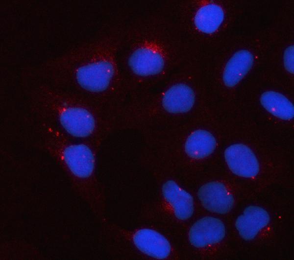
Secreted. Secreted, extracellular space. Secreted, extracellular space, extracellular matrix. In the plasma, APOE is associated with chylomicrons, chylomicrons remnants, VLDL, LDL and HDL lipoproteins (PubMed:1911868, PubMed:8340399). Lipid poor oligomeric APOE is associated with the extracellular matrix in a calcium- and heparan-sulfate proteoglycans-dependent manner (PubMed:9488694). Lipidation induces the release from the extracellular matrix (PubMed:9488694).


PMID: 6325438 by Zannis V.I., et al. Synthesis, intracellular processing, and signal peptide of human apolipoprotein E.
PMID: 6327682 by McLean J.W., et al. Human apolipoprotein E mRNA. cDNA cloning and nucleotide sequencing of a new variant.
*More publications can be found for each product on its corresponding product page