This website uses cookies to ensure you get the best experience on our website.
- Table of Contents
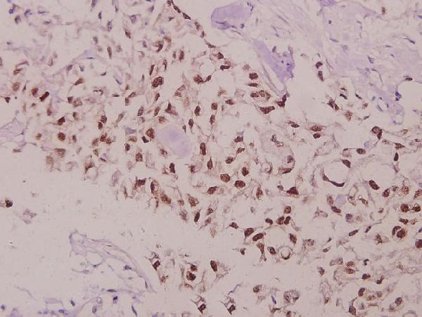
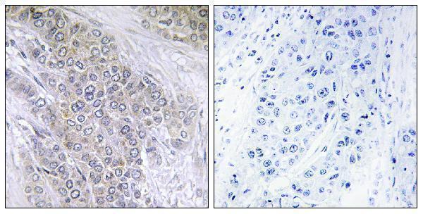
2 Citations 16 Q&As
22 Citations 16 Q&As
158 Citations 16 Q&As
1 Citations


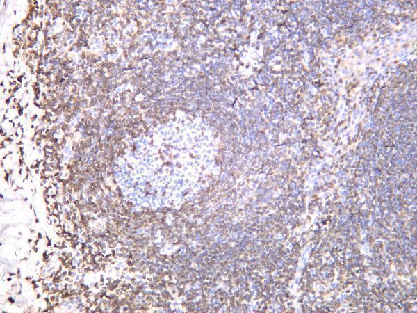
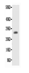
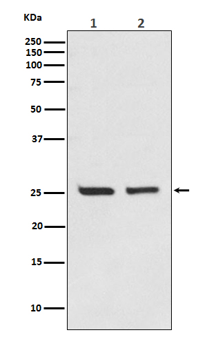
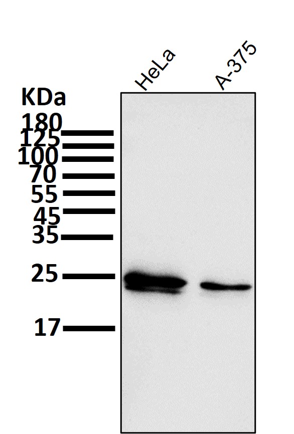
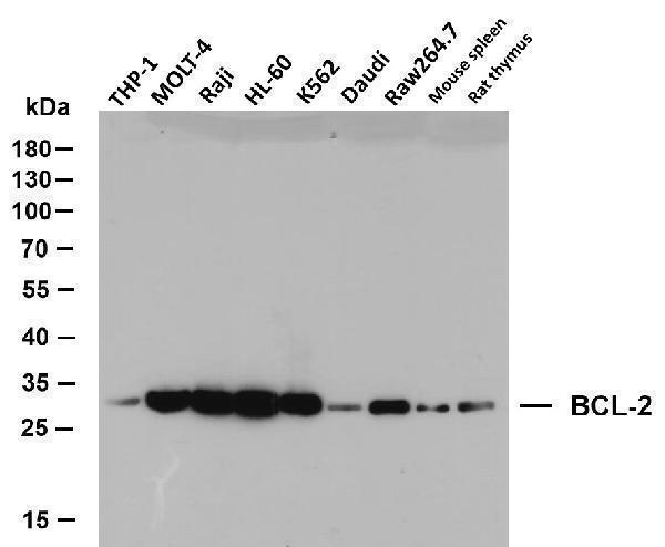
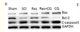
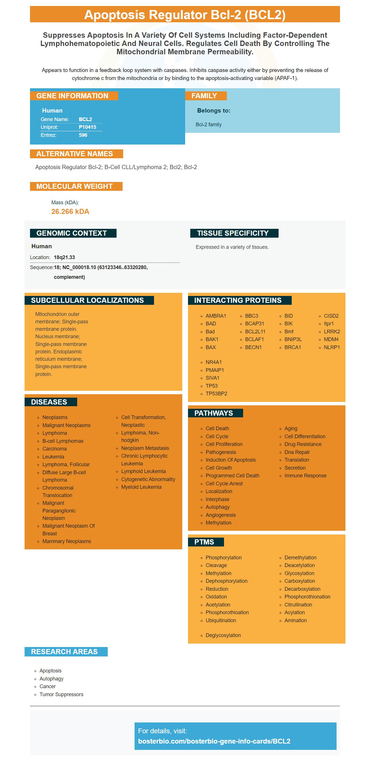
Facts about Apoptosis regulator Bcl-2.

Appears to function in a feedback loop system with caspases. Inhibits caspase activity either by preventing the release of cytochrome c from the mitochondria or by binding to the apoptosis-activating variable (APAF-1).
| Human | |
|---|---|
| Gene Name: | BCL2 |
| Uniprot: | P10415 |
| Entrez: | 596 |

| Belongs to: |
|---|
| Bcl-2 family |

apoptosis regulator Bcl-2; B-cell CLL/lymphoma 2; Bcl2; Bcl-2
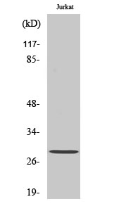
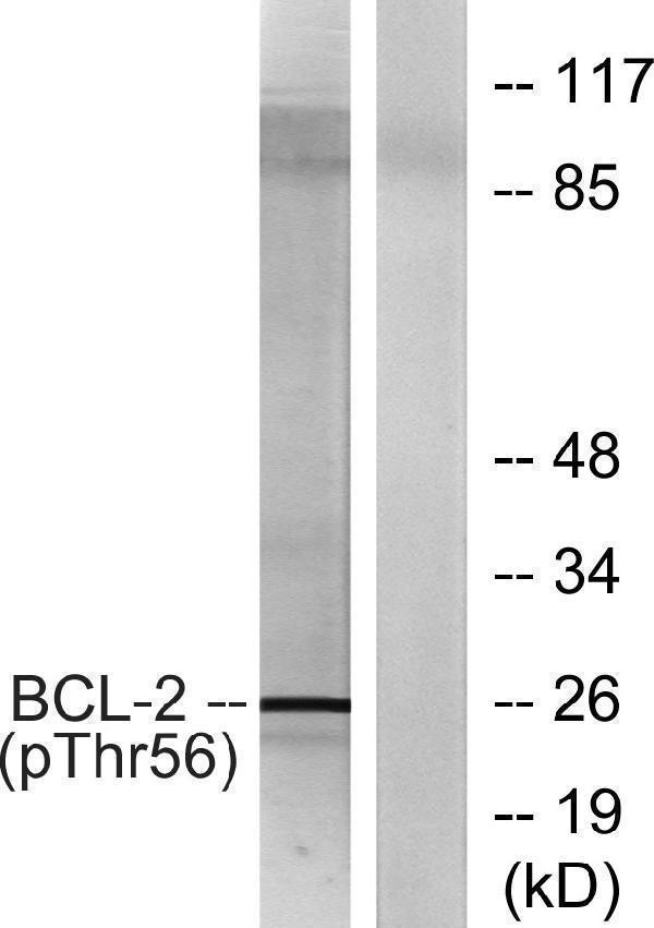
Mass (kDA):
26.266 kDA

| Human | |
|---|---|
| Location: | 18q21.33 |
| Sequence: | 18; NC_000018.10 (63123346..63320280, complement) |
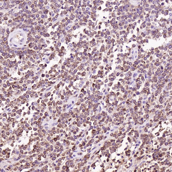
Expressed in a variety of tissues.
Mitochondrion outer membrane; Single-pass membrane protein. Nucleus membrane; Single-pass membrane protein. Endoplasmic reticulum membrane; Single-pass membrane protein.


PMID: 3523487 by Tsujimoto Y., et al. Analysis of the structure, transcripts, and protein products of bcl- 2, the gene involved in human follicular lymphoma.
PMID: 1508712 by Eguchi Y., et al. Isolation and characterization of the chicken bcl-2 gene: expression in a variety of tissues including lymphoid and neuronal organs in adult and embryo.
*Showing only the more recent 20. More publications can be found for each product on its corresponding product page