This website uses cookies to ensure you get the best experience on our website.


- Table of Contents
Plan chromogenic CDK2 IHC in paraffin sections using the nuclear staining observed in subsets of intestinal, squamous and germinal-center cells as a reference (HPA tissue IHC). Consider epitope coverage across the 2 annotated isoforms when selecting an antibody (UniProt).

Expected localisation, validated protocols, controls and antibodies — the at-a-glance facts below, then the full design guide.
| Expected localisation | Nuclear staining in subsets of cells (HPA tissue IHC) | |
| Staining pattern | Nuclear staining in intestinal, squamous and germinal-center cells (HPA tissue IHC) | |
| Antigen retrieval | EDTA pH 8.0 HIER, heat-mediated (datasheet M00166-4) | |
| Positive control | Appendix+4 more · see all | |
| Negative control | Adipose tissue+4 more · see all |
| Fixation | Keep paraffin-section fixation consistent (standard IHC practice; not target-specific); Matched tissue-IHC evidence does not establish the fixation claim. Validate the specimen-specific method before use. (selected-SKU IHC image M00166-4) | |
| Caveat | Staining and RNA show medium consistency (HPA tissue IHC) | |
| Regulation | No tissue-specific expression pattern is annotated (UniProt) | |
| Isoform / epitope | 2 isoforms; epitope coverage needs checking (UniProt) |
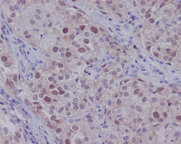
The catalog antibody protocol (datasheet: M00166-4) is followed by four published CDK2 IHC protocols (PMC11831524; PMC2816830; PMC3441142; PMC2668819).
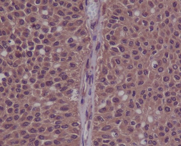
| Sample | Paraffin-embedded human laryngeal squamous cell carcinomas tissue; fixative not specified (datasheet M00166-4) |
| Fixation | Image fixative and duration unreported (datasheet M00166-4); verify before use. |
| Sectioning | 4–5 µm sections on charged slides (standard) |
| Deparaffinisation | Xylene, graded ethanol series to water (standard) |
| Antigen retrieval | Heat retrieval: EDTA pH 8.0 (datasheet M00166-4); 20 min, 95–100 °C (standard) |
| Peroxidase block | 3% H2O2, 10 min, room temperature (standard) |
| Blocking | 10% goat serum (datasheet M00166-4) |
| Primary antibody | Mouse monoclonal (clone 6D5B5) anti-CDK2, 2 μg/ml (datasheet M00166-4) |
| Primary incubation | Overnight at 4 °C (datasheet M00166-4) |
| Detection | HRP-conjugated secondary, DAB chromogen (datasheet M00166-4) |
| Counterstain | Hematoxylin, blue, dehydrate and mount (standard) |
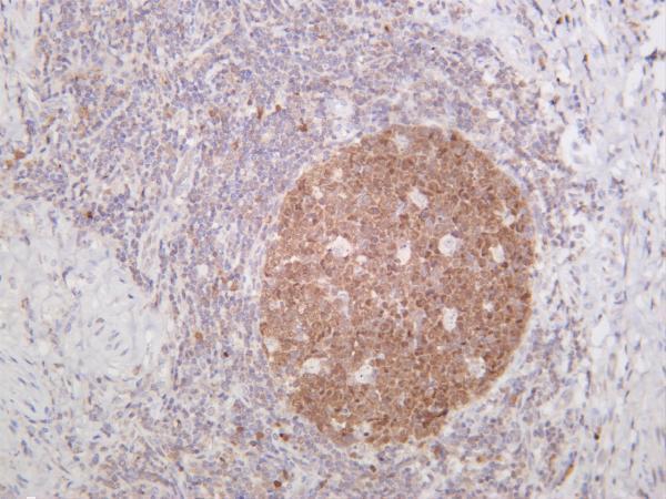
| Expected result | CDK2-positive staining in lymphoid tissue of appendix (HPA tissue IHC: Medium). HPA tissue profile: Nuclear expression in subsets of cells in the intestinal tract and squamous epithelia, cells in germinal centers and seminiferous ducts. No signal in the no-primary control. |
CDK2 staining in paraffin sections should be predominantly nuclear in subsets of intestinal glandular cells, squamous epithelial cells, germinal center cells and cells of seminiferous ducts (HPA tissue IHC). HPA rates the tissue pattern Enhanced, with medium consistency between staining and RNA expression (HPA tissue IHC). CDK2 can also occupy cytoplasm and centrosomes; it has no transmembrane segment (UniProt P24941 localization and topology).
| Subset-restricted nuclear staining in intestinal glands, squamous epithelium or germinal centers. | This matches the reported tissue pattern (HPA tissue IHC). Duodenal and rectal glandular cells, esophageal and oral squamous cells, and lymph-node germinal center cells each have Medium staining (HPA tissue IHC). Score the named cell population and its nuclear signal rather than treating the entire tissue section as uniformly positive (HPA tissue IHC; general IHC practice). |
| Strong, widespread membrane-only or extracellular staining with little nuclear signal. | This differs from the reported nuclear tissue pattern (HPA tissue IHC). CDK2 has no transmembrane segment (UniProt P24941 topology). Review morphology and controls before calling it CDK2: HPA also reports a plasma-membrane location in ICC-IF, but marks that location uncertain (HPA subcellular ICC-IF). |
| Prominent staining of an HPA-listed negative cell population, such as adipocytes. | Adipocytes in adipose tissue and breast are reported as Not detected (HPA tissue IHC). Check whether the signal follows tissue edges or appears in the negative control; cross-reactivity or detection background is possible (general IHC practice). A positive result elsewhere in the section does not validate staining in adipocytes (general IHC practice). |
| Diffuse color across nuclei, cytoplasm and surrounding tissue, obscuring cell boundaries. | The observed tissue pattern is nuclear in subsets, so an indiscriminate haze is difficult to interpret (HPA tissue IHC). Cytoplasmic CDK2 is biologically possible (UniProt P24941 localization); cytoplasmic color alone is therefore insufficient to label a section artefactual. Use background controls and preserved morphology to distinguish a cellular pattern from diffuse detection background (general IHC practice). |
| No nuclear staining in lymph-node germinal centers or esophageal squamous cells. | Both populations have Medium staining in the HPA tissue record (HPA tissue IHC). First check that the expected cells are present and interpretable, then assess assay performance with a known-positive section and detection controls (general IHC practice). Absence in one field cannot establish CDK2 absence throughout a heterogeneous tissue (HPA tissue IHC). |
| Cell population and intensity | HPA reports Medium staining in several named epithelial and lymphoid populations, Low staining in placental cytotrophoblasts, and Not detected in specified cells such as adipocytes (HPA tissue IHC). Compare like cell types, not whole organs: a negative call for one cell population does not describe every cell in that tissue (HPA tissue IHC). |
| Compartment and cell-cycle context | HPA's tissue profile is nuclear in subsets (HPA tissue IHC). UniProt also lists cytoplasm, Cajal bodies, endosomes and centrosomes, with centrosomal localization in late G2 (UniProt P24941 localization). A small extranuclear signal can merit review, but localization alone does not establish kinase activity or cell-cycle phase (UniProt P24941 localization and function; general IHC practice). |
| Antibody validation and scope | The HPA tissue record is Enhanced, while its stated antibody-staining/RNA consistency is medium (HPA tissue IHC). CAB013115 is listed as IHC Enhanced; HPA066915 is listed as ICC Enhanced (HPA antibody validation). Apply the IHC evidence to paraffin-section expectations without treating the ICC rating as IHC validation (HPA antibody validation). |
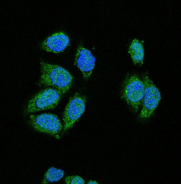
| Q: What should IF/ICC show? | A: Mainly nucleoplasmic CDK2; centrosome and basal-body locations are supported, calyx is approved, and plasma-membrane location is uncertain (HPA subcellular ICC-IF). Those annotations describe ICC-IF localization, not a paraffin-section staining protocol or a requirement that every IHC-positive cell show each additional compartment (HPA subcellular ICC-IF; HPA tissue IHC). |
| Situation | Likely cause | Next action |
|---|---|---|
| Positive-control tissue has no nuclear signal. | The expected cells may be absent from the field, or the staining run may have failed (HPA tissue IHC; general IHC practice). | Locate the named positive population, then verify retrieval, antibody incubation and chromogenic detection against the catalog antibody's IHC-P instructions (HPA tissue IHC; general IHC practice). |
| Every structure develops color. | Diffuse detection background or excess staining can mask the subset-restricted nuclear pattern (HPA tissue IHC; general IHC practice). | Compare a negative-control slide and review blocking, washes and chromogen development before interpreting cell localization (general IHC practice). |
| Adipocytes stain as strongly as germinal-center cells. | That distribution conflicts with the reported Not detected adipocytes and Medium germinal-center cells (HPA tissue IHC). | Inspect morphology and negative controls; report the unexpected population separately and confirm before calling it specific (general IHC practice). |
| Signal appears only at tissue edges or in extracellular spaces. | Such a distribution differs from the reported nuclear pattern (HPA tissue IHC). | Check for section-edge artefact and compare the same areas with a negative control; avoid scoring extracellular color as positive cells (general IHC practice). |
| Weak staining is seen in colon glands. | Colon glandular cells are listed as Low, whereas duodenal and rectal glandular cells are Medium (HPA tissue IHC). | Score the named cell type and compare with a listed medium-staining population on a separately controlled section before calling the run failed (HPA tissue IHC; general IHC practice). |
| Cytoplasmic signal accompanies nuclear staining. | Cytoplasm is a UniProt-listed CDK2 location, although HPA describes the tissue IHC pattern as nuclear in subsets (UniProt P24941 localization; HPA tissue IHC). | Record nuclear and cytoplasmic staining separately; use negative controls to assess the cytoplasmic component rather than assuming it is specific or artefactual (general IHC practice). |
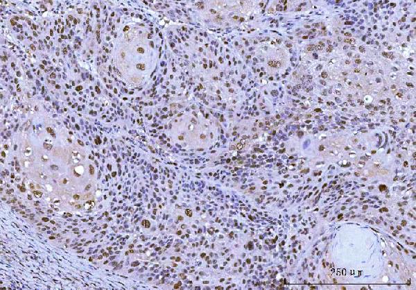
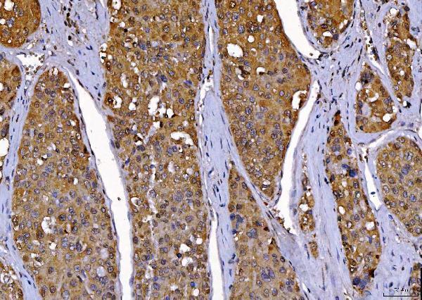
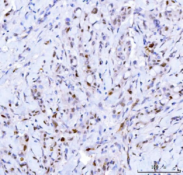
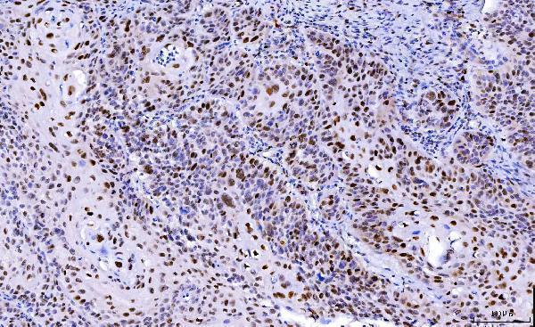




Comprehensive Human Protein Atlas IHC scoring per tissue (reliability: Enhanced — Medium consistency between antibody staining and RNA expression data.). Rows are taken directly from the HPA tissue chart — click any row's HPA link to view the source.
| Tissue | Cell type | Level | Evidence | Source |
|---|---|---|---|---|
| Appendix | Lymphoid tissue | Medium | Protein (IHC) | HPA → |
| Bone marrow | Hematopoietic cells | Medium | Protein (IHC) | HPA → |
| Duodenum | Glandular cells | Medium | Protein (IHC) | HPA → |
| Esophagus | Squamous epithelial cells | Medium | Protein (IHC) | HPA → |
| Lymph node | Germinal center cells | Medium | Protein (IHC) | HPA → |
| Tissue | Cell type | Level | Evidence | Source |
|---|---|---|---|---|
| Adipose tissue | Adipocytes | Not detected | Protein (IHC) | HPA → |
| Breast | Adipocytes | Not detected | Protein (IHC) | HPA → |
| Bronchus | Basal cells | Not detected | Protein (IHC) | HPA → |
| Cerebellum | Cells in granular layer | Not detected | Protein (IHC) | HPA → |
| Cerebral cortex | Endothelial cells | Not detected | Protein (IHC) | HPA → |
Troubleshoot CDK2 staining in paraffin section IHC by checking retrieval, cell type, compartment, and controls before interpreting signal.
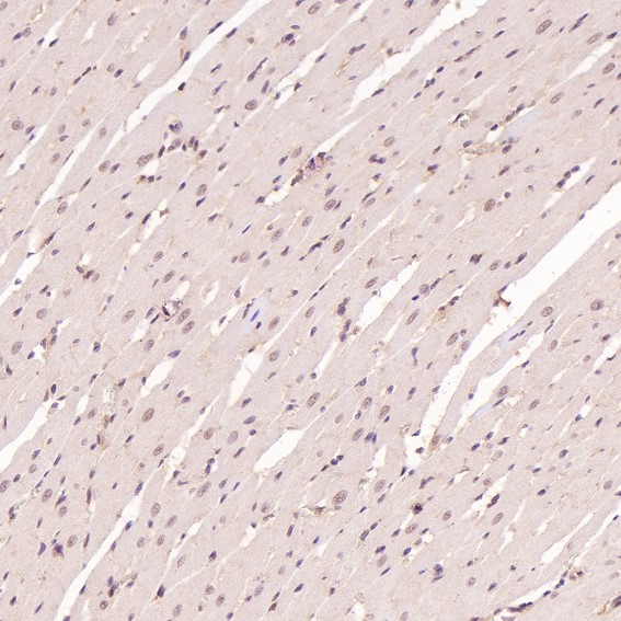
Anti-CDK2 antibodies have IHC images from human and rat tissue (catalog IHC captions) and IF/ICC images from human cells (M00166 and PB9534 IF captions).
M00166-4 was imaged in human laryngeal carcinoma, M00166-3 in human breast cancer, and PB9534 in human bladder carcinoma (respective IHC captions). M00166 was imaged in human breast carcinoma and rat heart, M00166-6 in human tonsil, and phospho-CDK2 (Y15) antibody MP00166 in human bladder cancer (respective IHC captions; MP00166 catalog title).
Which to pick: For paraffin-section IHC, choose mouse monoclonal M00166-4: its own caption documents EDTA retrieval at pH 8.0 and 2 μg/ml primary antibody; the fixative is unreported (M00166-4 catalog entry and IHC caption). For IF/ICC, consider rabbit monoclonal M00166 or rabbit PB9534; both list IF/ICC and have human-cell IF images (M00166 and PB9534 catalog entries and IF captions). For work spanning human and rat tissue, M00166 has IHC images in both, while mouse reactivity is listed in the catalog without a mouse IHC image in the supplied captions (M00166 catalog reactivity and IHC captions).