This website uses cookies to ensure you get the best experience on our website.
- Table of Contents
2 Citations 9 Q&As
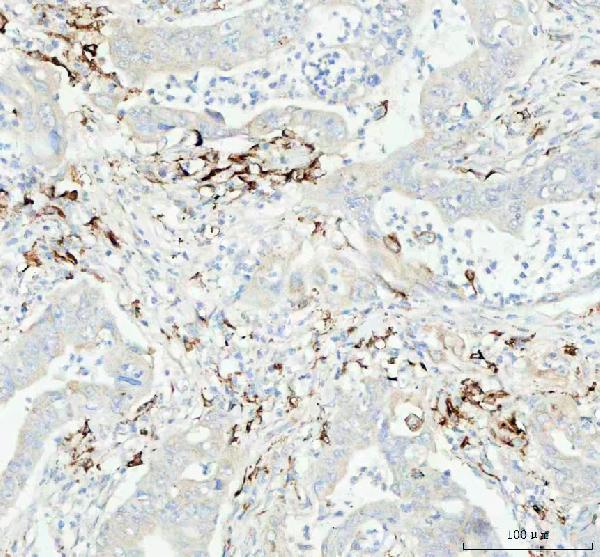
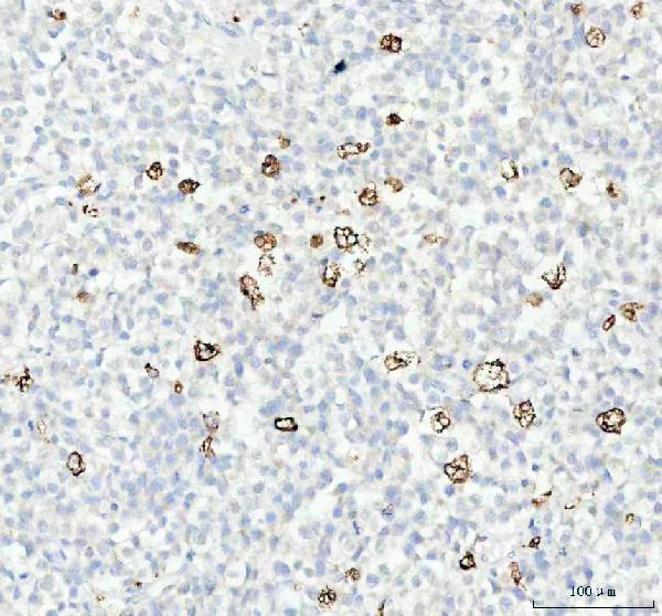
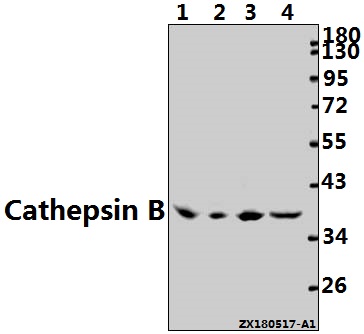
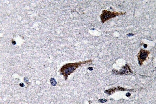
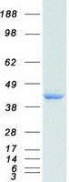
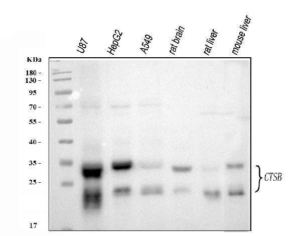
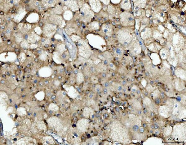
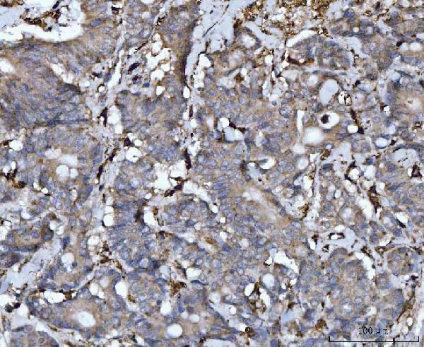
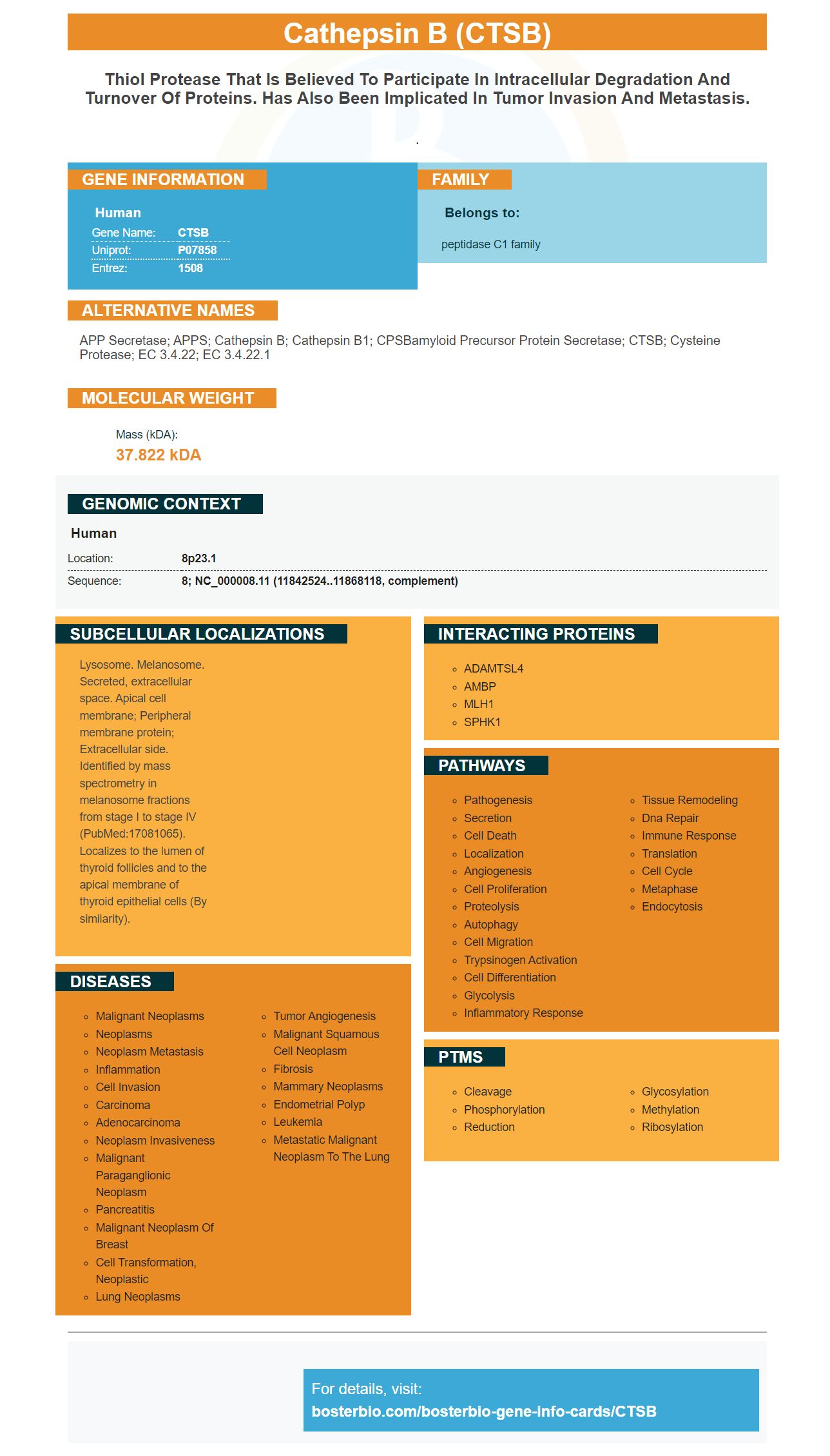
Facts about Cathepsin B.
.
Human | |
---|---|
Gene Name: | CTSB |
Uniprot: | P07858 |
Entrez: | 1508 |
Belongs to: |
---|
peptidase C1 family |
APP secretase; APPS; Cathepsin B; Cathepsin B1; CPSBamyloid precursor protein secretase; CTSB; cysteine protease; EC 3.4.22; EC 3.4.22.1
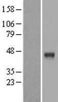
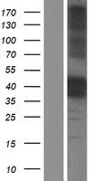
Mass (kDA):
37.822 kDA
Human | |
---|---|
Location: | 8p23.1 |
Sequence: | 8; NC_000008.11 (11842524..11868118, complement) |
Lysosome. Melanosome. Secreted, extracellular space. Apical cell membrane; Peripheral membrane protein; Extracellular side. Identified by mass spectrometry in melanosome fractions from stage I to stage IV (PubMed:17081065). Localizes to the lumen of thyroid follicles and to the apical membrane of thyroid epithelial cells (By similarity).
This article will discuss the advantages and methods behind Boster's Anti-Cathepsin B/CTSB marker. This article covers the benefits and applications, as well as the validation and the methods used by this new biomarker. Boster has a global network that includes scientists who work together to make this product accessible to researchers and developers. By following the procedures and benefits that are described here, you'll be well on your way to having a successful lab experience.
Boster Anti-Cathepsin Bo/CATSB Marker is a highly specific antibody, is used to identify human Cathepsin B using Western Blots or directly ELISAs. This antibody is compatible with IHC and WB applications and reacts with Cathepsin B from human, mice, and rats. It has been tested for specificity and high affinity. It can be stored at -20°C for a period of a year.
Cathepsin B is an important cancer therapeutic molecule. It plays an important part in many cancer-related processes and pathologies within human cells. Invasive cancer-related phenotypes are associated with the expression of this enzyme. The protein is typically associated with lysosomes with low pH. Cathepsin B facilitates autophagy and immune responses. Its ability to break down extracellular matrix molecules is essential in cancer research and treatment.
The USF-1 and USF-2 genes regulate transcription of cathepsin B. USF1c and USF2c inhibit the binding of microRNAs involved in the regulation of tumors which regulates cathepsin B. The USF1 protein is the only cathepsin B/CTSB marker that is RNA-based of its kind.
Boster's Anti-Catheptsin Marker B/CTSB is a powerful multi-faceted anti-cancer immunotherapy tool. It is particularly effective in clinical trials that require targeted chemotherapy or radiotherapy. Because this molecule plays such an important role in metastatic and cancer-related processes the targeting of it can have significant clinical benefits.
This condition can be treated by the application of the CTSB Marker for diagnosis. The CTSB marker is present in tumors, particularly in the invasive and neovascularized margins. This protein plays a crucial role in the immune system's response and invasion, and regulates the process of anti-apoptosis and tumor angiogenesis. This marker has been examined in a small amount of tumors, therefore the research on it is restricted.
The CTSB gene is found in every cell of the body which includes the brain and the prostate. In mice, CTSB significantly correlated with the glioma-related tumor markers like the p-ERK gene, c-Myc and the SNAP-A. The gene is also closely related to the TAM subtypes of Gliomas and MDSC. The use of this gene in gliomas is still in its early stages.
This gene plays a variety of roles in the body. CTSB is involved in numerous aspects hepatitis B virus infections and has been associated with a higher risk of hepatocellular carcinoma. When CTSB interacts with BPSP it can enhance the ability of HCC cells to invade and migrate. HCC cells in the laboratory. CTSB also interacts with HBV DNA, increasing its liver activity.
The protein is also implicated in the metastasis of cancer. Cathepsin B facilitates invasion and alters the extracellular matrix. While its role in the invasion of cells in a collective fashion is unclear, the mechanism through which this protein contributes to this phenomenon is still to be investigated. It was nevertheless discovered that CTSB is highly expressed in the invasive aspect of SACC and that its expression was associated with a poor prognosis of the disease. The significance of the protein was further examined and a 3D spheroid invasion test created to simulate the invasion of cells by collective cells.
In a mouse model of pancreatic neuroendocrine tumorigenesis, CTSB regulates the activation of the Wnt/b-catenin axis and also interacts with the NLRP3 inflammasome, which activates the pro-tumor immune response. CTSB is involved in antigen-presenting, immune cells differentiation, and homeostasis. It is tightly linked to cervical cancer-related immune suppression and can cause the death of T lymphocytes that are CD8+.
We also compared the expression of CTSB markers in differentiated L6 myoblast cultures and those treated with vehicle. After treatment with AICAR, levels of mRNA as well as protein of CTSB were significantly higher three hours later. CTSB levels in intracellular cells were similar in both conditions. These results indicate that CTSB is highly expressed in L6 myoblasts and is a reliable marker of CNS-related illnesses.
Furthermore in addition, the CTSB marker could also function as a biomarker for gliomas, and a practical target for gliomas. Future applications include targeted drug delivery, precise diagnosis and treatment, as well as surgical procedures that are guided by images. These studies indicate that the CTSB marker could be a powerful tool to fight gliomas. It has to be validated first. This means that more clinical studies are needed to validate this marker.
The targeted therapies for gliomas are still a new field. CTSB is still a issue. The CTSB Marker has been found to be highly expressed in gliomas . It is associated with malignant molecular characteristics as well as immune cell infiltration and immunosuppression processes. It could possibly serve as a therapeutic and biomarker for prognosis. This could assist doctors in deciding which cancer treatments are the most effective.
Another significant finding from the CTSB marker was the significant increase in plasma CTSB levels due to exercise. This finding was in correlation with aerobic fitness, which suggests that exercise could increase CTSB levels. The positive correlation between CTSB recall and CF recall was also dependent on the changes in hippocampal volume and aerobic fitness. These results suggest that CTSB can be used to measure the improvements in memory associated with exercise.
Interestingly, it is interesting to note that the CTSB marker is also expressed in gliomas of high grade with low grade tumor grade and IDH-wild-type gliomas. CTSB expression is associated with a poor prognosis for HGG. Further it is believed that the CTSB gene is associated with anti-apoptogenesis as well as tumor angiogenesis. However, previous research has focused on GBMs and not on large-scale clinical research.
The CTSB gene expression level is highly correlated with gliomas, a type of cancer, as well as with immunosuppression processes. CTSB is a promising biomarker. This study outlines the ways CTSB can be used to diagnose gliomas and how it can be used to predict the outcome of gliomas. This includes: the use of CTSB in cancer immunotherapy and identifying the tumor of the patient, and determining whether CTSB is present in the tumor.
The molecular and cellular functions of CTSB are not yet fully understood. CTSB regulates autophagy and the lysosome population, and plays a role in calcium efflux. CTSB also blocks the phosphorylation TFEB, a marker of the lysosomal process. As a result, CTSB may be involved in the regulation of autophagy as well as the biogenesis of lysosomes when under homeostatic conditions.
The CTSB gene was first assigned to the 8p22 chromosome by Wang et al. They then the gene was mapped to 8p23.1-p22 using Southern blot analysis. In addition, Fong et al. CTSB was mapped to 8p23.1 - p22 using hybridization signals from normal fibroblast interphase cells as well as cathepsin-containing cells. The gene was also discovered in mice by Deussing and al.
The molecular biology community has implicated cathepsin B in the process of removing tumors, enhancing invasion of malignant tumors by altering the extracellular matrix. However, the significance of cathepsin B in collective cell invasion remains unclear. SACC remains a cell that exhibits collective invasion and CTSB knockdown cells express the CTSB gene at the invasive end. SACC patients with CTSB overexpression in the invasive portion of SACC were less likely to survive. This research led to the creation of an 3D Spheroid Invasion test to recapitulate the movement of collective cells.
In a group of 61 patients suffering from CRC, serum CTSB and mRNA levels was measured. Serum CTSB expression was significantly higher in patients suffering from lymph node metastases compared to normal mucosal cells. CRC patients may be able detect lymph node metastases through the increase in CTSB levels in their serum. It may also help in the detection of lymph node metastases among patients with advanced cancer stages.
PMID: 3463996 by Chan S.J., et al. Nucleotide and predicted amino acid sequences of cloned human and mouse preprocathepsin B cDNAs.
PMID: 8112600 by Cao L., et al. Human gastric adenocarcinoma cathepsin B: isolation and sequencing of full-length cDNAs and polymorphisms of the gene.
*More publications can be found for each product on its corresponding product page