This website uses cookies to ensure you get the best experience on our website.
- Table of Contents
103 Citations 16 Q&As
137 Citations 16 Q&As
61 Citations 18 Q&As
43 Citations 16 Q&As
31 Citations 16 Q&As
44 Citations 4 Q&As
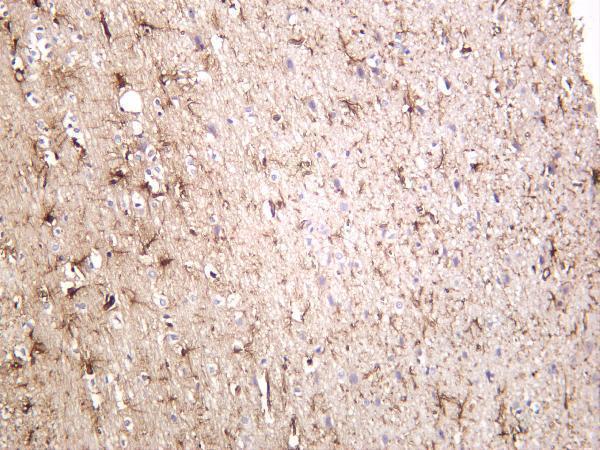
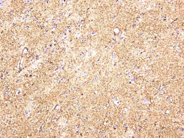
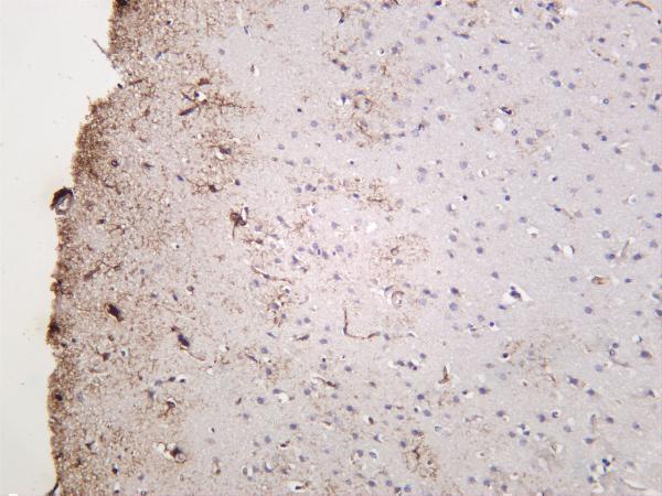
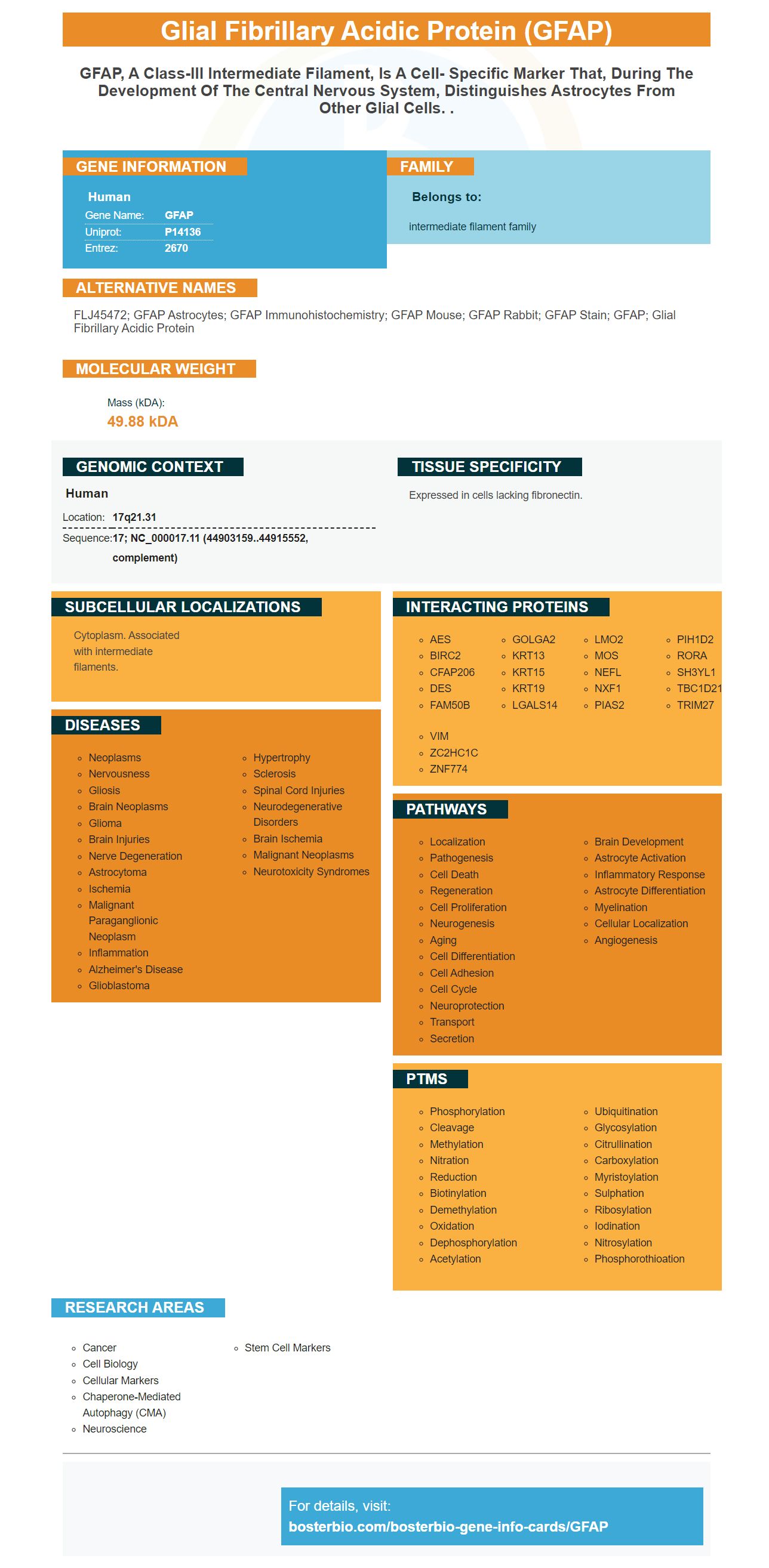
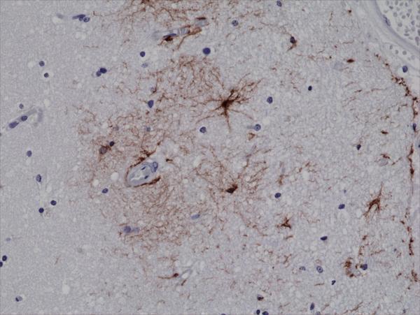
Facts about Glial fibrillary acidic protein.
Human | |
---|---|
Gene Name: | GFAP |
Uniprot: | P14136 |
Entrez: | 2670 |
Belongs to: |
---|
intermediate filament family |
FLJ45472; GFAP astrocytes; GFAP immunohistochemistry; GFAP mouse; GFAP rabbit; GFAP stain; GFAP; glial fibrillary acidic protein
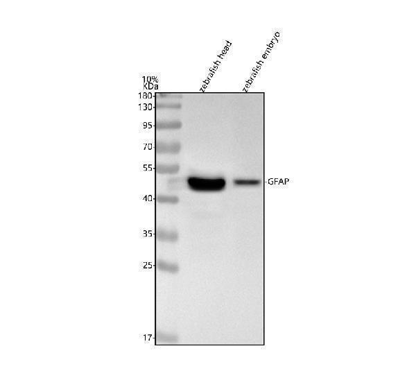
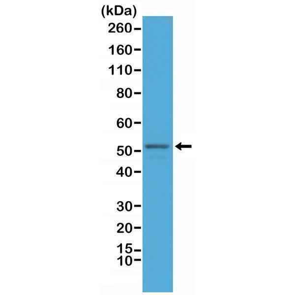
Mass (kDA):
49.88 kDA
Human | |
---|---|
Location: | 17q21.31 |
Sequence: | 17; NC_000017.11 (44903159..44915552, complement) |
Expressed in cells lacking fibronectin.
Cytoplasm. Associated with intermediate filaments.
The GFAP Marker is a biofluid biomarker with diagnostic, prognostic and theranostic value. Its clinical value includes monitoring the damage to astrocyte cells following an hemorrhagic stroke. However, a lot of research remains to be conducted in order to determine how this marker functions. The article summarizes the most promising uses of the GFAP marker. It also discusses how physicians can make use of its potential.
This article discusses the diagnostic, prognostic, and theranostic benefits of GFAP in the brain. Giza, C. C. and Bazarian, J. S. Ashwal, S. Kerwin, A. J. Winston, E. S. Rowell, S. E. and Dec, K. are the authors of this study.
This study proves that GFAP is a reliable biofluid marker with diagnostic, prognostic as well as theranostic utility. The optimal cutoff point for GFAP and UCH-L1 was calculated by looking at the two biofluid-based markers. The optimal cutoff was 1.559 ng/ml and showed an sensitivities of 84.6 percent with a specificity of 69.2 percent, and positive predictive value (PPV) of 64.7%.
GFAP is an brain-specific protein, which functions as a key part of the cytoskeleton of astrocytes. GFAP was recently identified as a biofluid marker in the peripheral blood because of disruptions to the blood-brain barrier. In neurological diseases, higher levels of GFAP may be diagnostic and predictive.
Six months after the traumatic brain injury A higher level of GFAP was associated with lower outcomes. This may enable early risk stratification. Additionally the serum GFAP levels are related to the nature of the brain injury, indicating that the biomarker can assist in predicting the outcomes of patients in the brain.
Glial fibrillary acidic proteins (GFAP), may be used as a prognostic or diagnostic biomarker for stroke patients. It could also be used in assessing the response of patients to treatments and monitor changes in biochemicals after traumatic brain injury. The temporal profile of GFAP is not completely known.
Researchers have identified a brand new gene, GFAP, as a potential autoantigen in the aftermath of brain injury. The gene is a precursor to a family of proteins called glial fibrillary acidic protein (GFAP). It is found in the brain and is present in a variety of organs including the cerebellum, skull and thalamus. It is used to predict the outcomes of brain injuries, strokes and PMS episodes.
The expression of GFAP has been implicated in various neurological disorders, such as stroke, traumatic brain injury and trauma to the brain. In the next article, we present an antibody that recognizes both the GFAP-BDP and human GFAP. This antibody recognizes the full length GFAP and its cleavage products with calpain. Additionally, it shows the specificity of GFAP-BDP and is able to detect the protein in the human brain.
There are many ways in which the protein of the brain that has been injured enters the blood. One of them is disruption of the BBB release without regard to BBB integrity or passive diffusion from cerebral spinal fluid to blood. It is possible that the protein manifests first in the extracellular area of the brain, and then is transferred to blood. After brain trauma, the dominant autoantigen is the GFAP Marker.
There are currently four main GFAP forms: GFAP–a, GFAP–b, and GFAP–c. The first is known as GFAP a, which is the most commonly used variant. The other is GFAP-d which has a non-neural exon. Both are distinct and have distinct functions.
The GFAP protein, also known as glial fibrillary acidic protein is released into biofluids after hemorrhagic stroke. This marker can be used as a biomarker that can be used to diagnose neurological diseases. Williams, C.K. and Vinters, H.V. conducted studies that revealed that GFAP is a strong candidate biomarker for neurological disorders.
Patients suffering from hemorhagic stroke produce this marker in their bloodstream. The levels in the blood of GFAP are elevated within three to four hours after the onset of stroke, however the release is delayed by 24 to 48 hrs following an stroke that is ischemic. Because GFAP is released early after hemorrhagic strokes, it could be a valuable biomarker for the condition.
GFAP has been observed to increase in patients with MMTBI (stroke without loss of consciousness). In a study where the cause of the patient's injury to the brain is unknown, GFAP levels were detected in patients four, eight twelve, and 16 hours after the start of hemorrhagic stroke. The study is still in progress, but future studies may provide more insight into the possible role of GFAP in neurological disease.
These findings also suggest that GFAP's function in predicting secondary insults following severe TBI. Because secondary insults can be difficult to detect and are difficult to detect, blood levels for GFAP could be used in emergency rooms to distinguish mild from moderate TBI. There are many possible applications of this marker, for instance the prediction of CT abnormalities and separating mild from moderate TBI.
GFAP, a neurobiomarker that can be used to detect brain trauma, could be used to determine a patient's response to treatment. While the temporal profile of serum GFAP remains unclear, it has been shown that it could improve the accuracy of IMPACT outcome calculators. It has not been confirmed clinically to be useful in predicting outcomes. Although the preliminary results aren't conclusive, the next step in the investigation into GFAP will be the identification of stroke victims.
Researchers have recently reported promising results with the GFAP marker for longitudinal measurement of astrocyte cell damage following brain injury. These markers can be used to evaluate the severity of brain injuries and determine the results of neurosurgical procedures. They can be used to enhance clinical data. Below are the major findings of these research studies. They suggest that s-GFAP could be utilized to monitor astrocyte damage after a stroke.
Recent studies have demonstrated that GFAP levels are elevated in a variety of brain tissues, including astrocytes. Moreover, increased levels of GFAP have been linked with the development of several degenerative conditions. In strokes that are ischemic, GFAP expression was found to increase over time; GFAP levels correlated with the size of infarcted brain areas. Foerch and colleagues6 also found that serum GFAP levels increased quickly and consistently in the same study. Vos et al7 also reported an association between the s GFAP levels upon admission to the hospital and the severity of the brain injury following SAH.
GFAP is not listed in any clinical guideline, but its presence in serum could help doctors monitor the progression of the development of a cancer called glioma. The results aren't conclusive. The next step is to study the GFAP marker's function in the progression of an cancer called a glioma. Its inflammatory role in brain damage and injury to astrocyte cells is unclear at the moment.
The GFAP marker is a highly sensitive marker for monitoring the damage done to astrocyte cells due to the stroke. It can detect damage to cells of astrocytes even if it has been for a few days after brain injury. In a study on rats, GFAP levels were observed immediately following an extremely severe TBI, even before the CT scan was carried out. However, GFAP levels in humans were elevated for seven days following injury. This suggests that the GFAP marker can be used to track the development and damage to the astrocyte cell.
Human brain tissue has revealed an intriguing marker for the GFAP protein, the Boster Bio GFAP Gene. This molecule is a protein that is known to regulate the filament network within the cell. It is produced in large quantities during mitosis and then moves to the cleavage furrow. Different sets of kinases play a role in mitosis. GFAP Kinases can be activated in the furrow of cleavage. This permits precise control of its distribution. Multiple degenerative processes are observed in GFAP knockout mice, such as impaired white matter structure and abnormal myelination.
Antisense oligonucleotides inhibit GFAP expression. The antisense oligonucleotides are designed to block GFAP expression, thereby reverse the pathology that has been established. ASOs have a long-lasting effect that lasts several months and can be noticed in a matter of weeks. Antisense oligonucleotide therapy causes an increase in Rosenthal fibers and microglia-related downstream markers and activated astrocytes are restored to normal.
GFAP is an intermediate filament protein that has a 50kDa protein that is expressed in maturing and developing astrocytes within the CNS. The release of GFAP-BDPs is closely associated with astrocyte damage and cell death. It is a possibility to be a biomarker for CNS injury and could be routinely used to treat patients suffering from brain injuries.
Researchers have identified a number of points of phosphorylation within the GFAP gene. Many of these sites are controlled by Kinases and PKC. Numerous phosphorylation sites are found in the N-terminal domains of GFAP. They include T7 (PKA) S8 (PKC) and S13 (CAMPKII, PK, and PKC.
PMID: 2740350 by Reeves S.A., et al. Molecular cloning and primary structure of human glial fibrillary acidic protein.
PMID: 2163003 by Brenner M., et al. Characterization of human cDNA and genomic clones for glial fibrillary acidic protein.
*Showing only the more recent 20. More publications can be found for each product on its corresponding product page