Click image to see more details
-

-
-
-
-
+11
Product Info Summary
| SKU: | MA1045 |
|---|---|
| Size: | 100 μg/vial |
| Reactive Species: | Human, Mouse, Pig, Rat |
| Host: | Mouse |
| Application: | IF, IHC, IHC-F, WB |
Customers Who Bought This Also Bought

Product info
Product Name
Anti-GFAP Antibody (Monoclonal, G-A-5)
SKU/Catalog Number
MA1045
BM0055 is an alternative SKU for this antibody, used in previous lots.
Size
100 μg/vial
Form
Lyophilized
Description
Boster Bio Anti-GFAP Antibody (Monoclonal, G-A-5) catalog # MA1045. Tested in IF, IHC, IHC-F, WB applications. This antibody reacts with Human, Mouse, Pig, Rat.
Storage & Handling
Store at -20˚C for one year from date of receipt. After reconstitution, at 4˚C for one month. It can also be aliquotted and stored frozen at -20˚C for six months. Avoid repeated freeze-thaw cycles.
Cite This Product
Anti-GFAP Antibody (Monoclonal, G-A-5) (Boster Biological Technology, Pleasanton CA, USA, Catalog # MA1045)
Host
Mouse
Contents
Mouse IgG in stabilizing components, 1.2% sodium acetate and 0.01mg NaN3.
Clonality
Monoclonal
Clone Number
G-A-5
Isotype
Mouse IgG1
Immunogen
GFAP from pig spinal cord.
Cross-reactivity
No cross-reactivity with other proteins
Reactive Species
MA1045 is reactive to Gfap in Human, Mouse, Pig, Rat
Observed Molecular Weight
50 kDa
Calculated molecular weight
50.0 kDa
Background of Gfap
Glial fibrillary acidic protein (GFAP) is an intermediate filament protein of 52Kda. GFAP gene is mapped to human 17q21. GFAP is a useful marker of astroglia in the brain. Mutations in GFAP, encoding glial fibrillary acidic protein, are associated with Alexander disease.
Antibody Validation
Boster validates all antibodies on WB, IHC, ICC, Immunofluorescence, and ELISA with known positive control and negative samples to ensure specificity and high affinity, including thorough antibody incubations.
Application & Images
Applications
MA1045 is guaranteed for IF, IHC, IHC-F, WB Boster Guarantee
Recommend Dilution
| Application | Dilution | Species |
|---|---|---|
| Western blot | 0.5-1μg/ml | Human, mouse, pig, rat |
| Immunohistochemistry (Paraffin-embedded Section) | 0.4-1μg/ml | Human, pig, rat |
| Immunohistochemistry (Frozen Section) | 0.5-1μg/ml | Human, pig, rat, - |
| Immunofluorescence | 2μg/ml | Rat |
Tested application
Suggested blocking solution with 5% non-fat milk or BSA; (*)Recommended protein loading: 20-40 µg per lane
Use TE buffer pH 9.0 for antigen retrieval; (*) citrate buffer pH 6.0 is an alternative.
Validation Images & Assay Conditions
Click image to see more details
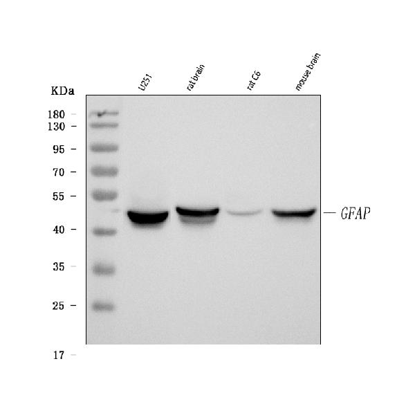
Western blot analysis of GFAP using anti-GFAP antibody (MA1045).
Electrophoresis was performed on a 5-20% SDS-PAGE gel at 70V (Stacking gel) / 90V (Resolving gel) for 2-3 hours. The sample well of each lane was loaded with 30 ug of sample under reducing conditions.
Lane 1: human U251 whole cell lysates,
Lane 2: rat brain tissue lysates,
Lane 3: rat C6 whole cell lysates,
Lane 4: mouse brain tissue lysates.
After electrophoresis, proteins were transferred to a nitrocellulose membrane at 150 mA for 50-90 minutes. Blocked the membrane with 5% non-fat milk/TBS for 1.5 hour at RT. The membrane was incubated with mouse anti-GFAP antigen affinity purified monoclonal antibody (Catalog # MA1045) at 1 μg/mL overnight at 4°C, then washed with TBS-0.1%Tween 3 times with 5 minutes each and probed with a goat anti-mouse IgG-HRP secondary antibody at a dilution of 1:10000 for 1.5 hour at RT. The signal is developed using an Enhanced Chemiluminescent detection (ECL) kit (Catalog # EK1001) with Tanon 5200 system. A specific band was detected for GFAP at approximately 50 kDa. The expected band size for GFAP is at 54 kDa.

Click image to see more details
IF analysis of GFAP using anti-GFAP antibody (MA1045) and anti-MBP antibody (PA1050)
GFAP was detected in paraffin-embedded section of rat brain tissues. Heat mediated antigen retrieval was performed in citrate buffer (pH6 epitope retrieval solution ) for 20 mins. The tissue section was blocked with 10% goat serum. The tissue section was then incubated with 1μg/mL mouse anti-GFAP Antibody (MA1045)and anti-MBP Antibody (PA1050) overnight at 4°C. DyLight®488 Conjugated Goat Anti-Mouse IgG (BA1126) and Cy3 Conjugated Goat Anti-Rabbit IgG (BA1032) were used as secondary antibody at 1:100 dilution and incubated for 30 minutes at 37°C. The section was counterstained with DAPI. Visualize using a fluorescence microscope and filter sets appropriate for the label used.

Click image to see more details
IHC analysis of GFAP using anti-GFAP antibody (MA1045).
GFAP was detected in a paraffin-embedded section of rat brain tissue. Heat mediated antigen retrieval was performed in EDTA buffer (pH 8.0, epitope retrieval solution). The tissue section was blocked with 10% goat serum. The tissue section was then incubated with 1 μg/ml mouse anti-GFAP Antibody (MA1045) overnight at 4°C. Peroxidase Conjugated Goat Anti-mouse IgG was used as secondary antibody and incubated for 30 minutes at 37°C. The tissue section was developed using HRP Conjugated Mouse IgG Super Vision Assay Kit (Catalog # SV0001) with DAB as the chromogen.

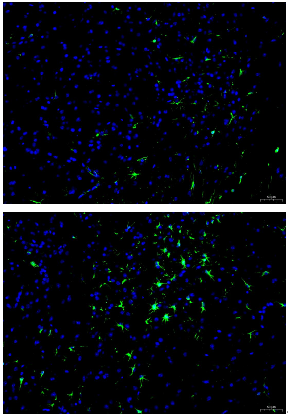
Click image to see more details
IF analysis of GFAP using anti-GFAP antibody (MA1045) .
GFAP was detected in an immunocytochemical section of rat C6 cells. The cells were fixed with 4% paraformaldehyde for 10 minutes and then treated with a membrane permeabilization agent (AR0205) for 5 minutes.The cells were blocked with 10% goat serum. And then incubated with mouse anti-GFAP Antibody (MA1045) at a dilution of 2 μg/mL overnight at 4°C. DyLight®488 Conjugated Goat Anti-mouse IgG (BA1126) was used as secondary antibody at 1:500 dilution and incubated for 30 minutes at 37°C. The section was counterstained with DAPI. Visualize using a fluorescence microscope and filter sets appropriate for the label used.

Click image to see more details
IF analysis of GFAP using anti-GFAP antibody (MA1045).
GFAP was detected in a paraffin-embedded section of rat spinal tissue. Heat mediated antigen retrieval was performed in EDTA buffer (pH 8.0, epitope retrieval solution). The tissue section was blocked with 10% goat serum. The tissue section was then incubated with 1:200 rabbit anti-GFAP Antibody (PA1050) overnight at 4°C. DyLight®488 Conjugated Goat Anti-Rabbit IgG (BA1127) was used as secondary antibody incubated for 45 minutes at 37°C. The section was counterstained with DAPI. Visualize using a fluorescence microscope and filter sets appropriate for the label used.

Click image to see more details
ALK5 is increased in the ischemic hemisphere in a MCAO/R rat model. a Representative images of ALK5 expression in the ischemic hemisphere 24 h after I/R ( n = 6 biological replicates). b Representative images of ALK5 expression in the ischemic hemisphere at 14 d after I/R ( n = 6 biological replicates). c Comparison of mean intensity ratios in Western blot analysis. d Representative images of immunohistochemical staining for ALK5 expression 24 h and 14 d after I/R ( n = 5 biological replicates) (scale bar = 100 μm). e Comparison of the mean density value in immunohistochemical analysis for ALK5 expression. f Representative images of immunofluorescence staining for ALK5 (green), beta-III tubulin (red)/GFAP (red) and cellular nuclei (blue). (scale bar = 100 μm). Arrows show the positive cells, and the inserted images show magnified images of representative cells. * P < 0.05, compared to the sham group at the same time point (Student’s t test)
Index in PubMed under a CC BY license. PMID: 31043581

Click image to see more details
Vagus nerve stimulation (VNS) decreases spinal cord tissue damage. (A–C) Representative Immunofluorescence (IF) staining images of each group post injure 28 days, and bar charts show the fluorescence intensity mean value (IMV) for GFAP and Laminin, n = 5 per group, scale bar = 500 or 200 μm. (D,E) Representative Hematoxylin-eosin (HE) staining images 28 days after injury and quantification data of cavity necrotic tissue in each group, n = 5 per group, scale bar = 500 μm. (F,G) Representative Nissl staining images 28 days after injury and quantification of the numbers of Nissl-stained dark neurons in each group, n = 5 per group, scale bar = 20 μm. * P < 0.05, ** P < 0.01, and **** P < 0.0001 between the Sham and Sham-VNS groups, # P < 0.05, ## P < 0.01 between the VNS and VNS-MLA groups.
Index in PubMed under a CC BY license. PMID: 35464311

Click image to see more details
Effects of MSCs treatment on the phenotype distribution of microglia and astrocytes. (A–D) Double immunofluorescence staining of Iba-1/iNOS-positive M1 microglia, Iba-1/Arg-1-positive M2 microglia, GFAP/C3-positive A1 astrocytes and GFAP/S100A10-positive A2 astrocytes in hippocampus. (E–H) Representative quantification showing that MSC significantly decreased the number of M1, A1 cells and increased the number of M2, A2 cells in hippocampus. (** P < 0.01, vs. Sham-operated group, ## P < 0.01, vs. RHRSP group; n = 6/group). scale bar = 50 μm.
Index in PubMed under a CC BY license. PMID: 35663575

Click image to see more details
Effects of MSCs treatment on the activation of microglia and astrocytes. (A,D) Representative images of immunofluorescence staining of Iba-1 and GFAP in the cortex and hippocampus. (B,C,E,F) Quantification of immunofluorescence staining for Iba-1 and GFPAP in the cortex and hippocampus (* P < 0.05, ** P < 0.01, vs. sham-operated group; # P < 0.05, ## P < 0.01, vs. RHRSP group; n = 6/group). scale bar = 50 μm.
Index in PubMed under a CC BY license. PMID: 35663575

Click image to see more details
Inhibition of NLRP3 inflammasome reversed CIH-related A1/A2 astrocyte phenotypic transformation. a GFAP immunohistochemical staining and GFAP + cell count. b Sholl analysis of morphological changes of astrocytes. c-d Expression of GFAP, C3d, and s100a10 detected by Western blotting and RT-qPCR. Scale bar = 50 μm. Data are presented as the mean ± SD. For RT-qPCR experiments, each group consisted of 4 rats, and the experiment was repeated 3 times; for Western blotting, the experiment was repeated 4 times and there were 5 rats per group. For statistical analysis, an unpaired t-test was used. * P < 0.05, ** P < 0.01, *** P < 0.001, **** P < 0.0001
Index in PubMed under a CC BY license. PMID: 36437451

Click image to see more details
CIH induced A1/A2 phenotype astrocyte transformation, with the increase of A1 type. a Immunofluorescence images of astrocytes. The astrocytes were labeled by anti-GFAP (green), anti-C3d (red), and anti-S100a10 (red). b Expression of GFAP, C3d, and S100a10 was detected by Western blotting. c The mRNA expression of GFAP, C3d, and S100a10. Scale bar = 50 μm. Data are presented as the mean ± SD. ANOVA with Tukey’s post hoc test was used to compare the control, CIH 2w, CIH 4w, and CIH 6w groups. For RT-qPCR experiments, each group consisted of 4 rats, and the experiment was repeated 3 times; for western blotting, which was repeated 4 times, there were 5 rats per group. * P < 0.05, ** P < 0.01, *** P < 0.001, **** P < 0.0001
Index in PubMed under a CC BY license. PMID: 36437451

Click image to see more details
CIH induced activation of astrocytes. a GFAP immunohistochemical staining and GFAP + cell count in each subregion of the hippocampus. b Sholl analysis of morphological changes of astrocytes under CIH. Scale bar = 50 μm. Data are presented as the mean ± SD. ANOVA with Tukey’s post hoc test was used to compare the control, CIH 2w, CIH 4w, and CIH 6w group. There were three rat brain slices in each group, and 5–6 visual fields were randomly selected according to different subregions for statistical analysis. * P < 0.05, *** P < 0.001, **** P < 0.0001
Index in PubMed under a CC BY license. PMID: 36437451

Click image to see more details
Minocycline inhibited microglia activation and the following astrocyte activation induced by hippocampal CNTN1 overexpression. (A-F) Representative images showed immunostaining against microglial marker Iba1 on brain sections in hippocampus of mice in different groups. (G) Quantiative analysis of Iba1 positive microglia in hippocampus of mice in different groups by integrated optical density (IOD). (H) Quantitative real time qPCR detection of mRNA expression of CD11b and CD68 in hippocampus. (I) Quantitative analyses of complexity with Iba1 positive microglia in hippocampus in different groups. (J) Quantitative real time qPCR detection of mRNA expression of IL1α, IL6 and CCL2 in hippocampus in different groups. n=6 per group. (K&L) Representative immunoblots (K) and quantitative analyses (L) of vimentin and GFAP expression in hippocampus in different groups. Data in , and L were analyzed by Kruskal-Wallis statistical test; Data in . I was analyzed by two-way ANOVA followed by Bonferroni’s multiple comparison tests. Data in , I and J were analyzed by one-way ANOVA followed by Bonferroni’s multiple comparison tests. Data were presented as mean ± sem. *p < 0.05, **p < 0.01, ***p < 0.001 and ****p < 0.0001. NS: Saline. Scale bar: 50 μm.
Index in PubMed under a CC BY license. PMID: 37196127

Click image to see more details
CNTN1 overexpression by AAV stereotactic injection activated microglia and astrocyte in the hippocampus. (A-C) Representative images showed immunostaining against microglial marker Iba1 on brain sections in the hippocampus of mice in different groups. (D&E) Quantiative analysis of Iba1 positive microglia of hippocampus in different groups by total number of microglia (D) and integrated optical density (IOD) (E). (F) Quantitative analysis complexity of Iba1 positive microglia in hippocampus with Sholl analysis in different groups. (G) Quantitative real time qPCR detection of mRNA expression levels of CD11b and CD68 in hippocampus in different groups. (H) Quantitative real time qPCR detection of mRNA expression levels of IL1α, IL6, iNOS and CCL2 in hippocampus in different groups. n=4 per group. (I&J) Representative immunoblot (I) and quantitative analysis (J) of vimentin and GFAP expression in hippocampus in different groups. Data in , E, H, J were analyzed by Kruskal-Wallis statistical test; Data in was analyzed by two-way ANOVA followed by Bonferroni’s multiple comparison test. Data in was analyzed by one-way ANOVA followed by Bonferroni’s multiple comparison tests. Data were presented as mean ± sem. *p < 0.05, **p < 0.01 and ****p < 0.0001. Scale bar: 50 μm.
Index in PubMed under a CC BY license. PMID: 37196127

Click image to see more details
EF-sEVs promoted neurogenesis at the injury site in the spinal cord. A Representative immunofluorescence images showing the staining of neurofilaments (NF, green) and glial fibrillary acidic protein (GFAP, red) in lesion sites in different groups. B Fluorescent immunostaining of choline acetyl transferase (ChAT, green) in lesion sites in different groups. Scale bar=500 μm ( A , B left line before magnification) and 100 μm ( A , B right lines after magnification)
Index in PubMed under a CC BY license. PMID: 38012570
Specific Publications For Anti-GFAP Antibody (Monoclonal, G-A-5) (MA1045)
Loading publications
Recommended Resources
Here are featured tools and databases that you might find useful.
- Boster's Pathways Library
- Protein Databases
- Bioscience Research Protocol Resources
- Data Processing & Analysis Software
- Photo Editing Software
- Scientific Literature Resources
- Research Paper Management Tools
- Molecular Biology Software
- Primer Design Tools
- Bioinformatics Tools
- Phylogenetic Tree Analysis
Customer Reviews
Have you used Anti-GFAP Antibody (Monoclonal, G-A-5)?
Share your experimental results or join a short interview to earn up to $1,000 in product credits or other rewards.
1 Reviews For Anti-GFAP Antibody (Monoclonal, G-A-5)
Immunofluorescence using Anti-GFAP antibody (MA1045) clearly labeled protoplasmic astrocytes in the spinal cord gray matter, showing dense, bushy processes surrounding neurons, with excellent specificity and staining.
Excellent
| SKU | MA1045 |
|---|---|
| Application | Immunofluorescence |
| Sample | rat spinal tissue |
| Sample Processing Description | Paraffin-embedded transverse sections of rat spinal cord were prepared after formalin fixation. |
| Other Reagents | Tris-EDTA Antigen Retrieval Buffer (50×, pH 9.0), DAPI |
| Primary Antibody | GFAP Antibody (Monoclonal, G-A-5) |
| Primary Incubation | 1:200, overnight at 4 ℃ |
| Secondary Antibody | Goat Anti-Mouse IgG (H+L) Secondary Antibody, Fluoro488 Conjugated |
| Secondary Incubation | 45 minutes in 37℃ |
| Detection | Imaging system:Leica DM2500 |
| Results Summary | GFAP is a marker of astrocytes. In this experiment, immunostaining for GFAP was used to label astrocytes in the gray matter of the spinal cord to observe their distribution, density, and morphology. The results showed that the labeled protoplasmic astrocytes in the gray matter had short, thick, and highly branched processes with rough surfaces, forming a dense “bushy” network tightly surrounding neuronal cell bodies and synapses, consistent with theoretical expectations and demonstrating excellent staining. |
Ruibo Zhao, South China Agricultural University
Verified customer
Submitted 2026-01-26
Customer Q&As
Have a question?
Find answers in Q&As, reviews.
Can't find your answer?
Submit your question
16 Customer Q&As for Anti-GFAP Antibody (Monoclonal, G-A-5)
Question
We are currently using anti-GFAP antibody (Monoclonal, G-A-5) MA1045 for pig tissue, and we are happy with the WB results. The species of reactivity given in the datasheet says human, pig, rat. Is it true that the antibody can work on dog tissues as well?
Verified Customer
Verified customer
Asked: 2020-01-31
Answer
The anti-GFAP antibody (Monoclonal, G-A-5) (MA1045) has not been validated for cross reactivity specifically with dog tissues, though there is a good chance of cross reactivity. We have an innovator award program that if you test this antibody and show it works in dog you can get your next antibody for free. Please contact me if I can help you with anything.
Boster Scientific Support
Answered: 2020-01-31
Question
Is this MA1045 anti-GFAP antibody (Monoclonal, G-A-5) reactive to the isotypes of GFAP?
Verified Customer
Verified customer
Asked: 2020-01-09
Answer
The immunogen of MA1045 anti-GFAP antibody (Monoclonal, G-A-5) is GFAP from pig spinal cord. Could you tell me which isotype you are interested in so I can help see if the immunogen is part of this isotype?
Boster Scientific Support
Answered: 2020-01-09
Question
Thank you for helping with my inquiry over the phone. Here are the WB image, lot number and protocol we used for blood using anti-GFAP antibody (Monoclonal, G-A-5) MA1045. Let me know if you need anything else.
Verified Customer
Verified customer
Asked: 2019-12-23
Answer
We appreciate the data. You have provided everything we needed. Our lab team are working to resolve your inquiry as quickly as possible, and we appreciate your patience and understanding! Please let me know if there is anything you need in the meantime.
Boster Scientific Support
Answered: 2019-12-23
Question
you antibody to test anti-GFAP antibody (Monoclonal, G-A-5) MA1045 on rat blood for research purposes, then I may be interested in using anti-GFAP antibody (Monoclonal, G-A-5) MA1045 for diagnostic purposes as well. Is the antibody suitable for diagnostic purposes?
Verified Customer
Verified customer
Asked: 2019-11-29
Answer
The products we sell, including anti-GFAP antibody (Monoclonal, G-A-5) MA1045, are only intended for research use. They would not be suitable for use in diagnostic work. If you have the means to develop a product into diagnostic use, and are interested in collaborating with us and develop our product into an IVD product, please contact us for more discussions.
Boster Scientific Support
Answered: 2019-11-29
Question
Is a blocking peptide available for product anti-GFAP antibody (Monoclonal, G-A-5) (MA1045)?
Verified Customer
Verified customer
Asked: 2019-10-03
Answer
We do provide the blocking peptide for product anti-GFAP antibody (Monoclonal, G-A-5) (MA1045). If you would like to place an order for it please contact support@bosterbio.com and make a special request.
Boster Scientific Support
Answered: 2019-10-03
Question
We have seen staining in rat brain thalamus. Any tips? Is anti-GFAP antibody (Monoclonal, G-A-5) supposed to stain brain thalamus positively?
Verified Customer
Verified customer
Asked: 2019-09-26
Answer
From what I have seen in literature brain thalamus does express GFAP. From what I have seen in Uniprot.org, GFAP is expressed in dorsal motor nucleus of vagus nerve, brain thalamus, brain, kidney, fetal brain cortex, fetal brain, blood, among other tissues. Regarding which tissues have GFAP expression, here are a few articles citing expression in various tissues:
Blood, Pubmed ID: 12837269
Brain, Pubmed ID: 15489334
Brain, and Thalamus, Pubmed ID: 14702039
Fetal brain, Pubmed ID: 12058025
Fetal brain cortex, Pubmed ID: 2780570
Kidney, Pubmed ID: 17974005
Boster Scientific Support
Answered: 2019-09-26
Question
I was wanting to use your anti-GFAP antibody (Monoclonal, G-A-5) for IHC-P for rat blood on frozen tissues, but I want to know if it has been validated for this particular application. Has this antibody been validated and is this antibody a good choice for rat blood identification?
Verified Customer
Verified customer
Asked: 2019-08-05
Answer
It shows on the product datasheet, MA1045 anti-GFAP antibody (Monoclonal, G-A-5) has been tested for IF, IHC-P, IHC-F, WB on human, pig, rat tissues. We have an innovator award program that if you test this antibody and show it works in rat blood in IHC-frozen, you can get your next antibody for free.
Boster Scientific Support
Answered: 2019-08-05
Question
I have attached the WB image, lot number and protocol we used for blood using anti-GFAP antibody (Monoclonal, G-A-5) MA1045. Please let me know if you require anything else.
Verified Customer
Verified customer
Asked: 2019-07-03
Answer
Thank you very much for the data. Our lab team are working to resolve this as quickly as possible, and we appreciate your patience and understanding! You have provided everything we needed. Please let me know if there is anything you need in the meantime.
Boster Scientific Support
Answered: 2019-07-03
Question
We bought anti-GFAP antibody (Monoclonal, G-A-5) for IF on kidney a few years ago. I am using rat, and We intend to use the antibody for IHC-P next. My question regards examining kidney as well as blood in our next experiment. Could you please give me some suggestion on which antibody would work the best for IHC-P?
H. Roberts
Verified customer
Asked: 2018-10-25
Answer
I have checked the website and datasheets of our anti-GFAP antibody (Monoclonal, G-A-5) and it seems that MA1045 has been validated on rat in both IF and IHC-P. Thus MA1045 should work for your application. Our Boster satisfaction guarantee will cover this product for IHC-P in rat even if the specific tissue type has not been validated. We do have a comprehensive range of products for IHC-P detection and you can check out our website bosterbio.com to find out more information about them.
Boster Scientific Support
Answered: 2018-10-25
Question
My question regarding product MA1045, anti-GFAP antibody (Monoclonal, G-A-5). I was wondering if it would be possible to conjugate this antibody with biotin. I would need it to be without BSA or sodium azide. I am planning on using a buffer exchange of sodium azide with PBS only. Would there be problems for me to conjugate the antibody and store it in -20 degrees in small aliquots?
Verified Customer
Verified customer
Asked: 2017-10-05
Answer
We suggest not storing this antibody with PBS buffer only in -20 degrees. If you want to store it in -20 degrees it is best to add some cryoprotectant like glycerol. If you want carrier free MA1045 anti-GFAP antibody (Monoclonal, G-A-5), we can provide it to you in a special formula with trehalose and/or glycerol. These molecules will not interfere with conjugation chemistry and provide a good level of protection for the antibody from degradation. Please be sure to specify this in your purchase order.
Boster Scientific Support
Answered: 2017-10-05
Question
Is there a BSA free version of anti-GFAP antibody (Monoclonal, G-A-5) MA1045 available?
Verified Customer
Verified customer
Asked: 2017-09-18
Answer
I appreciate your recent telephone inquiry. I can confirm that some lots of this anti-GFAP antibody (Monoclonal, G-A-5) MA1045 are BSA free. For now, these lots are available and we can make a BSA free formula for you free of charge. It will take 3 extra days to prepare. If you require this antibody BSA free again in future, please do not hesitate to contact me and I will be pleased to check which lots we have in stock that are BSA free.
Boster Scientific Support
Answered: 2017-09-18
Question
We are interested in using your anti-GFAP antibody (Monoclonal, G-A-5) for r-rno-9613829; chaperone mediated autophagy studies. Has this antibody been tested with western blotting on rat brain tissue? We would like to see some validation images before ordering.
Verified Customer
Verified customer
Asked: 2017-08-03
Answer
I appreciate your inquiry. This MA1045 anti-GFAP antibody (Monoclonal, G-A-5) is validated on rat brain tissue. It is guaranteed to work for IF, IHC-P, IHC-F, WB in human, pig, rat. Our Boster guarantee will cover your intended experiment even if the sample type has not been be directly tested.
Boster Scientific Support
Answered: 2017-08-03
Question
Would MA1045 anti-GFAP antibody (Monoclonal, G-A-5) work on parafin embedded sections? If so, which fixation method do you recommend we use (PFA, paraformaldehyde, other)?
T. Jackson
Verified customer
Asked: 2017-07-13
Answer
It shows on the product datasheet, MA1045 anti-GFAP antibody (Monoclonal, G-A-5) as been validated on IHC-P. It is best to use PFA for fixation because it has better tissue penetration ability. PFA needs to be prepared fresh before use. Long term stored PFA turns into formalin, as the PFA molecules congregate and become formalin.
Boster Scientific Support
Answered: 2017-07-13
Question
I see that the anti-GFAP antibody (Monoclonal, G-A-5) MA1045 works with IHC-P, what is the protocol used to produce the result images on the product page?
Verified Customer
Verified customer
Asked: 2017-05-17
Answer
You can find protocols for IHC-P on the "support/technical resources" section of our navigation menu. If you have any further questions, please send an email to support@bosterbio.com
Boster Scientific Support
Answered: 2017-05-17
Question
My colleagues were happy with the WB result of your anti-GFAP antibody (Monoclonal, G-A-5). However we have seen positive staining in fetal brain cortex cytoplasm. using this antibody. Is that expected? Could you tell me where is GFAP supposed to be expressed?
A. Li
Verified customer
Asked: 2016-10-11
Answer
From literature, fetal brain cortex does express GFAP. Generally GFAP expresses in cytoplasm. Regarding which tissues have GFAP expression, here are a few articles citing expression in various tissues:
Blood, Pubmed ID: 12837269
Brain, Pubmed ID: 15489334
Brain, and Thalamus, Pubmed ID: 14702039
Fetal brain, Pubmed ID: 12058025
Fetal brain cortex, Pubmed ID: 2780570
Kidney, Pubmed ID: 17974005
Boster Scientific Support
Answered: 2016-10-11
Question
Will anti-GFAP antibody (Monoclonal, G-A-5) MA1045 work for IHC-P with blood?
L. Wu
Verified customer
Asked: 2015-12-30
Answer
According to the expression profile of blood, GFAP is highly expressed in blood. So, it is likely that anti-GFAP antibody (Monoclonal, G-A-5) MA1045 will work for IHC-P with blood.
Boster Scientific Support
Answered: 2015-12-30
