This website uses cookies to ensure you get the best experience on our website.
- Table of Contents
4 Citations 5 Q&As
2 Citations 10 Q&As
2 Citations 5 Q&As
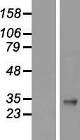
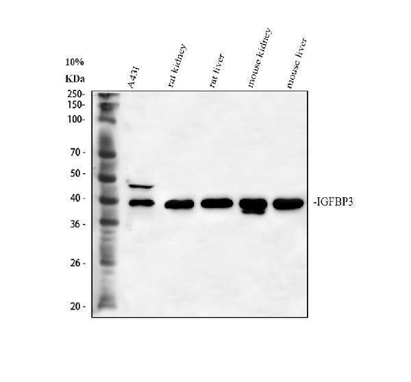
Facts about Insulin-like growth factor-binding protein 3.
Also exhibits IGF-independent antiproliferative and apoptotic effects mediated by its receptor TMEM219/IGFBP-3R. .
Human | |
---|---|
Gene Name: | IGFBP3 |
Uniprot: | P17936 |
Entrez: | 3486 |
Belongs to: |
---|
No superfamily |
acid stable subunit of the 140 K IGF complex; binding protein 29; binding protein 53; growth hormone-dependent binding protein; IBP-3; IBP3BP-53; IGF-binding protein 3; IGFBP3; IGFBP-3; insulin-like growth factor binding protein 3; insulin-like growth factor-binding protein 3
Mass (kDA):
31.674 kDA
Human | |
---|---|
Location: | 7p12.3 |
Sequence: | 7; NC_000007.14 (45912245..45921272, complement) |
Expressed by most tissues. Present in plasma.
Secreted.
The IGFBP3 gene encodes an enzyme that regulates a variety of cell processes. Its ability to bind to potential partners allows it to regulate many cellular functions, including cell proliferative. The gene is linked to various metabolic diseases which include cancer. It also has autocrine and paracrine functions. Continue reading to learn more about the IGFBP3 gene's best uses and advantages.
IGFBP3 is involved in a variety of biological processes. Its extracellular levels are increased by DNA damage and oxidative insults and are believed to be a factor in senescence. Additionally, it could mediate oxidative stress-induced cellular senescence. However, it is unclear whether this protein has an autocrine or paracrine role in cells.
IGFBP3 is able to stop tumor cells from growing. These effects are independent of IGF action and could be mediated by specific IGFBP3-binding proteins. Weinzimer and co-workers identified one of these proteins in a study conducted in 2005. Biosensor interactions confirmed that the binding site was specific. The treatment with Transferrin slowed the prostate cancer growth as well as bladder smooth muscle cells.
IGF-1 is an endocrine and paracrine signal for adjacent epithelial tumor cells. IGF-1's activity is a key factor in the progression of disease. A low IGF-1 level in BrCa can also support the disease's progression. However, the precise role of IGF-1 in invasive breast cancer is not yet clear. It is essential to continue research into this cellular process to determine whether it has a paracrine or autocrine function.
IGFBP-3 is activated by mitogens and is dissociated with Erk1/2. This activation leads to the expression of Egr-1 a protein that regulates the transcription of genes associated with angiogenesis and tumor growth. Its decreased activity prevents tumor growth by reducing the paracrine and autocrine mechanisms. However, further research needs to be carried out to confirm these findings.
A Mendelian randomization study discovered a link between an increase in IGFBP3 and a lower BrCa risk. This suggests that the protein could have an autocrine or paracrine role in regulating cell response and angiogenesis in humans. Although there is not enough evidence to support this idea however, it could be a promising option for treatment. IGFBP3 is a potent inflammatory mediator.
Although it isn't clear whether IGFBP3 is a paracrin with a similar function, a number of studies suggest it has both. Its autocrine function promotes tissue growth by promoting cell division, which in turn regulates the metabolism of the tissue. It is a key factor in angiogenesis. IGFBP3 is a major player in the regulation and promotion of cellular growth.
IGFBP3 is a glycoprotein involved in the transportation of growth factors from their cell source to their receptors. Its roles in the extracellular environment as well as inside cells have long been recognized. It is the most important IGF transport protein in the bloodstream. It is the growth factor in complexes. IGF-2 and IGFBP-3 each contain the binding protein as well as an acid labile subunit.
Apoptosis is the term used to describe a process that causes cells to die. It happens through the operation of two pathways that are cross-linked. One involves cell surface receptors known as death receptors, and the second one involves mitochondria. Apoptosis can occur via any of these pathways and IGFBP-3 is involved in both. IGFBP3's purpose is to block the proliferation of cancer cells.
IGFBP3 plays two distinct roles. It can activate IGF-dependent signals and also perform IGF-independent activities. It can also associate with plasma membrane proteins. It then translocates across the plasma membrane to the cytosol. It then interacts with a variety of proteins, which are listed in Figure 2.
IGFBP-3 may be downregulated in certain cancers. There have been many instances in which the expression of IGFBP-3 has been linked to poor outcomes for patients. While it has been shown that IGFBP-3 has multiple roles in the cell culture but in vivo it's unclear how it controls these different effects. It has dual inhibitory and stimulating effect on cells, however it is not known how it works in normal cells.
IGFBP-3, a protein containing the IGF-1R receptor that regulates cellular functions and is associated with the regulation of IGF-1R. Its role in the regulation of IGF-independent processes isn't clear, but it is known to boost IGF-1R signaling. IGFBP-3 expression, as an example, is associated with cell proliferation as well as immortalization of cells.
IGFBP-3 can be absorbed by cells from bloodstream via secretion. Its central domain is extremely varied, with only 15% of the IGFBPs. IGFBP-3 interacts with various serum proteins such as transferrin and lactoferrin. Its binding affinity is around twice that of apo-transferrin.
The inter-acting partners of IGFBP-3 were identified using the yeast two-hybrid system. In this system, two different IGFBP-3 deletion mutants were utilized as baits. IGFBP-3 binds with Rpb3 one of the twelve subunits in RNA polymerase 2. Rpb3 is a transcription factor that recruits DNA-binding spots.
IGFBP-3's nuclear localization signal is vital for its transfer from nucleus to cytoplasm. In mice, IGFBP-3 mutants at Leu 197 and 200 were found to have higher levels of IGFBP-3 in the nuclear compartment and cytoplasm but without the ability to export to the cytoplasm. This indicates that the IGFBP3 marker boosts IGF-1R signaling in these cells.
IGFBP-3 binding with humanin a secretory protein found in nature, results in increased levels of IGF-1R and a decrease in the expression of humanin. The results of this study confirmed the interaction between humanin and IGFBP-3 and a peptide containing MBD has been shown to induce apoptosis in stressed HEK293 cells.
IGFBP-3 plays an important role in helping to facilitate IGF-1R-dependent cell surface receptor binding and activating the signaling pathway. The IGFs then are responsible for their action through binding to the IGF-1R. IGFBP-3 interacts with a variety of other proteins and performs numerous functions. See Figure 2 for a list of IGFBP3 binding cell partners.
IGFBP-3 is directly linked to the glucose-regulated proteins 78 (GRP78), and Binding Immunoglobulin protein (BIP). IGFBP-3 reduces cell proliferation by preventing Apoptosis. It also blocks the interaction between GRP78 and caspase-7. This results in controlling apoptosis as well as cell survival.
Immunotherapy, with its many benefits, is a wonderful method of fighting cancer as well as protecting healthy cells. The drugs target cancer cells and trigger the death of cells programmed to die. In reality, cancer cells can be found in any organ, including the lungs, and they can also adopt the characteristics of other organs. This treatment could be beneficial to patients with lung cancer. This research could help to develop more effective treatments for cancer.
Chemotherapy aims to kill cancer cells that are rapidly multiplying and can trigger a variety of negative side negative effects. The targeted therapies are based on drugs that target specific proteins and genes to tackle this issue. They search for specific elements in cancer cells. Cancer patients may experience side effects from immunotherapy if the treatment fails to perform. However many cancer patients have found that targeted therapies reduce adverse effects and extends the duration of their treatment. If they're not able to tolerate chemotherapy, they may choose to undergo immunotherapy.
Repurposing existing drugs to treat cancer is another kind of targeted therapy. NSCLC can be treated with drugs that target Ras/Raf/MEK/ERK signaling pathways. Additionally, targeted therapy offers a viable alternative to surgery and chemotherapy. This technique focuses on specific mutations and pathways within cancer cells. Targeted drugs have revolutionized treatment of NSCLC.
To ensure that a novel cancer drug is safe, researchers must carefully design its preclinical studies. Testing in mice for preclinical safety is the most common way to evaluate drug candidates. However, before they are tested on humans they must have proven effective in animal models. Certain drug candidates, like ErSO were tested on dogs suffering from cancer. The dogs with cancer are typically genetically similar to humans. They respond to the same medications that they do in humans. Additionally, dogs are comparable in size to humans, meaning they can be used as an excellent model for larger tumors.
PMID: 2464130 by Wood W.I., et al. Cloning and expression of the growth hormone-dependent insulin-like growth factor-binding protein.
PMID: 1695633 by Cubbage M.L., et al. Insulin-like growth factor binding protein-3. Organization of the human chromosomal gene and demonstration of promoter activity.
*More publications can be found for each product on its corresponding product page