This website uses cookies to ensure you get the best experience on our website.
- Table of Contents
1 Citations 9 Q&As
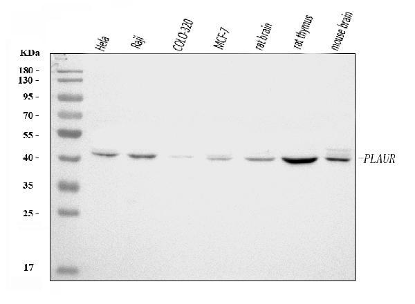
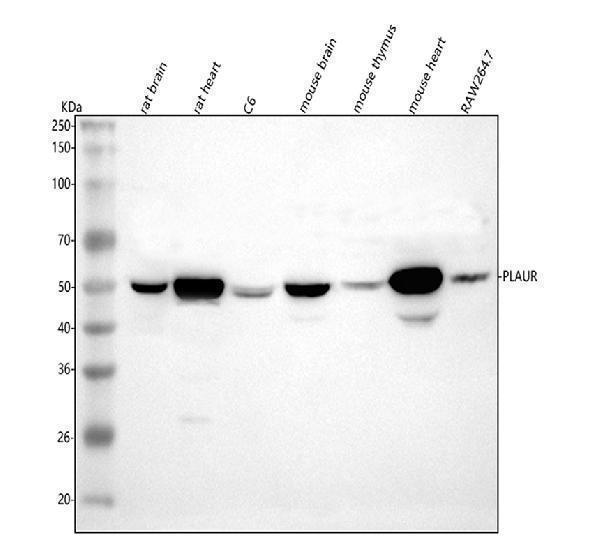
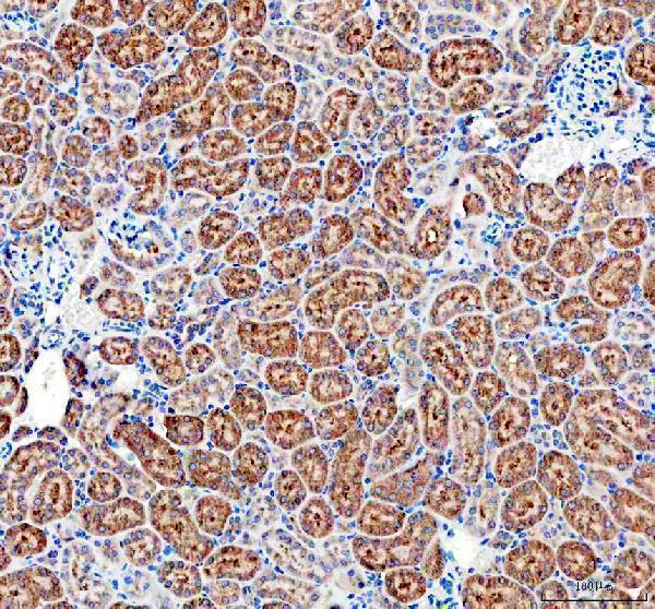
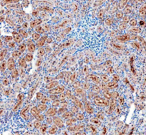
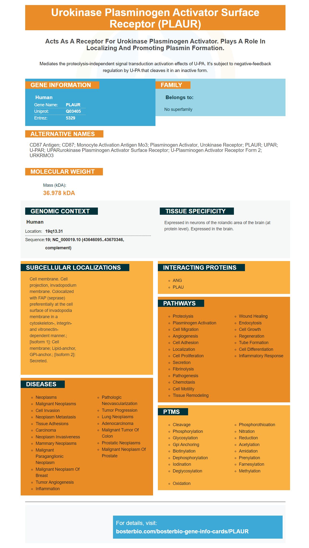
Facts about Urokinase plasminogen activator surface receptor.
Mediates the proteolysis-independent signal transduction activation effects of U-PA. It's subject to negative-feedback regulation by U-PA that cleaves it in an inactive form.
Human | |
---|---|
Gene Name: | PLAUR |
Uniprot: | Q03405 |
Entrez: | 5329 |
Belongs to: |
---|
No superfamily |
CD87 antigen; CD87; Monocyte activation antigen Mo3; plasminogen activator, urokinase receptor; PLAUR; uPAR; U-PAR; UPARurokinase plasminogen activator surface receptor; u-plasminogen activator receptor form 2; URKRMO3
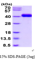
Mass (kDA):
36.978 kDA
Human | |
---|---|
Location: | 19q13.31 |
Sequence: | 19; NC_000019.10 (43646095..43670346, complement) |
Expressed in neurons of the rolandic area of the brain (at protein level). Expressed in the brain.
Cell membrane. Cell projection, invadopodium membrane. Colocalized with FAP (seprase) preferentially at the cell surface of invadopodia membrane in a cytoskeleton-, integrin- and vitronectin-dependent manner.; [Isoform 1]: Cell membrane; Lipid-anchor, GPI-anchor.; [Isoform 2]: Secreted.
You've come to the right place if you are a scientist interested in Anti-uPA receptor/PLAUR Marker. We will discuss Boster Bio's Anti uPA Receptor/PLAUR Marker as well as its applications, safety, and validation. Learn more about it. We hope you will find this article useful! If you have any queries or concerns, do not hesitate to contact us. We're always ready to answer any questions you may have.
Antibodies to uPAR and PLAUR are available from a variety of suppliers. These antibodies target the uPAR gene that encodes the plasminogen activater receptor, also known as uPAR. Numerous orthologs have been discovered in other species, including canines and porcines. The severity of various forms of cancers can be assessed by molecular markers for uPAR.
Many cancers have uPA/uPAR genes, which makes them a valuable target for diagnostic and therapeutic purposes. Numerous studies have been done to discover the role played by uPAR in cancer development. Monoclonal antibodies against uPAR like ATN-658, have also been developed. These antibodies are currently being examined in clinical settings to determine their potential application in cancer diagnosis or treatment.
The inborn immune response is a key component of the uPA-receptors. They regulate cell adhesion and movement. When a cell has too little uPA, it fails in its ability to effectively respond to infection. The anti-uPA receptor (PLAR) is a potent inhibitor of uPA. In a study using mice that were deficient in the uPA receptor, uPA deficient cells failed to attract macrophages or neutrophils to the area of infection.
Prognostic factors for head and neck cancer include factors include PAI-1 and uPA. Patients with neck and head cancer had elevated levels PAI-1 and uPA. This suggests that these molecules play a key role in the development and progression. Both PAI-1 and uPA1 could play a role in metastasis and could be used as targets for diagnostic purposes.
uPA-receptors are proteins which activate other enzymes. P-selectin, for instance, is one of these proteins. It is located primarily in the cytoplasm. Both uPA receptors are able to be inhibited by diminuting their activity. They can be used to identify the presence of uPA in various tissues or cells. Boster Bio's Anti uPA Receptor/PLAUR Marker can be used to target this protein.
Tumor cells can attach to a UPA-uPAR to trigger its activity. This can trigger cancer cells to migrate and invade. In the end, the uPA-uPAR mechanism is crucial to the development of proteolysis during cancer-related invasion. However, uPA-uPAR also plays a key role in multiple stages of cancer.
The PLAUR Marker has been used to detect pancreatic cancer. The PLAUR gene is closely linked to the human epidermal Growth Factor Receptor 2, also known as HER2. Pierga et al. reported the first observation of a connection between PLAU gene expression and HER2 in breast cancer patients. Ming and. al. followed up by conducting a study. Ming and co. discovered a co-amplification between PLAU and HER2 in patients suffering from recurrent and advanced breast cancer.
The confirmed PLAUR gene expression in glioma patients was retrieved from TCGA and CGGA databases. Cox regression analysis was used to determine the expression of PLAUR in glioma patients. PLAUR expression was a significant independent predictor and positively correlated with radiotherapy intensity, grade and overall survival. The results of these studies support the use of PLAUR as a possible marker in patients with glioma.
The expression of PLAUR in glioblastoma was confirmed with an open single-cell sequence data set. The tumors showed high PLAUR expression, suggesting that they contain macrophages. This is in line with previous studies. The validation cohort showed a good correlation with disease severity and overall survival. Both CHAT and PLAUR expression were significant in determining the relationship with the stage of disease. These results are crucial in the assessment of the role of PLAUR in the glioblastoma.
PLAUR expression was linked to diffuse glioma with malignant phenotype. A high level of PLAUR expression was associated with an increase in the number of M2 macrophages, and less CD8 T cells. PLAUR expression was also associated to immunosuppressive marker. These results provide clues for immunotherapy and further clarify the clinical significance of PLAUR in patients suffering from gliomas.
PLAUR expression in patients with gliomas is affected by the World Health Organization grade and subtype. Patients with IDH mutations and 1p/19q codeletion (IDH-wt) tumors have higher PLAUR expression. PLAUR immunostaining has been conducted on gliomas that have different IDH statuses and grades. Grade IV gliomas showed high levels of PLAUR-expressing cells, while grade II tumors had lower levels.
In a number of studies the security of the PLAUR marker has been challenged. This is mostly because of the linkage with the chance of developing pancreatic carcinoma. A new study, however, aims to investigate the correlation between the marker and the risk of pancreatic cancer. The PLAUR Marker is an essential instrument for diagnosing the disease and may be used to determine the risk of pancreatic cancer. Although the exact mechanism of co-amplification is not yet determined, it suggests that the simultaneous targeting of uPAR and Her2 is beneficial for patients suffering from breast cancer.
PMID: 1689240 by Roldan A.L., et al. Cloning and expression of the receptor for human urokinase plasminogen activator, a central molecule in cell surface, plasmin dependent proteolysis.
PMID: 1316922 by Min H.Y., et al. cDNA for Mo3, a monocyte activation antigen, encodes the human receptor for urokinase plasminogen activator.
*More publications can be found for each product on its corresponding product page