This website uses cookies to ensure you get the best experience on our website.
- Table of Contents
4 Citations 5 Q&As
3 Citations 4 Q&As
3 Citations 16 Q&As
2 Citations 16 Q&As
3 Citations 16 Q&As
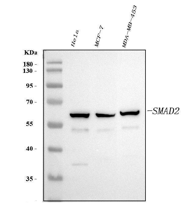
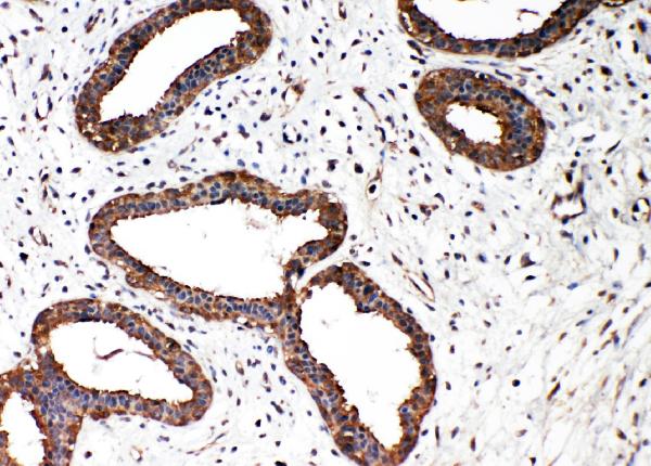
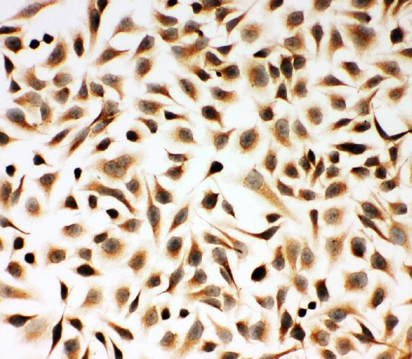
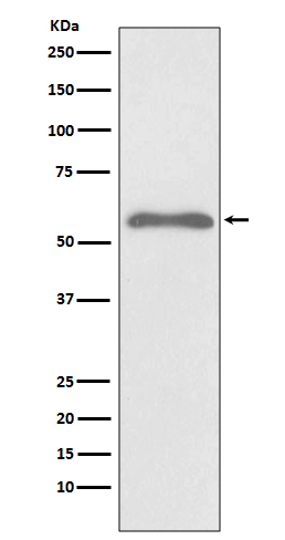
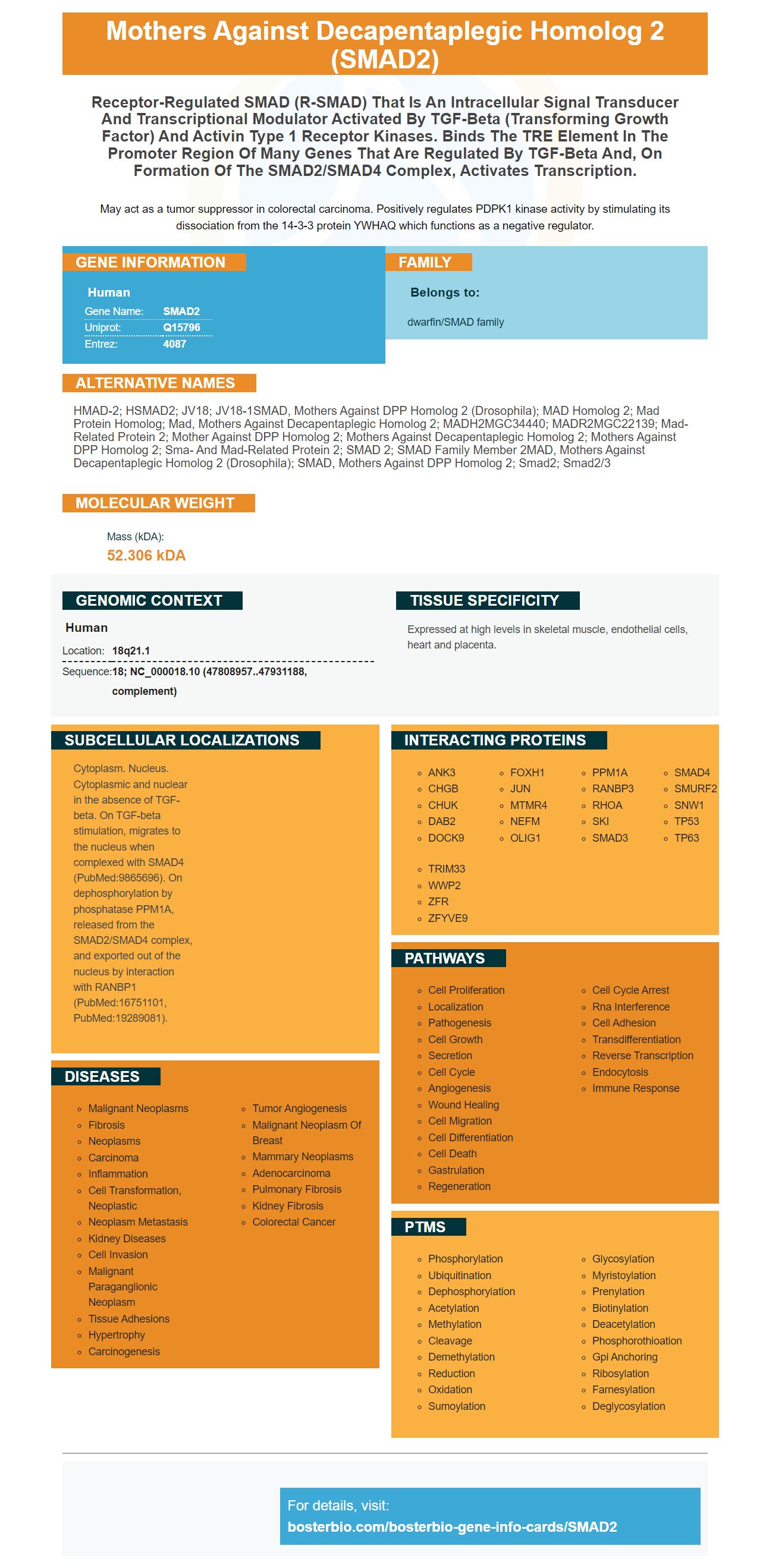
Facts about Mothers against decapentaplegic homolog 2.
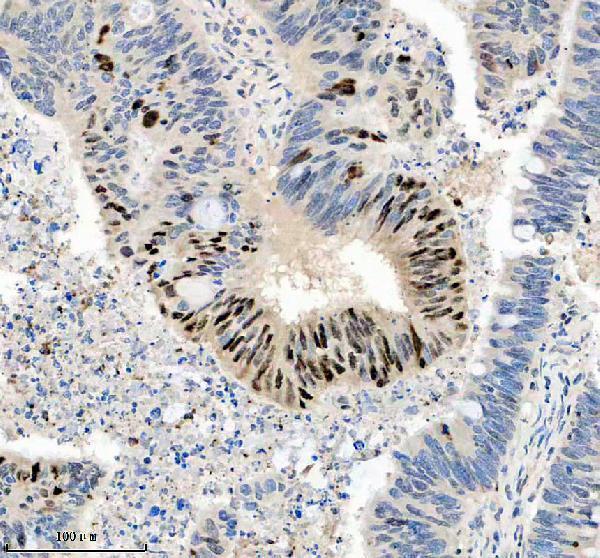
May act as a tumor suppressor in colorectal carcinoma. Positively regulates PDPK1 kinase activity by stimulating its dissociation from the 14-3-3 protein YWHAQ which functions as a negative regulator.
Human | |
---|---|
Gene Name: | SMAD2 |
Uniprot: | Q15796 |
Entrez: | 4087 |
Belongs to: |
---|
dwarfin/SMAD family |
hMAD-2; hSMAD2; JV18; JV18-1SMAD, mothers against DPP homolog 2 (Drosophila); MAD homolog 2; Mad protein homolog; Mad, mothers against decapentaplegic homolog 2; MADH2MGC34440; MADR2MGC22139; Mad-related protein 2; mother against DPP homolog 2; mothers against decapentaplegic homolog 2; Mothers against DPP homolog 2; Sma- and Mad-related protein 2; SMAD 2; SMAD family member 2MAD, mothers against decapentaplegic homolog 2 (Drosophila); SMAD, mothers against DPP homolog 2; Smad2; Smad2/3
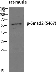
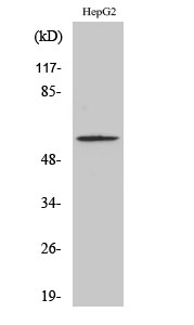
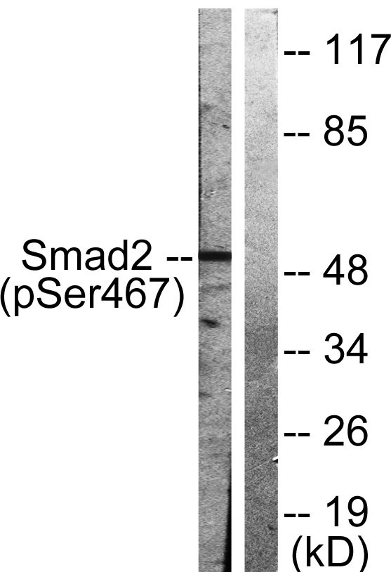
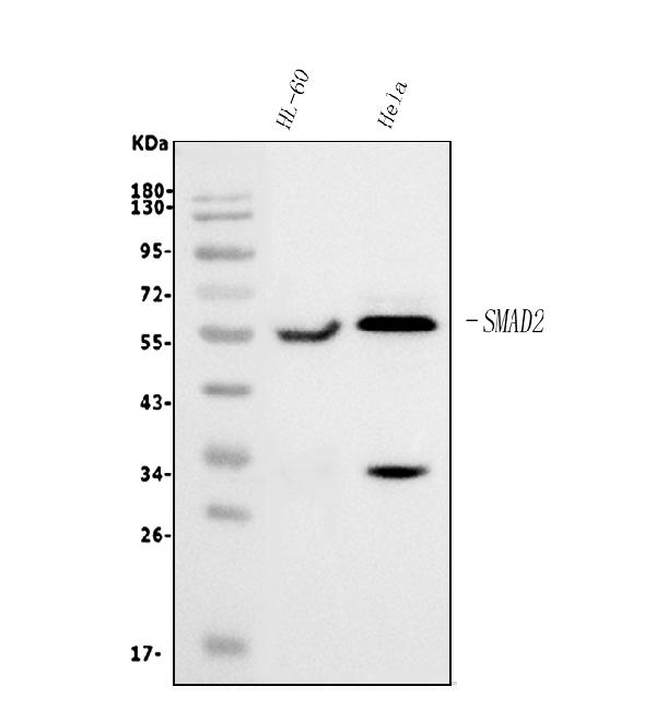
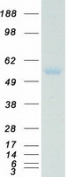
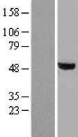
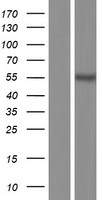
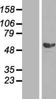
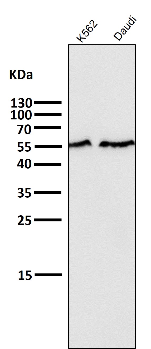
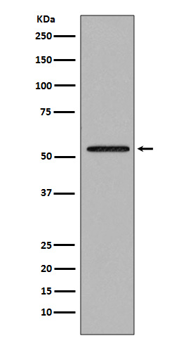
Mass (kDA):
52.306 kDA
Human | |
---|---|
Location: | 18q21.1 |
Sequence: | 18; NC_000018.10 (47808957..47931188, complement) |
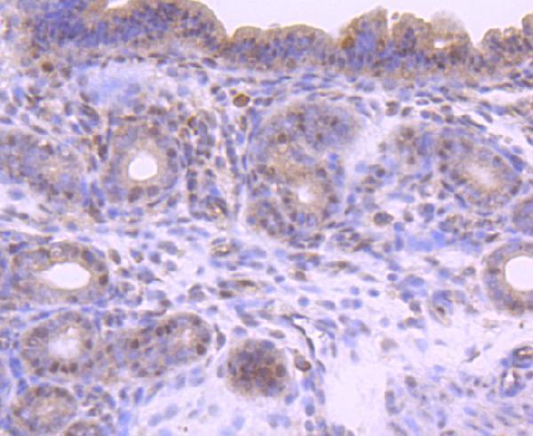
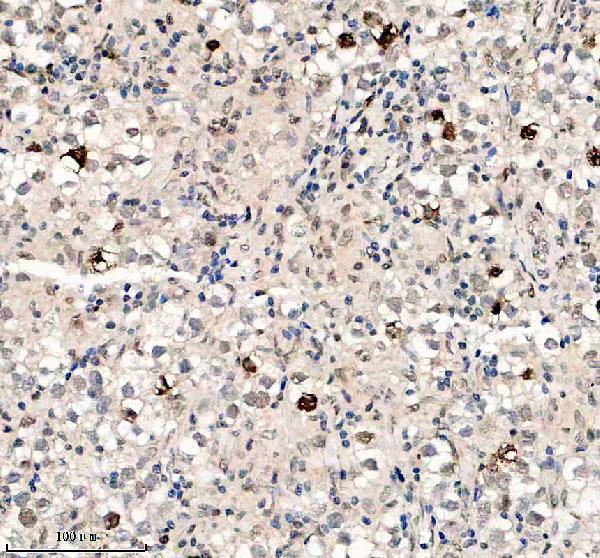
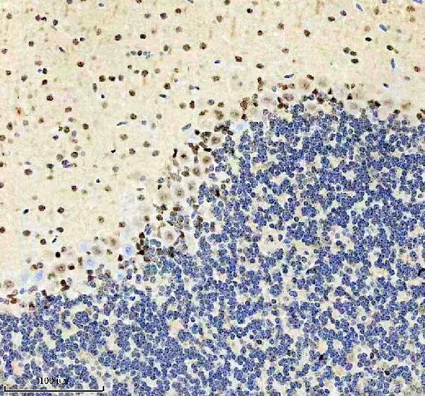
Expressed at high levels in skeletal muscle, endothelial cells, heart and placenta.
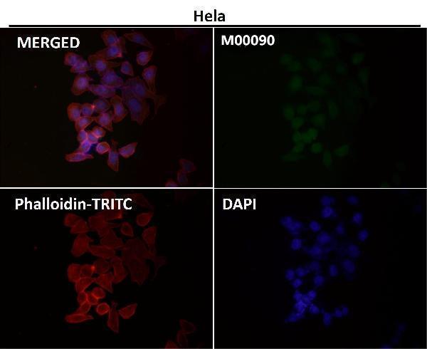
Cytoplasm. Nucleus. Cytoplasmic and nuclear in the absence of TGF-beta. On TGF-beta stimulation, migrates to the nucleus when complexed with SMAD4 (PubMed:9865696). On dephosphorylation by phosphatase PPM1A, released from the SMAD2/SMAD4 complex, and exported out of the nucleus by interaction with RANBP1 (PubMed:16751101, PubMed:19289081).
If you are looking for a monoclonal antibody to the SMAD2 Marker, you may be wondering what Boster Bio has to offer. If you are interested in the protein that makes up the SMAD2 receptor, then you should look for a Boster Bio antibody that is high-affinity and validated for Western Blotting, Immunohistochemistry, and ELISA. The antibodies from Boster Bio are validated for use in the research community and provide high-quality results.
This monoclonal antibody is designed to detect CD68, a protein that is highly expressed on macrophages, osteoclasts, and myeloid dendritic cells. It is also expressed to a lesser extent on lymphoid cells, human umbilical cord mesenchymal stem cells, and platelets. In addition, CD68 is expressed in a variety of other cell types, including non-hemorrhagic cancer cell lines and granulocyte diseased states. In addition, it has been detected in intestinal neutrophils from inflammatory bowel disease.
CD68 has various roles in the body and is important for tissue macrophage function. It binds to organ and tissue-specific lectins and may enable macrophages to crawl over selectin-bearing substrates. In addition, CD68 is quickly re-circulated from lysosomes and endosomes.
This monoclonal antibody is designed to recognize mouse CD68. CD68, also known as macrosialin, is a heavily glycosylated type I integral membrane protein. It is closely related to Lysosomal/endosomal membrane glycoproteins and is a reliable marker of the macrophage lineage. It has been widely used as a pan-macrophage marker. Although it is present in low levels on conventional dendritic cells, the CD68 gene is located on 17p13.1. CD68 is expressed on the cell surface by macrophages and is also found on monocytes and dendritic cells.
This antibody is available for purchase from BosterBio, catalog number A00602-1. This monoclonal antibody reacted with Human, Non-Human Primate, and Rabbit. The product has been extensively tested for its ability to detect this cell antigen. Its effectiveness has been confirmed by numerous studies using immunofluorescence and immunohistochemistry.
CD68 is highly expressed on macrophages. The levels of CD68 in CAD are higher than in non-CAD patients. CD68 may play a role in macrophage infiltration into adipose tissue. FFA, a fat-derived fatty acid, is released by adipocytes and contributes to adipocyte lipolysis. In addition, adipocytes are characterized by increased amounts of CD68+ cells in the epicardial adipose tissue. IL-6 and TNF-a are also elevated in CAD patients. This may also play a role in macrophage aggregation.
To determine whether Boster Bio Anti-CD68 (Mactophage Marker) is effective, we first examined the immunostaining of human adeno-smooth muscle actin and DiI-acLDL-coated sections. We used an Olympic MX-50 optical microscope, and immunostaining was assessed quantitatively in five fields of the lesion area, using a graded grid.
In addition to performing multiple immunohistochemistry, Boster also validates its antibodies. This includes testing them on a variety of platforms using known positive and negative samples. Additionally, Boster rewards its first reviewers with product credits. As a thank you, Boster recognizes the work of scientists around the world. It is the first choice of many leading researchers.
Research on this monoclonal anti-CD68 mAb indicates that it is very effective in preventing inflammation. Anti-CD68 mAbs have also been shown to inhibit lipid peroxidation in vitro, a hallmark of inflammation. However, these studies were in mice, which are not human. These studies show that boster Bio Anti-CD68 mAbs are effective in reducing pain associated with rheumatoid arthritis and osteoarthritis.
The protein CD68 is a component of the Scavenger receptor family. When inflammatory stimuli are present in macrophages, it is upregulated and is capable of binding to modified LDL, phosphatidylserine, and apoptotic cells. It is also involved in the internalization of oxLDL and subsequently contributes to atherogenesis.
PMID: 8673135 by Riggins G.J., et al. Mad-related genes in the human.
PMID: 8774881 by Zhang Y., et al. Receptor-associated Mad homologues synergize as effectors of the TGF- beta response.
*More publications can be found for each product on its corresponding product page