Click image to see more details
-

-
-
-
-
+1
Product Info Summary
| SKU: | P00090 |
|---|---|
| Size: | 100 μl |
| Reactive Species: | Human, Mouse, Rat |
| Host: | Rabbit |
| Application: | IP, IHC, WB |
Customers Who Bought This Also Bought

Product info
Product Name
Anti-Phospho-Smad2 (S255) Rabbit Monoclonal Antibody
SKU/Catalog Number
P00090
BM4695 is an alternative SKU for this antibody, used in previous lots.
Size
100 μl
Form
Liquid
Description
Boster Bio Anti-Phospho-Smad2 (S255) Rabbit Monoclonal Antibody catalog # P00090. Tested in WB, IHC, IP applications. This antibody reacts with Human, Mouse, Rat.
Storage & Handling
Store at -20°C for one year. For short term storage and frequent use, store at 4°C for up to one month. Avoid repeated freeze-thaw cycles.
Cite This Product
Anti-Phospho-Smad2 (S255) Rabbit Monoclonal Antibody (Boster Biological Technology, Pleasanton CA, USA, Catalog # P00090)
Host
Rabbit
Contents
Rabbit IgG in stabilizing components, phosphate buffered saline, pH 7.4, 150mM NaCl, 0.02% sodium azide and 50% glycerol.
*This antibody is supplied in a stabilized formulation.
Compatibility with conjugation reactions depends on the chemistry of the conjugation method used.
For conjugation methods that are not compatible with the stabilizing components present in this formulation, a carrier-free antibody format is required.
Clonality
Monoclonal
Clone Number
HBE-19
Isotype
Rabbit IgG
Immunogen
A synthesized peptide derived from human Smad2
Reactive Species
P00090 is reactive to SMAD2 in Human, Mouse, Rat
Observed Molecular Weight
52 kDa
Calculated molecular weight
52.3 kDa
Antibody Validation
Boster validates all antibodies on WB, IHC, ICC, Immunofluorescence, and ELISA with known positive control and negative samples to ensure specificity and high affinity, including thorough antibody incubations.
Application & Images
Applications
P00090 is guaranteed for IP, IHC, WB Boster Guarantee
Recommend Dilution
WB 1:1000-1:2000
IHC 1:50-1:200
IP 1:50
Tested application
Suggested blocking solution with 5% non-fat milk or BSA; (*)Recommended protein loading: 20-40 µg per lane
Use TE buffer pH 9.0 for antigen retrieval; (*) citrate buffer pH 6.0 is an alternative.
Validation Images & Assay Conditions
Click image to see more details
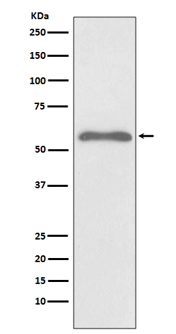
Western blot analysis of Phospho-Smad2 (S255) expression in Hela cell treated with Okadaic acid and Calyculin A lysate.

Click image to see more details
SRPX2 is elevated in fibroblasts in a TGF-β/SMADs manner. A : Western blot analysis of SRPX2 expression in HPFs following different dose of TGF-β1 induction for 24 h. B : Results for time-course Western blot analysis of SRPX2 expression in HPFs following TGF-β1 (10 ng/ml). C : Results for co-immunostaining of SRPX2 and p-SMAD2/3 in HPFs following TGF-β1 induction for 1h (up), lung sections of pulmonary fibrosis mice (middle), and lung sections from IPF patients (down). The nuclei were stained blue by DAPI, and the images were taken under original magnification ×400. D-E : Western blot (D) and RT-PCR (E) analysis of SRPX2 expression in HPFs pre-treated with SB431542 treatment following TGF-β1 induction. F-G : Western blot (F) and RT-PCR (G) analysis of SRPX2 expression in HPFs pre-treated with SIS3-HCL following TGF-β1 induction. The data are represented as the mean ± SEM of three independent experiments. ***, p < 0.001.
Index in PubMed under a CC BY license. PMID: 34093874

Click image to see more details
SRPX2 regulated TGF-β/SMADs signaling pathways by AP1 and SMAD7. A: Results for Western blot analysis of p-SMAD2, SMAD2, p-SMAD3 and SMAD3 expression in HPFs following TGF-β1 stimulation. B-C : Western blot (B) and RT-PCR (C) analysis of SMAD7 expression in HPFs following TGF-β1 induction. D : Expression of AP1 in HPFs after TGF-β1 stimulation. E : Western blot results for analysis of the levels of P-SMAD2, P-SMAD3 and SMAD7 in HPFs pre-treated with T-5224 (an inhibitor for AP-1) treatment following TGF-β1 induction. The data are represented as the mean ± SEM of three independent experiments. *, p < 0.05; **, p < 0.01; ***, p < 0.001.
Index in PubMed under a CC BY license. PMID: 34093874

Click image to see more details
Srpx2 promoted FMT in BLM-induced pulmonary fibrosis. A : Western blot analysis of Fibronectin, Col1a1, α-SMA and Srpx2 expression in mice after BLM induction with Scrambled or Srpx2 siRNA-loaded liposomes. B : Representative images of immunostaining of Fibronectin, Col1a1 and α-SMA in the mice lung sections. The nuclei were stained blue by DAPI, and the images were taken under original magnification ×400. C : Western blot analysis of p-Smad2, p-Smad3 and Smad7 expression in mice after BLM induction. D : RT-PCR analysis of AP-1 expression in mice in each group. Six mice were included in each study group. The data are represented as the mean ± SEM. *, p < 0.05; **, p < 0.01; ***, p < 0.001.
Index in PubMed under a CC BY license. PMID: 34093874

Click image to see more details
Immunohistochemical analysis of paraffin-embedded human transitional cell carcinoma of bladder, using Phospho-Smad2 (S255) Antibody.
Specific Publications For Anti-Phospho-Smad2 (S255) Rabbit Monoclonal Antibody (P00090)
Loading publications
Recommended Resources
Here are featured tools and databases that you might find useful.
- Boster's Pathways Library
- Protein Databases
- Bioscience Research Protocol Resources
- Data Processing & Analysis Software
- Photo Editing Software
- Scientific Literature Resources
- Research Paper Management Tools
- Molecular Biology Software
- Primer Design Tools
- Bioinformatics Tools
- Phylogenetic Tree Analysis
Customer Reviews
Have you used Anti-Phospho-Smad2 (S255) Rabbit Monoclonal Antibody?
Share your experimental results or join a short interview to earn up to $1,000 in product credits or other rewards.
0 Reviews For Anti-Phospho-Smad2 (S255) Rabbit Monoclonal Antibody
Customer Q&As
Have a question?
Find answers in Q&As, reviews.
Can't find your answer?
Submit your question
4 Customer Q&As for Anti-Phospho-Smad2 (S255) Rabbit Monoclonal Antibody
Question
We have been able to see staining in human kidney. Any tips? Is anti-Phospho-Smad2 (S255) Rabbit Monoclonal antibody supposed to stain kidney positively?
Verified Customer
Verified customer
Asked: 2020-04-17
Answer
According to literature kidney does express SMAD2. According to Uniprot.org, SMAD2 is expressed in kidney, placenta, kidney, pancreas spleen, chronic myeloid leukemia cell, colon adenocarcinoma, cervix carcinoma, cervix carcinoma erythroleukemia, among other tissues. Regarding which tissues have SMAD2 expression, here are a few articles citing expression in various tissues:
Cervix carcinoma, Pubmed ID: 18669648, 18691976, 20068231
Cervix carcinoma, and Erythroleukemia, Pubmed ID: 23186163
Chronic myeloid leukemia cell, Pubmed ID: 8980228
Colon adenocarcinoma, Pubmed ID: 9702198
Kidney, Pubmed ID: 8752209
Kidney, Pancreas, and Spleen, Pubmed ID: 15489334
Placenta, Pubmed ID: 8774881
Boster Scientific Support
Answered: 2020-04-17
Question
We are currently using anti-Phospho-Smad2 (S255) Rabbit Monoclonal antibody P00090 for human tissue, and we are satisfied with the IP results. The species of reactivity given in the datasheet says human, mouse, rat. Is it true that the antibody can work on feline tissues as well?
Verified Customer
Verified customer
Asked: 2019-10-23
Answer
The anti-Phospho-Smad2 (S255) Rabbit Monoclonal antibody (P00090) has not been validated for cross reactivity specifically with feline tissues, but there is a good chance of cross reactivity. We have an innovator award program that if you test this antibody and show it works in feline you can get your next antibody for free. Please contact me if I can help you with anything.
Boster Scientific Support
Answered: 2019-10-23
Question
Our team were happy with the WB result of your anti-Phospho-Smad2 (S255) Rabbit Monoclonal antibody. However we have been able to see positive staining in chronic myeloid leukemia cell cytoplasm using this antibody. Is that expected? Could you tell me where is SMAD2 supposed to be expressed?
Verified Customer
Verified customer
Asked: 2019-06-21
Answer
From what I have seen in literature, chronic myeloid leukemia cell does express SMAD2. Generally SMAD2 expresses in cytoplasm. Regarding which tissues have SMAD2 expression, here are a few articles citing expression in various tissues:
Cervix carcinoma, Pubmed ID: 18669648, 18691976, 20068231
Cervix carcinoma, and Erythroleukemia, Pubmed ID: 23186163
Chronic myeloid leukemia cell, Pubmed ID: 8980228
Colon adenocarcinoma, Pubmed ID: 9702198
Kidney, Pubmed ID: 8752209
Kidney, Pancreas, and Spleen, Pubmed ID: 15489334
Placenta, Pubmed ID: 8774881
Boster Scientific Support
Answered: 2019-06-21
Question
We have tried in the past anti-Phospho-Smad2 (S255) Rabbit Monoclonal antibody for IP on pancreas spleen in a previous experiment. I am using rat, and We want to use the antibody for WB next. We want examining pancreas spleen as well as cervix carcinoma in our next experiment. Could you please give me some suggestion on which antibody would work the best for WB?
Verified Customer
Verified customer
Asked: 2017-12-15
Answer
I have checked the website and datasheets of our anti-Phospho-Smad2 (S255) Rabbit Monoclonal antibody and I see that P00090 has been validated on rat in both IP and WB. Thus P00090 should work for your application. Our Boster satisfaction guarantee will cover this product for WB in rat even if the specific tissue type has not been validated. We do have a comprehensive range of products for WB detection and you can check out our website bosterbio.com to find out more information about them.
Boster Scientific Support
Answered: 2017-12-15
