Click image to see more details
Product Info Summary
| SKU: | P00090-1 |
|---|---|
| Size: | 100 μl |
| Reactive Species: | Human, Mouse, Rat |
| Host: | Rabbit |
| Application: | Flow Cytometry, WB |
Customers Who Bought This Also Bought

Product info
Product Name
Anti-Phospho-Smad2 (S250) Rabbit Monoclonal Antibody
SKU/Catalog Number
P00090-1
BM4693 is an alternative SKU for this antibody, used in previous lots.
Size
100 μl
Form
Liquid
Description
Boster Bio Anti-Phospho-Smad2 (S250) Rabbit Monoclonal Antibody catalog # P00090-1. Tested in WB, Flow Cytometry applications. This antibody reacts with Human, Mouse, Rat.
Storage & Handling
Store at -20°C for one year. For short term storage and frequent use, store at 4°C for up to one month. Avoid repeated freeze-thaw cycles.
Cite This Product
Anti-Phospho-Smad2 (S250) Rabbit Monoclonal Antibody (Boster Biological Technology, Pleasanton CA, USA, Catalog # P00090-1)
Host
Rabbit
Contents
Rabbit IgG in stabilizing components, phosphate buffered saline, pH 7.4, 150mM NaCl, 0.02% sodium azide and 50% glycerol.
*This antibody is supplied in a stabilized formulation.
Compatibility with conjugation reactions depends on the chemistry of the conjugation method used.
For conjugation methods that are not compatible with the stabilizing components present in this formulation, a carrier-free antibody format is required.
Clonality
Monoclonal
Clone Number
HBC-19
Isotype
Rabbit IgG
Immunogen
A synthesized peptide derived from human Smad2
Reactive Species
P00090-1 is reactive to SMAD2 in Human, Mouse, Rat
Observed Molecular Weight
58 kDa
Calculated molecular weight
52.3 kDa
Antibody Validation
Boster validates all antibodies on WB, IHC, ICC, Immunofluorescence, and ELISA with known positive control and negative samples to ensure specificity and high affinity, including thorough antibody incubations.
Application & Images
Applications
P00090-1 is guaranteed for Flow Cytometry, WB Boster Guarantee
Recommend Dilution
WB 1:500-2000
FC 1:20
Tested application
Suggested blocking solution with 5% non-fat milk or BSA; (*)Recommended protein loading: 20-40 µg per lane
Validation Images & Assay Conditions
Click image to see more details
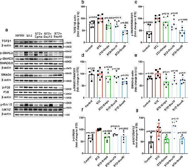
Scutellarin Inhibits TGF-β1 and Its Downstream Signalling Pathway. a Representative images of Western blotting samples for TGF-β1, p-SMAD2, p-SMAD3, SMAD2/3, SMAD4, p-p38, p38 and p-Erk and Erk1/2 of the mice treated with vehicle, scutellarin or empagliflozin. b – g Quantifications of the protein as indicated. All data are normalized to the STZ group and presented as the mean ± S.D.; n = 4–6 for each group, “n” stands for the number of animals; p vs. the model group (STZ)
Index in PubMed under a CC BY license. PMID: 38656633

Click image to see more details
Recombinant HE4 promoted fibroblast-myofibroblast transition and fibroblastic proliferation. A, B Different concentrations of rHE4 facilitated fibronectin, collagen I and α-SMA expression both in mRNA level (n = 3) and protein level (n = 4). The representative western blot images were shown and the band intensity was quantified by ImageJ. C Treatment with rHE4 in HPF cells enhanced phosphorylation of Smad2 (n = 4). D Typical images of EdU-staining HPFs followed by rHE4 treatment for 48 h and the quantitative graph were shown (n = 3). magnification = × 200. E CCK8 proliferation curve signified the pro-proliferative effect of rHE4 (n = 4). HPF was stimulated by rHE4 for 0 h, 24 h, 48 h and 72 h. F Representative western blot images of PCNA and Survivin were displayed and quantified by ImageJ (n = 3). Data are presented as mean ± SEM. P-values were calculated using one-way ANOVA followed by Newman–Keuls test. *P < 0.05, **P < 0.01, and ***P < 0.001 represent significant differences. rHE4, recombinant human epididymis protein 4; EdU, 5-ethynyl-2′-deoxyuridine; CCK8, Cell Counting Kit-8; PCNA, proliferating cell nuclear antigen; HPF, human pulmonary fibroblast
Index in PubMed under a CC BY license. PMID: 35550579

Click image to see more details
Western blot analysis of Phospho-Smad2 (S250) using anti-Phospho-Smad2 (S250) antibody (P00090-1).
Electrophoresis was performed on a 5-20% SDS-PAGE gel at 70V (Stacking gel) / 90V (Resolving gel) for 2-3 hours. The sample well of each lane was loaded with 30 ug of sample under reducing conditions.
Lane 1: human Hela whole cell lysates,
Lane 2: human HepG2 whole cell lysates,
Lane 3: human HT-1080 whole cell lysates,
Lane 4: rat heart tissue lysates,
Lane 5: rat skeletal muscle tissue lysates,
Lane 6: mouse heart tissue lysates,
Lane 7: mouse skeletal muscle tissue lysates.
After electrophoresis, proteins were transferred to a nitrocellulose membrane at 150 mA for 50-90 minutes. Blocked the membrane with 5% non-fat milk/TBS for 1.5 hour at RT. The membrane was incubated with rabbit anti-Phospho-Smad2 (S250) antigen affinity purified monoclonal antibody (Catalog # P00090-1) at 1:1000 overnight at 4°C, then washed with TBS-0.1%Tween 3 times with 5 minutes each and probed with a goat anti-rabbit IgG-HRP secondary antibody at a dilution of 1:1000 for 1.5 hour at RT. The signal is developed using an Enhanced Chemiluminescent detection (ECL) kit (Catalog # EK1002) with Tanon 5200 system. A specific band was detected for Phospho-Smad2 (S250) at approximately 58 kDa. The expected band size for Phospho-Smad2 (S250) is at 52 kDa.
Specific Publications For Anti-Phospho-Smad2 (S250) Rabbit Monoclonal Antibody (P00090-1)
Loading publications
Recommended Resources
Here are featured tools and databases that you might find useful.
- Boster's Pathways Library
- Protein Databases
- Bioscience Research Protocol Resources
- Data Processing & Analysis Software
- Photo Editing Software
- Scientific Literature Resources
- Research Paper Management Tools
- Molecular Biology Software
- Primer Design Tools
- Bioinformatics Tools
- Phylogenetic Tree Analysis
Customer Reviews
Have you used Anti-Phospho-Smad2 (S250) Rabbit Monoclonal Antibody?
Share your experimental results or join a short interview to earn up to $1,000 in product credits or other rewards.
0 Reviews For Anti-Phospho-Smad2 (S250) Rabbit Monoclonal Antibody
Customer Q&As
Have a question?
Find answers in Q&As, reviews.
Can't find your answer?
Submit your question
5 Customer Q&As for Anti-Phospho-Smad2 (S250) Rabbit Monoclonal Antibody
Question
We ordered your anti-Phospho-Smad2 (S250) Rabbit Monoclonal antibody for WB on pancreas spleen a few years ago. I am using mouse, and We want to use the antibody for Flow Cytometry next. We want examining pancreas spleen as well as cervix carcinoma erythroleukemia in our next experiment. Could give a recommendation on which antibody would work the best for Flow Cytometry?
Verified Customer
Verified customer
Asked: 2020-04-30
Answer
I have checked the website and datasheets of our anti-Phospho-Smad2 (S250) Rabbit Monoclonal antibody and it appears that P00090-1 has been validated on mouse in both WB and Flow Cytometry. Thus P00090-1 should work for your application. Our Boster satisfaction guarantee will cover this product for Flow Cytometry in mouse even if the specific tissue type has not been validated. We do have a comprehensive range of products for Flow Cytometry detection and you can check out our website bosterbio.com to find out more information about them.
Boster Scientific Support
Answered: 2020-04-30
Question
My colleagues were satisfied with the WB result of your anti-Phospho-Smad2 (S250) Rabbit Monoclonal antibody. However we have seen positive staining in colon adenocarcinoma cytoplasm using this antibody. Is that expected? Could you tell me where is SMAD2 supposed to be expressed?
Verified Customer
Verified customer
Asked: 2020-02-21
Answer
From literature, colon adenocarcinoma does express SMAD2. Generally SMAD2 expresses in cytoplasm. Regarding which tissues have SMAD2 expression, here are a few articles citing expression in various tissues:
Cervix carcinoma, Pubmed ID: 18669648, 18691976, 20068231
Cervix carcinoma, and Erythroleukemia, Pubmed ID: 23186163
Chronic myeloid leukemia cell, Pubmed ID: 8980228
Colon adenocarcinoma, Pubmed ID: 9702198
Kidney, Pubmed ID: 8752209
Kidney, Pancreas, and Spleen, Pubmed ID: 15489334
Placenta, Pubmed ID: 8774881
Boster Scientific Support
Answered: 2020-02-21
Question
We are currently using anti-Phospho-Smad2 (S250) Rabbit Monoclonal antibody P00090-1 for mouse tissue, and we are happy with the Flow Cytometry results. The species of reactivity given in the datasheet says human, mouse, rat. Is it likely that the antibody can work on bovine tissues as well?
Verified Customer
Verified customer
Asked: 2019-10-03
Answer
The anti-Phospho-Smad2 (S250) Rabbit Monoclonal antibody (P00090-1) has not been validated for cross reactivity specifically with bovine tissues, but there is a good chance of cross reactivity. We have an innovator award program that if you test this antibody and show it works in bovine you can get your next antibody for free. Please contact me if I can help you with anything.
Boster Scientific Support
Answered: 2019-10-03
Question
My question regards using your anti-Phospho-Smad2 (S250) Rabbit Monoclonal antibody for smad protein signal transduction studies. Has this antibody been tested with western blotting on hela cell lysate? We would like to see some validation images before ordering.
Verified Customer
Verified customer
Asked: 2019-08-09
Answer
I appreciate your inquiry. This P00090-1 anti-Phospho-Smad2 (S250) Rabbit Monoclonal antibody is tested on hela cell lysate. It is guaranteed to work for Flow Cytometry, WB in human, mouse, rat. Our Boster guarantee will cover your intended experiment even if the sample type has not been be directly tested.
Boster Scientific Support
Answered: 2019-08-09
Question
We have been able to see staining in mouse colon adenocarcinoma. Any tips? Is anti-Phospho-Smad2 (S250) Rabbit Monoclonal antibody supposed to stain colon adenocarcinoma positively?
Verified Customer
Verified customer
Asked: 2018-06-19
Answer
From what I have seen in literature colon adenocarcinoma does express SMAD2. From what I have seen in Uniprot.org, SMAD2 is expressed in kidney, placenta, kidney, pancreas spleen, chronic myeloid leukemia cell, colon adenocarcinoma, cervix carcinoma, cervix carcinoma erythroleukemia, among other tissues. Regarding which tissues have SMAD2 expression, here are a few articles citing expression in various tissues:
Cervix carcinoma, Pubmed ID: 18669648, 18691976, 20068231
Cervix carcinoma, and Erythroleukemia, Pubmed ID: 23186163
Chronic myeloid leukemia cell, Pubmed ID: 8980228
Colon adenocarcinoma, Pubmed ID: 9702198
Kidney, Pubmed ID: 8752209
Kidney, Pancreas, and Spleen, Pubmed ID: 15489334
Placenta, Pubmed ID: 8774881
Boster Scientific Support
Answered: 2018-06-19
