



Click image to see more details
Product Info Summary
| SKU: | M00239 |
|---|---|
| Size: | 100 μl |
| Reactive Species: | Human, Mouse, Rat |
| Host: | Rabbit |
| Application: | Flow Cytometry, IP, WB |
Customers Who Bought This Also Bought

Product info
Product Name
Anti-LGR5/GPR49 Rabbit Monoclonal Antibody
SKU/Catalog Number
M00239
BM4244 is an alternative SKU for this antibody, used in previous lots.
Size
100 μl
Form
Liquid
Description
Boster Bio Anti-LGR5/GPR49 Rabbit Monoclonal Antibody catalog # M00239. Tested in WB, IP, Flow Cytometry applications. This antibody reacts with Human, Mouse, Rat.
Storage & Handling
Store at -20°C for one year. For short term storage and frequent use, store at 4°C for up to one month. Avoid repeated freeze-thaw cycles.
Cite This Product
Anti-LGR5/GPR49 Rabbit Monoclonal Antibody (Boster Biological Technology, Pleasanton CA, USA, Catalog # M00239)
Host
Rabbit
Contents
Rabbit IgG in stabilizing components, phosphate buffered saline, pH 7.4, 150mM NaCl, 0.02% sodium azide and 50% glycerol.
*This antibody is supplied in a stabilized formulation.
Compatibility with conjugation reactions depends on the chemistry of the conjugation method used.
For conjugation methods that are not compatible with the stabilizing components present in this formulation, a carrier-free antibody format is required.
Clonality
Monoclonal
Clone Number
CHF-12
Isotype
Rabbit IgG
Immunogen
A synthesized peptide derived from human LGR5/GPR49
Reactive Species
M00239 is reactive to LGR5 in Human, Mouse, Rat
Observed Molecular Weight
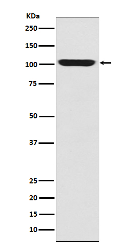
100 kDa
Calculated molecular weight
100.0 kDa
Antibody Validation
Boster validates all antibodies on WB, IHC, ICC, Immunofluorescence, and ELISA with known positive control and negative samples to ensure specificity and high affinity, including thorough antibody incubations.
Application & Images
Applications
M00239 is guaranteed for Flow Cytometry, IP, WB Boster Guarantee
Recommend Dilution
WB 1:500-2000
IP 1:20
FC 1:20
Tested application
Suggested blocking solution with 5% non-fat milk or BSA; (*)Recommended protein loading: 20-40 µg per lane
Validation Images & Assay Conditions
Click image to see more details
Western blot analysis of GPR49 expression in Human fetal skeletal muscle lysate.
Click image to see more details
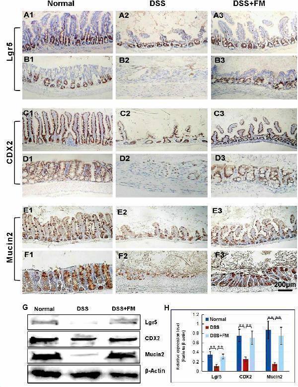
The distribution of Lgr5 + ISCs in the intestinalmucosa and the subcellular localization and relative expression level detection of epithelial function proteins CDX2 and villin in the intestinal mucosa of IBD at 7 days after termination of DSS administration. (A) The Lgr5 + ISCs (brown) in the small intestinal mucosa: (A1) the normal group, the villi and the crypts were arranged compactly, and Lgr5 + ISCs were observed in the crypts; (A2) the DSS group, the villi and the crypts were scattered, with few Lgr5 + ISCs; (A3) the DSS + B. subtilis -fermented milk group, there were more Lgr5 + ISCs in villi and crypts compared with those in the DSS group. (B) The Lgr5 + ISCs (brown) in the colonic mucosa: (B1) the normal group, the glands were arranged compactly, and there were large amounts of Lgr5 + ISCs at the bottom of the glands; (B2) the DSS group: the ulcers were replaced by scars. No Lgr5 + ISCs were observed in the scars; (B3) the DSS + B. subtilis -fermented milk group: the colonic epithelium was integrated, with some regenerated glands. A number of Lgr5 + ISCs were observed at the bottom of the regenerated glands. (C) The CDX2 was localized in the epithelial cellular nuclei (brown) by immunohistochemistry staining in the small intestinal mucosa: (C1) the normal group: the villi and the crypts were arranged compactly, and CDX2 + epithelial cells were observed on the surface of the villi and the crypts; (C2) the DSS group: the villi and the crypts were scattered, and few CDX2 + epithelial cells were observed on the surface of the crypt and the villi; (C3) the DSS + B. subtilis -fermented milk group: more villi and crypts were observed in comparison with the DSS group, and there were more CDX2 + epithelial cells covering the villi and crypts. (D) The CDX2 was localized in the epithelial cellular nuclei (brown) by immunohistochemistry staining in the colonic mucosa: (D1) the normal group: the colonic glands were arranged compactly, and CDX2 + epithelial cells were observed on the surface of the glands; (D2) the DSS group: the glands were scattered, and few CDX2 + epithelial cells were observed in the scar; (D3) the DSS + B. subtilis -fermented milk group: more colonic glands were observed in comparison with the DSS group, and there were more CDX2 + epithelial cells in the glands. (E) The Mucin2 was localized in the cytoplasm of the goblet cells (brown) by immunohistochemistry staining in the small intestinal mucosa: (E1) the normal group, a number of Mucin2 + goblet cells observed in the epithelium; (E2) the DSS group: only few Mucin2 + goblet cells were observed in the remaining villi and crypts; (E3) the DSS + B. subtilis -fermented milk group: more Mucin2 + goblet cells were observed in the recovered mucosa. (F) The Mucin2 was localized in the cytoplasm of the goblet cells (brown) by immunohistochemistry staining in the colonic mucosa: (F1) the normal group, large amounts of Mucin2 + goblet cells were observed in the mucosa; (F2) the DSS group: only few Mucin2 + goblet cells were observed in the scars; (F3) the DSS + B. subtilis -fermented milk group: more Mucin2 + goblet cells were observed in the recovered colonic mucosa. (G,H) Western blotting was applied for detection of the relative expression level of Lgr5, CDX2, and Mucin2 in the samples containing equivalent ileum and colon. The expression level of Lgr5, CDX2, and Mucin2 in the DSS group was significantly lower than that of the normal (control) group. The expression level of Lgr5, CDX2, and Mucin2 in the DSS + B. subtilis -fermented milk (FM) group was significantly higher than that of the DSS group ( n = 5, ** represents p < 0.01).
Index in PubMed under a CC BY license. PMID: 33519783
Specific Publications For Anti-LGR5/GPR49 Rabbit Monoclonal Antibody (M00239)
Loading publications
Recommended Resources
Here are featured tools and databases that you might find useful.
- Boster's Pathways Library
- Protein Databases
- Bioscience Research Protocol Resources
- Data Processing & Analysis Software
- Photo Editing Software
- Scientific Literature Resources
- Research Paper Management Tools
- Molecular Biology Software
- Primer Design Tools
- Bioinformatics Tools
- Phylogenetic Tree Analysis
Customer Reviews
Have you used Anti-LGR5/GPR49 Rabbit Monoclonal Antibody?
Share your experimental results or join a short interview to earn up to $1,000 in product credits or other rewards.
0 Reviews For Anti-LGR5/GPR49 Rabbit Monoclonal Antibody
Customer Q&As
Have a question?
Find answers in Q&As, reviews.
Can't find your answer?
Submit your question
1 Customer Q&As for Anti-LGR5/GPR49 Rabbit Monoclonal Antibody
Question
We are currently using anti-LGR5/GPR49 Rabbit Monoclonal antibody M00239 for rat tissue, and we are content with the WB results. The species of reactivity given in the datasheet says human, mouse, rat. Is it possible that the antibody can work on monkey tissues as well?
J. Jackson
Verified customer
Asked: 2017-01-05
Answer
The anti-LGR5/GPR49 Rabbit Monoclonal antibody (M00239) has not been tested for cross reactivity specifically with monkey tissues, but there is a good chance of cross reactivity. We have an innovator award program that if you test this antibody and show it works in monkey you can get your next antibody for free. Please contact me if I can help you with anything.
Boster Scientific Support
Answered: 2017-01-05

