Click image to see more details
Product Info Summary
| SKU: | PB9133 |
|---|---|
| Size: | 100 μg/vial |
| Reactive Species: | Human, Mouse, Rat |
| Host: | Rabbit |
| Application: | IF, ICC, WB |
Customers Who Bought This Also Bought

Product info
Product Name
Anti-Bmi1 Antibody Picoband®
SKU/Catalog Number
PB9133
PB0102 is an alternative SKU for this antibody, used in previous lots.
Size
100 μg/vial
Form
Lyophilized
Description
Boster Bio Anti-Bmi1 Antibody Picoband® catalog # PB9133. Tested in ICC/IF, WB applications. This antibody reacts with Human, Mouse, Rat. The brand Picoband indicates this is a premium antibody that guarantees superior quality, high affinity, and strong signals with minimal background in Western blot applications. Only our best-performing antibodies are designated as Picoband, ensuring unmatched performance.
Storage & Handling
Store at -20˚C for one year from date of receipt. After reconstitution, at 4˚C for one month. It can also be aliquotted and stored frozen at -20˚C for six months. Avoid repeated freeze-thaw cycles.
Cite This Product
Anti-Bmi1 Antibody Picoband® (Boster Biological Technology, Pleasanton CA, USA, Catalog # PB9133)
Host
Rabbit
Contents
Each vial contains 4 mg Trehalose, 0.9 mg NaCl and 0.2 mg Na2HPO4.
Clonality
Polyclonal
Isotype
Rabbit IgG
Immunogen
A synthetic peptide corresponding to a sequence in the middle region of human Bmi1, different from the related mouse sequence by four amino acids.
Cross-reactivity
No cross-reactivity with other proteins
Reactive Species
PB9133 is reactive to BMI1 in Human, Mouse, Rat
Observed Molecular Weight
40 kDa
Calculated molecular weight
36.9 kDa
Background of BMI1
BMI1 (BMI1 polycomb ring finger oncogene), also known as RNF51, is a protein which in humans is encoded by the BMI1 gene. The Bmi1 gene is highly conserved in evolution as indicated by zoo blot hybridization with Bmi1 probes corresponding to the protein-encoding domain. By fluorescence in situ hybridization, the human BMI1 gene is assigned to chromosome 10p13. BMI1 has a key role in regulating the proliferative activity of normal stem and progenitor cells. Most importantly, they provided evidence that the proliferative potential of leukemic stem and progenitor cells lacking BMI1 is compromised because they eventually undergo proliferation arrest and show signs of differentiation and apoptosis, leading to transplant failure of the leukemia. Complementation studies showed that BMI1 completely rescues these proliferative defects. Deletion analysis showed that the RING finger and helix-turn-helix domains of BMI1 were required for life span extension and repression of the tumor suppressor p16 (INK4). BMI1 selectively extended the life span of these cultures. Confocal microscopy showed that BMI1 transiently colocalized with centromeres during interphase in HeLa cells.
Antibody Validation
Boster validates all antibodies on WB, IHC, ICC, Immunofluorescence, and ELISA with known positive control and negative samples to ensure specificity and high affinity, including thorough antibody incubations.
Application & Images
Applications
PB9133 is guaranteed for IF, ICC, WB Boster Guarantee
Recommend Dilution
| Application | Dilution | Species |
|---|---|---|
| Western blot | 0.1-0.5μg/ml | Human, Mouse, Rat |
| Immunocytochemistry/Immunofluorescence | 5 μg/ml | Human |
Tested application
Suggested blocking solution with 5% non-fat milk or BSA; (*)Recommended protein loading: 20-40 µg per lane
Validation Images & Assay Conditions
Click image to see more details
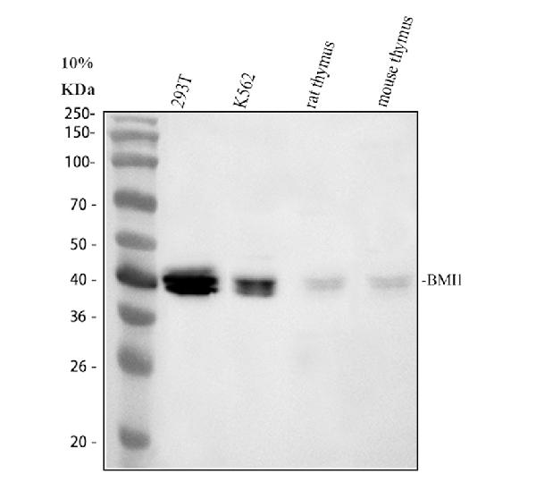
Western blot analysis of Bmi1 using anti-Bmi1 antibody (PB9133).
Electrophoresis was performed on a 10% SDS-PAGE gel at 80V (Stacking gel) / 120V (Resolving gel) for 2 hours. The sample well of each lane was loaded with 30 ug of sample under reducing conditions.
Lane 1: human 293T whole cell lysates,
Lane 2: human K562 whole cell lysates,
Lane 3: rat thymus tissue lysates,
Lane 4: mouse thymus tissue lysates.
After electrophoresis, proteins were transferred to a nitrocellulose membrane at 150 mA for 50-90 minutes. Blocked the membrane with 5% non-fat milk/TBS for 1.5 hour at RT. The membrane was incubated with rabbit anti-Bmi1 antigen affinity purified polyclonal antibody (PB9133) at 0.5 μg/mL overnight at 4°C, then washed with TBS-0.1%Tween 3 times with 5 minutes each and probed with a goat anti-rabbit IgG-HRP secondary antibody at a dilution of 1:5000 for 1.5 hour at RT. The signal is developed using an ECL Plus Western Blotting Substrate (Catalog # AR1196-200) with Tanon 5200 system. A specific band was detected for Bmi1 at approximately 40 kDa. The expected band size for Bmi1 is at 37 kDa.

Click image to see more details
IF analysis of Bmi1 using anti-Bmi1 antibody (PB9133) and anti-Alpha Tubulin antibody (M03989-3).
Bmi1 was detected in an immunocytochemical section of U2OS cells. Enzyme antigen retrieval was performed using IHC enzyme antigen retrieval reagent (AR0022) for 15 mins. The cells were blocked with 10% goat serum. And then incubated with 5 μg/mL rabbit anti-Bmi1 Antibody (PB9133) and mouse anti-Alpha Tubulin antibody (M03989-3) overnight at 4°C. Cy3 Conjugated Goat Anti-Rabbit IgG (BA1032) and Fluoro488 Conjugated Goat Anti-Mouse IgG (BA1126) were used as secondary antibody at 1:500 dilution and incubated for 30 minutes at 37°C. Visualize using a fluorescence microscope and filter sets appropriate for the label used.
Specific Publications For Anti-Bmi1 Antibody Picoband® (PB9133)
Loading publications
Recommended Resources
Here are featured tools and databases that you might find useful.
- Boster's Pathways Library
- Protein Databases
- Bioscience Research Protocol Resources
- Data Processing & Analysis Software
- Photo Editing Software
- Scientific Literature Resources
- Research Paper Management Tools
- Molecular Biology Software
- Primer Design Tools
- Bioinformatics Tools
- Phylogenetic Tree Analysis
Customer Reviews
Have you used Anti-Bmi1 Antibody Picoband®?
Share your experimental results or join a short interview to earn up to $1,000 in product credits or other rewards.
0 Reviews For Anti-Bmi1 Antibody Picoband®
Customer Q&As
Have a question?
Find answers in Q&As, reviews.
Can't find your answer?
Submit your question
1 Customer Q&As for Anti-Bmi1 Antibody Picoband®
Question
We are currently using anti-Bmi1 antibody PB9133 for human tissue, and we are well pleased with the WB results. The species of reactivity given in the datasheet says human, mouse, rat. Is it true that the antibody can work on pig tissues as well?
L. Bhatt
Verified customer
Asked: 2016-06-24
Answer
The anti-Bmi1 antibody (PB9133) has not been tested for cross reactivity specifically with pig tissues, though there is a good chance of cross reactivity. We have an innovator award program that if you test this antibody and show it works in pig you can get your next antibody for free. Please contact me if I can help you with anything.
Boster Scientific Support
Answered: 2016-06-24
