Click image to see more details
Product Info Summary
| SKU: | A01141-1 |
|---|---|
| Size: | 100 μg/vial |
| Reactive Species: | Human, Mouse, Rat |
| Host: | Rabbit |
| Application: | IF, ICC, WB |
Customers Who Bought This Also Bought

Product info
Product Name
Anti-14-3-3 zeta/delta/YWHAZ Antibody Picoband®
SKU/Catalog Number
A01141-1
Size
100 μg/vial
Form
Lyophilized
Description
Boster Bio Anti-14-3-3 zeta/delta/YWHAZ Antibody Picoband® catalog # A01141-1. Tested in IF, ICC, WB applications. This antibody reacts with Human, Mouse, Rat. The brand Picoband indicates this is a premium antibody that guarantees superior quality, high affinity, and strong signals with minimal background in Western blot applications. Only our best-performing antibodies are designated as Picoband, ensuring unmatched performance.
Storage & Handling
Store at -20˚C for one year from date of receipt. After reconstitution, at 4˚C for one month. It can also be aliquotted and stored frozen at -20˚C for six months. Avoid repeated freeze-thaw cycles.
Cite This Product
Anti-14-3-3 zeta/delta/YWHAZ Antibody Picoband® (Boster Biological Technology, Pleasanton CA, USA, Catalog # A01141-1)
Host
Rabbit
Contents
Each vial contains 4mg Trehalose, 0.9mg NaCl, 0.2mg Na2HPO4, 0.01mg NaN3.
Clonality
Polyclonal
Isotype
Rabbit IgG
Immunogen
A synthetic peptide corresponding to a sequence in the middle region of human 14-3-3 zeta/delta, which shares 97.8% amino acid (aa) sequence identity with both mouse and rat 14-3-3 zeta/delta.
Cross-reactivity
No cross-reactivity with other proteins.
Reactive Species
A01141-1 is reactive to YWHAZ in Human, Mouse, Rat
Observed Molecular Weight
28 kDa
Calculated molecular weight
27.7 kDa
Background of YWHAZ
14-3-3 protein zeta/delta (14-3-3ζ) is a protein that in humans is encoded by the YWHAZ gene on chromosome 8. This gene product belongs to the 14-3-3 family of proteins which mediate signal transduction by binding to phosphoserine-containing proteins. This highly conserved protein family is found in both plants and mammals, and this protein is 99% identical to the mouse, rat and sheep orthologs. The encoded protein interacts with IRS1 protein, suggesting a role in regulating insulin sensitivity. Several transcript variants that differ in the 5' UTR but that encode the same protein have been identified for this gene.
Antibody Validation
Boster validates all antibodies on WB, IHC, ICC, Immunofluorescence, and ELISA with known positive control and negative samples to ensure specificity and high affinity, including thorough antibody incubations.
Application & Images
Applications
A01141-1 is guaranteed for IF, ICC, WB Boster Guarantee
Recommend Dilution
| Application | Dilution | Species |
|---|---|---|
| Western blot | 0.1-0.5μg/ml | |
| Immunocytochemistry/Immunofluorescence | 5μg/ml |
Tested application
Suggested blocking solution with 5% non-fat milk or BSA; (*)Recommended protein loading: 20-40 µg per lane
Validation Images & Assay Conditions
Click image to see more details
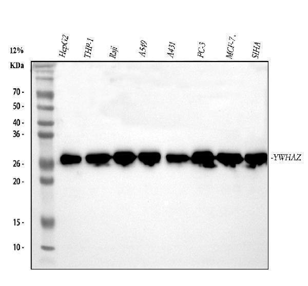
Western blot analysis of 14-3-3 zeta/delta using anti-14-3-3 zeta/delta antibody (A01141-1).
Electrophoresis was performed on a 5-20% SDS-PAGE gel at 70V (Stacking gel) / 90V (Resolving gel) for 2-3 hours. The sample well of each lane was loaded with 30 ug of sample under reducing conditions.
Lane 1: human HepG2 whole cell lysates,
Lane 2: human THP-1 whole cell lysates,
Lane 3: human Raji whole cell lysates,
Lane 4: human A549 whole cell lysates,
Lane 5: human A431 whole cell lysates,
Lane 6: human PC-3 whole cell lysates,
Lane 7: human MCF-7 whole cell lysates,
Lane 8: human SiHa whole cell lysates.
After electrophoresis, proteins were transferred to a nitrocellulose membrane at 150 mA for 50-90 minutes. Blocked the membrane with 5% non-fat milk/TBS for 1.5 hour at RT. The membrane was incubated with rabbit anti-14-3-3 zeta/delta antigen affinity purified polyclonal antibody (Catalog # A01141-1) at 0.5 μg/mL overnight at 4°C, then washed with TBS-0.1%Tween 3 times with 5 minutes each and probed with a goat anti-rabbit IgG-HRP secondary antibody at a dilution of 1:5000 for 1.5 hour at RT. The signal is developed using an Enhanced Chemiluminescent detection (ECL) kit (Catalog # EK1002) with Tanon 5200 system. A specific band was detected for 14-3-3 zeta/delta at approximately 28 kDa. The expected band size for 14-3-3 zeta/delta is at 28 kDa.

Click image to see more details
Western blot analysis of 14-3-3 zeta/delta using anti-14-3-3 zeta/delta antibody (A01141-1).
Electrophoresis was performed on a 5-20% SDS-PAGE gel at 70V (Stacking gel) / 90V (Resolving gel) for 2-3 hours. The sample well of each lane was loaded with 30 ug of sample under reducing conditions.
Lane 1: rat brain tissue lysates,
Lane 2: rat kidney tissue lysates,
Lane 3: rat RH35 whole cell lysates,
Lane 4: rat PC-12 whole cell lysates,
Lane 5: mouse brain tissue lysates,
Lane 6: mouse kidney tissue lysates,
Lane 7: mouse ANA-1 whole cell lysates,
Lane 8: mouse Neuro-2a whole cell lysates.
After electrophoresis, proteins were transferred to a nitrocellulose membrane at 150 mA for 50-90 minutes. Blocked the membrane with 5% non-fat milk/TBS for 1.5 hour at RT. The membrane was incubated with rabbit anti-14-3-3 zeta/delta antigen affinity purified polyclonal antibody (Catalog # A01141-1) at 0.5 μg/mL overnight at 4°C, then washed with TBS-0.1%Tween 3 times with 5 minutes each and probed with a goat anti-rabbit IgG-HRP secondary antibody at a dilution of 1:5000 for 1.5 hour at RT. The signal is developed using an Enhanced Chemiluminescent detection (ECL) kit (Catalog # EK1002) with Tanon 5200 system. A specific band was detected for 14-3-3 zeta/delta at approximately 28 kDa. The expected band size for 14-3-3 zeta/delta is at 28 kDa.

Click image to see more details
IF analysis of 14-3-3 zeta/delta using anti-14-3-3 zeta/delta antibody (A01141-1).
14-3-3 zeta/delta was detected in an immunocytochemical section of U2OS cells. Enzyme antigen retrieval was performed using IHC enzyme antigen retrieval reagent (AR0022) for 15 mins. The cells were blocked with 10% goat serum. And then incubated with 5 μg/mL rabbit anti-14-3-3 zeta/delta Antibody (A01141-1) overnight at 4°C. DyLight488 Conjugated Goat Anti-Rabbit IgG (BA1127) was used as secondary antibody at 1:500 dilution and incubated for 30 minutes at 37°C. The section was counterstained with DAPI. Visualize using a fluorescence microscope and filter sets appropriate for the label used.
Specific Publications For Anti-14-3-3 zeta/delta/YWHAZ Antibody Picoband® (A01141-1)
Loading publications
Recommended Resources
Here are featured tools and databases that you might find useful.
- Boster's Pathways Library
- Protein Databases
- Bioscience Research Protocol Resources
- Data Processing & Analysis Software
- Photo Editing Software
- Scientific Literature Resources
- Research Paper Management Tools
- Molecular Biology Software
- Primer Design Tools
- Bioinformatics Tools
- Phylogenetic Tree Analysis
Customer Reviews
Have you used Anti-14-3-3 zeta/delta/YWHAZ Antibody Picoband®?
Share your experimental results or join a short interview to earn up to $1,000 in product credits or other rewards.
0 Reviews For Anti-14-3-3 zeta/delta/YWHAZ Antibody Picoband®
Customer Q&As
Have a question?
Find answers in Q&As, reviews.
Can't find your answer?
Submit your question
5 Customer Q&As for Anti-14-3-3 zeta/delta/YWHAZ Antibody Picoband®
Question
I was wanting to use your anti-14-3-3 zeta/delta/YWHAZ antibody for IHC-P for rat skin on frozen tissues, but I want to know if it has been tested for this particular application. Has this antibody been tested and is this antibody a good choice for rat skin identification?
Verified Customer
Verified customer
Asked: 2020-01-08
Answer
You can see on the product datasheet, A01141-1 anti-14-3-3 zeta/delta/YWHAZ antibody has been validated for Flow Cytometry, IF, IHC-P, IHC-F, ICC, WB on human, mouse, rat tissues. We have an innovator award program that if you test this antibody and show it works in rat skin in IHC-frozen, you can get your next antibody for free.
Boster Scientific Support
Answered: 2020-01-08
Question
I have attached the WB image, lot number and protocol we used for skin using anti-14-3-3 zeta/delta/YWHAZ antibody A01141-1. Please let me know if you require anything else.
Verified Customer
Verified customer
Asked: 2019-12-23
Answer
Thank you very much for the data. Our lab team are working to resolve this as quickly as possible, and we appreciate your patience and understanding! You have provided everything we needed. Please let me know if there is anything you need in the meantime.
Boster Scientific Support
Answered: 2019-12-23
Question
Does anti-14-3-3 zeta/delta/YWHAZ antibody A01141-1 work for IHC-P with skin?
L. Rodriguez
Verified customer
Asked: 2019-12-16
Answer
According to the expression profile of skin, YWHAZ is highly expressed in skin. So, it is likely that anti-14-3-3 zeta/delta/YWHAZ antibody A01141-1 will work for IHC-P with skin.
Boster Scientific Support
Answered: 2019-12-16
Question
We are currently using anti-14-3-3 zeta/delta/YWHAZ antibody A01141-1 for rat tissue, and we are happy with the WB results. The species of reactivity given in the datasheet says human, mouse, rat. Is it true that the antibody can work on goat tissues as well?
Verified Customer
Verified customer
Asked: 2019-10-21
Answer
The anti-14-3-3 zeta/delta/YWHAZ antibody (A01141-1) has not been validated for cross reactivity specifically with goat tissues, though there is a good chance of cross reactivity. We have an innovator award program that if you test this antibody and show it works in goat you can get your next antibody for free. Please contact me if I can help you with anything.
Boster Scientific Support
Answered: 2019-10-21
Question
We appreciate helping with my inquiry over the phone. Here are the WB image, lot number and protocol we used for skin using anti-14-3-3 zeta/delta/YWHAZ antibody A01141-1. Let me know if you need anything else.
W. Krishna
Verified customer
Asked: 2016-07-25
Answer
Thanks for the data. You have provided everything we needed. Our lab team are working to resolve your inquiry as quickly as possible, and we appreciate your patience and understanding! Please let me know if there is anything you need in the meantime.
Boster Scientific Support
Answered: 2016-07-25
