Click image to see more details
-

-
-
-
-
+5
Product Info Summary
| SKU: | PB10026 |
|---|---|
| Size: | 100 μg/vial |
| Reactive Species: | Human, Mouse, Rat |
| Host: | Rabbit |
| Application: | Flow Cytometry, IF, IHC, WB |
Customers Who Bought This Also Bought

Product info
Product Name
Anti-ACTN3 Antibody Picoband®
SKU/Catalog Number
PB10026
Size
100 μg/vial
Form
Lyophilized
Description
Boster Bio Anti-ACTN3 Antibody Picoband® catalog # PB10026. Tested in Flow Cytometry, IF, IHC, WB applications. This antibody reacts with Human, Mouse, Rat. The brand Picoband indicates this is a premium antibody that guarantees superior quality, high affinity, and strong signals with minimal background in Western blot applications. Only our best-performing antibodies are designated as Picoband, ensuring unmatched performance.
Storage & Handling
Store at -20˚C for one year from date of receipt. After reconstitution, at 4˚C for one month. It can also be aliquotted and stored frozen at -20˚C for six months. Avoid repeated freeze-thaw cycles.
Cite This Product
Anti-ACTN3 Antibody Picoband® (Boster Biological Technology, Pleasanton CA, USA, Catalog # PB10026)
Host
Rabbit
Contents
Each vial contains antibody formulated with stabilizing components, 0.9 mg NaCl, 0.2 mg Na2HPO4, and 0.05 mg NaN3.
*This antibody is supplied in a stabilized formulation.
Compatibility with conjugation reactions depends on the chemistry of the conjugation method used.
For conjugation methods that are not compatible with the stabilizing components present in this formulation, a carrier-free antibody format is required.
Clonality
Polyclonal
Isotype
Rabbit IgG
Immunogen
A synthetic peptide corresponding to a sequence at the C-terminus of human ACTN3, different from the related mouse sequence by five amino acids.
Cross-reactivity
No cross-reactivity with other proteins
Reactive Species
PB10026 is reactive to ACTN3 in Human, Mouse, Rat
Observed Molecular Weight
103 kDa
Calculated molecular weight
103.2 kDa
Background of ACTN3
Alpha-actinin-3, also known as alpha-actinin skeletal muscle isoform 3 or F-actin cross-linking protein, is a protein that in humans is encoded by the ACTN3 gene. This gene encodes a member of the alpha-actin binding protein gene family. The encoded protein is primarily expressed in skeletal muscle and functions as a structural component of sarcomeric Z line. This protein is involved in crosslinking actin containing thin filaments. An allelic polymorphism in this gene results in both coding and non-coding variants; the reference genome represents the coding allele. The non-functional allele of this gene is associated with elite athlete status.
Antibody Validation
Boster validates all antibodies on WB, IHC, ICC, Immunofluorescence, and ELISA with known positive control and negative samples to ensure specificity and high affinity, including thorough antibody incubations.
Application & Images
Applications
PB10026 is guaranteed for Flow Cytometry, IF, IHC, WB Boster Guarantee
Recommend Dilution
| Application | Dilution | Species |
|---|---|---|
| Western blot | 0.1-0.5μg/ml | Human, Mouse, Rat |
| Immunohistochemistry (Paraffin-embedded Section) | 0.5-1μg/ml | Human, Mouse, Rat |
| Immunofluorescence | 5 μg/ml | Mouse |
| Flow Cytometry (Fixed) | 1-3μg/1x106 cells | Human |
Tested application
Suggested blocking solution with 5% non-fat milk or BSA; (*)Recommended protein loading: 20-40 µg per lane
Use TE buffer pH 9.0 for antigen retrieval; (*) citrate buffer pH 6.0 is an alternative.
Validation Images & Assay Conditions
Click image to see more details
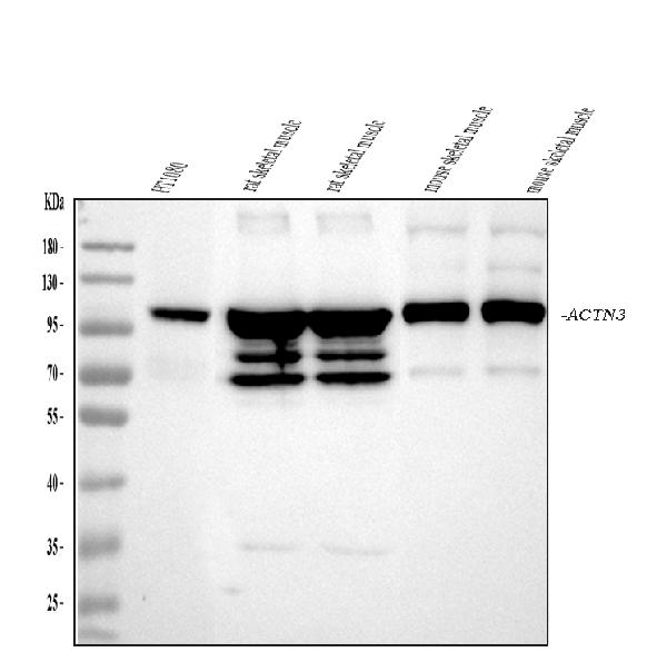
Western blot analysis of ACTN3/Alpha Actinin 3 using anti-ACTN3/Alpha Actinin 3 antibody (PB10026).
Electrophoresis was performed on a 5-20% SDS-PAGE gel at 70V (Stacking gel) / 90V (Resolving gel) for 2-3 hours. The sample well of each lane was loaded with 30 ug of sample under reducing conditions.
Lane 1: human HT1080 whole cell lysates,
Lane 2: rat skeletal muscle tissue lysates,
Lane 3: rat skeletal muscle tissue lysates,
Lane 4: mouse skeletal muscle tissue lysates,
Lane 5: mouse skeletal muscle tissue lysates.
After electrophoresis, proteins were transferred to a nitrocellulose membrane at 150 mA for 50-90 minutes. Blocked the membrane with 5% non-fat milk/TBS for 1.5 hour at RT. The membrane was incubated with rabbit anti-ACTN3/Alpha Actinin 3 antigen affinity purified polyclonal antibody (Catalog # PB10026) at 0.5 μg/mL overnight at 4°C, then washed with TBS-0.1%Tween 3 times with 5 minutes each and probed with a goat anti-rabbit IgG-HRP secondary antibody at a dilution of 1:5000 for 1.5 hour at RT. The signal is developed using an Enhanced Chemiluminescent detection (ECL) kit (Catalog # EK1002) with Tanon 5200 system. A specific band was detected for ACTN3/Alpha Actinin 3 at approximately 103 kDa. The expected band size for ACTN3/Alpha Actinin 3 is at 103 kDa.

Click image to see more details
Major patterns of protein distribution according to fiber type in human skeletal muscle. The fiber type distribution of representative proteins (indicated by gene name) is shown for each pattern, with values expressed as per cent of the maximal value. ACTN3 , α-actinin-3; CYCS , cytochrome c; MYL2 , MLC-2 slow; MYLK2 , myosin light chain kinase 2; RYR1 , ryanodine receptor 1; TNNC2 , fast troponin C
Index in PubMed under a CC BY license. PMID: 34727990

Click image to see more details
Myofibrillar proteins: fiber type distribution of representative proteins. Proteins showing similar distribution in the different types are not shown in the figure. Values are expressed as per cent of the maximal value. ACTN2 , α-actinin-2; ACTN3 , α-actinin-3; CAPZA1 , CapZ α1; LMOD2 , leiomodin-2; LRRC39 , Myomasp; MYL1 , MLC1/3-fast; MYL2 , MLC-2slow; MYL3 , MLC-1 slow; MYLPF , MLC2-fast; MYL6B , MLC-1sa; MYBPC1 , myosin-binding protein C1; MYBPC2 , myosin-binding protein C2; MYBPH , myosin-binding protein H; MYOM2 , myomesin 2; MYOM3 , myomesin 3; MYOZ2 , myozenin 2; MYOZ3 , myozenin 3; TNNC1 , slow troponin C; TNNC2 , fast troponin C; TNNI1 , slow troponin I; TNNI2 , fast troponin I; TNNT1 , slow troponin T; TNNT2 , fast troponin T; TPM1 , α-tropomyosin; TPM3 , γ-tropomyosin
Index in PubMed under a CC BY license. PMID: 34727990

Click image to see more details
Fiber type–specific distribution of selected proteins revealed by immunofluorescence analysis. Fiber types are labeled as 1 (type 1), 2A (type 2A), or 2X (type 2X). Hybrid 2A–2X fibers are labeled by asterisks. A – D Type 1–specific proteins. Each panel shows on the left a section stained for PGM5/aciculin ( A ), PDLIM1 ( B ), MCU ( C ), or IDH2 ( D ) and on the right a serial section stained with MYH-specific antibodies to reveal the three fiber types. No pure type 2X fibers is present in A and B . E – F Type 2–specific proteins. Each panel shows on the left a section stained for ACTN3 ( E ) or XIRP2 ( F ), on the center a serial section stained for MYH-specific antibodies to reveal the three fiber types, and on the right a section stained with anti-MYH antibody BF-35, which reacts with MYH7 and MYH2 but not with MYH1, thus stains all fiber types, except pure type 2X fibers. Note that whereas ACTN3 is especially abundant in type 2X fibers, as well as in hybrid 2A–2X fibers, XIRP2 is present at higher levels in both 2A and 2X fibers. WGA counterstain was applied to all sections, except those processed with the three anti-MYHs antibodies
Index in PubMed under a CC BY license. PMID: 34727990

Click image to see more details
IHC analysis of ACTN3/Alpha Actinin 3 using anti-ACTN3/Alpha Actinin 3 antibody (PB10026).
ACTN3/Alpha Actinin 3 was detected in paraffin-embedded section of mouse skeletal muscle tissues. Heat mediated antigen retrieval was performed in citrate buffer (pH6, epitope retrieval solution) for 20 mins. The tissue section was blocked with 10% goat serum. The tissue section was then incubated with 1μg/ml rabbit anti-ACTN3/Alpha Actinin 3 Antibody (PB10026) overnight at 4°C. Biotinylated goat anti-rabbit IgG was used as secondary antibody and incubated for 30 minutes at 37°C. The tissue section was developed using Strepavidin-Biotin-Complex (SABC)(Catalog # SA1022) with DAB as the chromogen.

Click image to see more details
IHC analysis of ACTN3/Alpha Actinin 3 using anti-ACTN3/Alpha Actinin 3 antibody (PB10026).
ACTN3/Alpha Actinin 3 was detected in paraffin-embedded section of rat skeletal muscle tissues. Heat mediated antigen retrieval was performed in citrate buffer (pH6, epitope retrieval solution) for 20 mins. The tissue section was blocked with 10% goat serum. The tissue section was then incubated with 1μg/ml rabbit anti-ACTN3/Alpha Actinin 3 Antibody (PB10026) overnight at 4°C. Biotinylated goat anti-rabbit IgG was used as secondary antibody and incubated for 30 minutes at 37°C. The tissue section was developed using Strepavidin-Biotin-Complex (SABC)(Catalog # SA1022) with DAB as the chromogen.

Click image to see more details
IHC analysis of ACTN3/Alpha Actinin 3 using anti-ACTN3/Alpha Actinin 3 antibody (PB10026).
ACTN3/Alpha Actinin 3 was detected in paraffin-embedded section of human lung cancer tissues. Heat mediated antigen retrieval was performed in citrate buffer (pH6, epitope retrieval solution) for 20 mins. The tissue section was blocked with 10% goat serum. The tissue section was then incubated with 1μg/ml rabbit anti-ACTN3/Alpha Actinin 3 Antibody (PB10026) overnight at 4°C. Biotinylated goat anti-rabbit IgG was used as secondary antibody and incubated for 30 minutes at 37°C. The tissue section was developed using Strepavidin-Biotin-Complex (SABC)(Catalog # SA1022) with DAB as the chromogen.

Click image to see more details
IF analysis of ACTN3/Alpha Actinin 3 using anti-ACTN3/Alpha Actinin 3 antibody (PB10026).
ACTN3/Alpha Actinin 3 was detected in a paraffin-embedded section of mouse skeletal muscle tissue. Heat mediated antigen retrieval was performed in EDTA buffer (pH 8.0, epitope retrieval solution). The tissue section was blocked with 10% goat serum. The tissue section was then incubated with 5 μg/mL rabbit anti-ACTN3/Alpha Actinin 3 Antibody (PB10026) overnight at 4°C. FITC Conjugated Goat Anti-Rabbit IgG (BA1105) was used as secondary antibody at 1:500 dilution and incubated for 30 minutes at 37°C. The section was counterstained with DAPI. Visualize using a fluorescence microscope and filter sets appropriate for the label used.

Click image to see more details
Flow Cytometry analysis of WISH cells using anti-ACTN3 antibody (PB10026).
Overlay histogram showing WISH cells stained with PB10026 (Blue line). To facilitate intracellular staining, cells were fixed with 4% paraformaldehyde and permeabilized with permeabilization buffer. The cells were blocked with 10% normal goat serum. And then incubated with rabbit anti-ACTN3 Antibody (PB10026,1μg/1x106 cells) for 30 min at 20°C. DyLight®488 conjugated goat anti-rabbit IgG (BA1127, 5-10μg/1x106 cells) was used as secondary antibody for 30 minutes at 20°C. Isotype control antibody (Green line) was rabbit IgG (1μg/1x106) used under the same conditions. Unlabelled sample without incubation with primary antibody and secondary antibody (Red line) was used as a blank control.
Specific Publications For Anti-ACTN3 Antibody Picoband® (PB10026)
Loading publications
Recommended Resources
Here are featured tools and databases that you might find useful.
- Boster's Pathways Library
- Protein Databases
- Bioscience Research Protocol Resources
- Data Processing & Analysis Software
- Photo Editing Software
- Scientific Literature Resources
- Research Paper Management Tools
- Molecular Biology Software
- Primer Design Tools
- Bioinformatics Tools
- Phylogenetic Tree Analysis
Customer Reviews
Have you used Anti-ACTN3 Antibody Picoband®?
Share your experimental results or join a short interview to earn up to $1,000 in product credits or other rewards.
0 Reviews For Anti-ACTN3 Antibody Picoband®
Customer Q&As
Have a question?
Find answers in Q&As, reviews.
Can't find your answer?
Submit your question
1 Customer Q&As for Anti-ACTN3 Antibody Picoband®
Question
We are currently using anti-ACTN3 antibody PB10026 for rat tissue, and we are satisfied with the WB results. The species of reactivity given in the datasheet says human, mouse, rat. Is it likely that the antibody can work on bovine tissues as well?
Verified Customer
Verified customer
Asked: 2018-09-26
Answer
The anti-ACTN3 antibody (PB10026) has not been tested for cross reactivity specifically with bovine tissues, but there is a good chance of cross reactivity. We have an innovator award program that if you test this antibody and show it works in bovine you can get your next antibody for free. Please contact me if I can help you with anything.
Boster Scientific Support
Answered: 2018-09-26
