Click image to see more details
Product Info Summary
| SKU: | PB10027 |
|---|---|
| Size: | 100 μg/vial |
| Reactive Species: | Human, Mouse, Rat |
| Host: | Rabbit |
| Application: | Flow Cytometry, IF, ICC, WB |
Customers Who Bought This Also Bought

Product info
Product Name
Anti-Activin Receptor Type IIA/ACVR2A Antibody Picoband®
SKU/Catalog Number
PB10027
PB1080 is an alternative SKU for this antibody, used in previous lots.
Size
100 μg/vial
Form
Lyophilized
Description
Boster Bio Anti-Activin Receptor Type IIA/ACVR2A Antibody Picoband® catalog # PB10027. Tested in Flow Cytometry, IF, ICC, WB applications. This antibody reacts with Human, Mouse, Rat. The brand Picoband indicates this is a premium antibody that guarantees superior quality, high affinity, and strong signals with minimal background in Western blot applications. Only our best-performing antibodies are designated as Picoband, ensuring unmatched performance.
Storage & Handling
Store at -20˚C for one year from date of receipt. After reconstitution, at 4˚C for one month. It can also be aliquotted and stored frozen at -20˚C for six months. Avoid repeated freeze-thaw cycles.
Cite This Product
Anti-Activin Receptor Type IIA/ACVR2A Antibody Picoband® (Boster Biological Technology, Pleasanton CA, USA, Catalog # PB10027)
Host
Rabbit
Contents
Each vial contains 4 mg Trehalose, 0.9 mg NaCl and 0.2 mg Na2HPO4.
Clonality
Polyclonal
Isotype
Rabbit IgG
Immunogen
E. coli-derived human ACVR2A recombinant protein (Position: Q421-L513). Human ACVR2A shares 100% and 98.9% amino acid (aa) sequence identity with mouse and rat ACVR2A, respectively.
Cross-reactivity
No cross-reactivity with other proteins
Reactive Species
PB10027 is reactive to ACVR2A in Human, Mouse, Rat
Observed Molecular Weight
65-90 kDa
Calculated molecular weight
57.8 kDa
Background of ACVR2A
Activin receptor type-2A is a protein that in humans is encoded by the ACVR2A gene. ACVR2A is an activin type 2 receptor. This gene encodes a receptor that mediates the functions of activins, which are members of the transforming growth factor-beta (TGF-beta) superfamily involved in diverse biological processes. The encoded protein is a transmembrane serine-threonine kinase receptor which mediates signaling by forming heterodimeric complexes with various combinations of type I and type II receptors and ligands in a cell-specific manner. The encoded type II receptor is primarily involved in ligand-binding and includes an extracellular ligand-binding domain, a transmembrane domain and a cytoplasmic serine-threonine kinase domain. This gene may be associated with susceptibility to preeclampsia, a pregnancy-related disease which can result in maternal and fetal morbidity and mortality. Alternative splicing results in multiple transcript variants of this gene.
Antibody Validation
Boster validates all antibodies on WB, IHC, ICC, Immunofluorescence, and ELISA with known positive control and negative samples to ensure specificity and high affinity, including thorough antibody incubations.
Application & Images
Applications
PB10027 is guaranteed for Flow Cytometry, IF, ICC, WB Boster Guarantee
Recommend Dilution
| Application | Dilution | Species |
|---|---|---|
| Western blot | 0.1-0.5μg/ml | Human, Mouse, Rat |
| Immunocytochemistry/Immunofluorescence | 5μg/ml | Human |
| Flow Cytometry (Fixed) | 1-3μg/1x106 cells | Human |
Tested application
Suggested blocking solution with 5% non-fat milk or BSA; (*)Recommended protein loading: 20-40 µg per lane
Validation Images & Assay Conditions
Click image to see more details
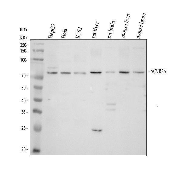
Western blot analysis of ACVR2A using anti-ACVR2A antibody (PB10027).
Electrophoresis was performed on a 10% SDS-PAGE gel at 80V (Stacking gel) / 120V (Resolving gel) for 2 hours. The sample well of each lane was loaded with 30 ug of sample under reducing conditions.
Lane 1: human HepG2 whole cell lysates,
Lane 2: human Hela whole cell lysates,
Lane 3: human K562 whole cell lysates,
Lane 4: rat liver tissue lysates,
Lane 5: rat brain tissue lysates,
Lane 6: mouse liver tissue lysates,
Lane 7: mouse brain tissue lysates.
After electrophoresis, proteins were transferred to a nitrocellulose membrane at 150 mA for 50-90 minutes. Blocked the membrane with 5% non-fat milk/TBS for 1.5 hour at RT. The membrane was incubated with rabbit anti-ACVR2A antigen affinity purified polyclonal antibody (PB10027) at 0.5 μg/mL overnight at 4°C, then washed with TBS-0.1%Tween 3 times with 5 minutes each and probed with a goat anti-rabbit IgG-HRP secondary antibody (Catalog # BA1054) at a dilution of 1:5000 for 1.5 hour at RT. The signal is developed using an ECL Plus Western Blotting Substrate (Catalog # AR1196-200) with Tanon 5200 system. A specific band was detected for ACVR2A at approximately 65-90 kDa. The expected band size for ACVR2A is at 58 kDa.

Click image to see more details
IF analysis of ACVR2A using anti-ACVR2A antibody (PB10027).
ACVR2A was detected in an immunocytochemical section of U2OS cells. Enzyme antigen retrieval was performed using IHC enzyme antigen retrieval reagent (AR0022) for 15 mins. The cells were blocked with 10% goat serum. And then incubated with 5 μg/mL rabbit anti-ACVR2A Antibody (PB10027) overnight at 4°C. Fluoro594 Conjugated Goat Anti-Rabbit IgG (BA1142) was used as secondary antibody at 1:500 dilution and incubated for 30 minutes at 37°C. The section was counterstained with DAPI. Visualize using a fluorescence microscope and filter sets appropriate for the label used.

Click image to see more details
Flow Cytometry analysis of HepG2 cells using anti-ACVR2A antibody (PB10027).
Overlay histogram showing HepG2 cells stained with PB10027 (Blue line). The cells were fixed with 4% paraformaldehyde and blocked with 10% normal goat serum. And then incubated with rabbit anti-ACVR2A Antibody (PB10027, 1 μg/1x106 cells) for 30 min at 20°C. Fluoro488 conjugated goat anti-rabbit IgG (BA1127, 5-10 μg/1x106 cells) was used as secondary antibody for 30 minutes at 20°C. Isotype control antibody (Green line) was rabbit IgG (1 μg/1x106) used under the same conditions. Unlabelled sample without incubation with primary antibody and secondary antibody (Red line) was used as a blank control.
Specific Publications For Anti-Activin Receptor Type IIA/ACVR2A Antibody Picoband® (PB10027)
Loading publications
Recommended Resources
Here are featured tools and databases that you might find useful.
- Boster's Pathways Library
- Protein Databases
- Bioscience Research Protocol Resources
- Data Processing & Analysis Software
- Photo Editing Software
- Scientific Literature Resources
- Research Paper Management Tools
- Molecular Biology Software
- Primer Design Tools
- Bioinformatics Tools
- Phylogenetic Tree Analysis
Customer Reviews
Have you used Anti-Activin Receptor Type IIA/ACVR2A Antibody Picoband®?
Share your experimental results or join a short interview to earn up to $1,000 in product credits or other rewards.
0 Reviews For Anti-Activin Receptor Type IIA/ACVR2A Antibody Picoband®
Customer Q&As
Have a question?
Find answers in Q&As, reviews.
Can't find your answer?
Submit your question
5 Customer Q&As for Anti-Activin Receptor Type IIA/ACVR2A Antibody Picoband®
Question
I see that the anti-Activin Receptor Type IIA/ACVR2A antibody PB10027 works with WB, what is the protocol used to produce the result images on the product page?
Verified Customer
Verified customer
Asked: 2020-03-31
Answer
You can find protocols for WB on the "support/technical resources" section of our navigation menu. If you have any further questions, please send an email to support@bosterbio.com
Boster Scientific Support
Answered: 2020-03-31
Question
We are currently using anti-Activin Receptor Type IIA/ACVR2A antibody PB10027 for rat tissue, and we are satisfied with the WB results. The species of reactivity given in the datasheet says human, rat. Is it true that the antibody can work on zebrafish tissues as well?
Verified Customer
Verified customer
Asked: 2019-11-13
Answer
The anti-Activin Receptor Type IIA/ACVR2A antibody (PB10027) has not been validated for cross reactivity specifically with zebrafish tissues, though there is a good chance of cross reactivity. We have an innovator award program that if you test this antibody and show it works in zebrafish you can get your next antibody for free. Please contact me if I can help you with anything.
Boster Scientific Support
Answered: 2019-11-13
Question
I am looking for to test anti-Activin Receptor Type IIA/ACVR2A antibody PB10027 on human colon hippocampus for research purposes, then I may be interested in using anti-Activin Receptor Type IIA/ACVR2A antibody PB10027 for diagnostic purposes as well. Is the antibody suitable for diagnostic purposes?
Verified Customer
Verified customer
Asked: 2019-09-24
Answer
The products we sell, including anti-Activin Receptor Type IIA/ACVR2A antibody PB10027, are only intended for research use. They would not be suitable for use in diagnostic work. If you have the means to develop a product into diagnostic use, and are interested in collaborating with us and develop our product into an IVD product, please contact us for more discussions.
Boster Scientific Support
Answered: 2019-09-24
Question
Is a blocking peptide available for product anti-Activin Receptor Type IIA/ACVR2A antibody (PB10027)?
Verified Customer
Verified customer
Asked: 2018-12-11
Answer
We do provide the blocking peptide for product anti-Activin Receptor Type IIA/ACVR2A antibody (PB10027). If you would like to place an order for it please contact support@bosterbio.com and make a special request.
Boster Scientific Support
Answered: 2018-12-11
Question
My question regarding product PB10027, anti-Activin Receptor Type IIA/ACVR2A antibody. I was wondering if it would be possible to conjugate this antibody with biotin. I would need it to be without BSA or sodium azide. I am planning on using a buffer exchange of sodium azide with PBS only. Would there be problems for me to conjugate the antibody and store it in -20 degrees in small aliquots?
Verified Customer
Verified customer
Asked: 2018-06-28
Answer
It is not recommended storing this antibody with PBS buffer only in -20 degrees. If you want to store it in -20 degrees it is best to add some cryoprotectant like glycerol. If you want carrier free PB10027 anti-Activin Receptor Type IIA/ACVR2A antibody, we can provide it to you in a special formula with trehalose and/or glycerol. These molecules will not interfere with conjugation chemistry and provide a good level of protection for the antibody from degradation. Please be sure to specify this in your purchase order.
Boster Scientific Support
Answered: 2018-06-28
