Click image to see more details
-

-
-
-
-
+11
Product Info Summary
| SKU: | A00566-2 |
|---|---|
| Size: | 0.1 mg |
| Reactive Species: | Human, Mouse, Rat |
| Host: | Rabbit |
| Application: | ELISA, IF, IHC-P, ICC, WB |
Customers Who Bought This Also Bought

Product info
Product Name
Anti-ADAM10 Antibody
SKU/Catalog Number
A00566-2
Size
0.1 mg
Form
Liquid
Description
Boster Bio Anti-ADAM10 Antibody (Catalog # A00566-2). Tested in ELISA, IHC-P, IF, ICC/IF, WB applications. This antibody reacts with Human, Mouse, Rat.
Storage & Handling
ADAM10 antibody can be stored at 4°C for three months and -20°C, stable for up to one year. Avoid repeated freeze-thaw cycles. Antibodies should not be exposed to prolonged high temperatures.
Cite This Product
Anti-ADAM10 Antibody (Boster Biological Technology, Pleasanton CA, USA, Catalog # A00566-2)
Host
Rabbit
Contents
ADAM10 Antibody is supplied in PBS containing 0.02% sodium azide.
Clonality
Polyclonal
Isotype
IgG
Immunogen
Anti-ADAM10 antibody was raised against a peptide corresponding to 17 amino acids near the carboxy terminus of human ADAM10. The immunogen is located within the last 50 amino acids of ADAM10.
Reactive Species
A00566-2 is reactive to ADAM10 in Human, Mouse, Rat
Observed Molecular Weight
68 kDa
Calculated molecular weight
84.1 kDa
Background of ADAM10
Proinflammatory cytokine tumor necrosis factor-alpha (TNF-α) contributes to a variety of inflammatory responses and programmed cell death. Notch receptor and its ligand participate in cell fate decisions during vertebrate development and are associated with several human disorders, including a T-cell lymphoma. TNF-α, notch and its ligand delta are all membrane-bond molecules, which are cleaved by proteases to release mature proteins or functional receptor. ADAM10, a metalloprotease-disintegrin in the family of mammalian ADAM (for a disintegrin and metalloprotease), was recently identified to cleave TNF-α, notch and its ligand delta. The genes encoding human, mouse, and bovine ADAM10 were recently cloned and designated ADAM 10, kuzbanian (KUZ), and MADM, respectively. ADAM10 mRNA is expressed in a variety of human and bovine tissues.
Antibody Validation
Boster validates all antibodies on WB, IHC, ICC, Immunofluorescence, and ELISA with known positive control and negative samples to ensure specificity and high affinity, including thorough antibody incubations.
Application & Images
Applications
A00566-2 is guaranteed for ELISA, IF, IHC-P, ICC, WB Boster Guarantee
Recommend Dilution
| Application | Dilution | Species |
|---|---|---|
| Antibody validated: Western Blot in human | mouse and rat samples; Immunofluorescence in human | mouse and rat samples; Immunohistochemistry in human and mouse samples. All other applications and species not yet tested. |
Validation Images & Assay Conditions
Click image to see more details
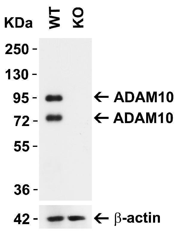
ADAM10 KO Validation in MEF Cells
Loading: 10 μg of lysate
Antibodies: ADAM10 A00566-2, 1 μg/mL and beta-actin 3779-1301, 1μg/mL, 1 h incubation at RT in 5% NFDM/TBST.
Secondary: Goat Anti-Rabbit IgG HRP conjugate at 1:10000 dilution.
A00566-2 detected both precursor ADAM10 (94KD) and mature ADAM10 (68kD).

Click image to see more details
ADAM10 KO Validation in 293 Cells
Loading: 15 μg of lysate
Antibodies: ADAM10 A00566-2, 2 μg/mL and beta-actin 3779-1301, 1μg/mL, 1 h incubation at RT in 5% NFDM/TBST.
Secondary: Goat Anti-Rabbit IgG HRP conjugate at 1:10000 dilution.
A00566-2 detected both precursor ADAM10 (94KD) and mature ADAM10 (68kD).

Click image to see more details
Independent Antibody Validation (IAV) via Protein Expression Profile in Human and Mouse Cell Lines
Loading: 15 μg of lysates per lane.
Antibodies: ADAM10 A00566-2, 0.5 μg/mL, ADAM10 24-024, 1 μg/mL, and -actin 3779, 1 μg/mL, 1h incubation at RT in 5% NFDM/TBST.
Secondary: Goat anti-rabbit IgG HRP conjugate at 1:10000 dilution.

Click image to see more details
WB Validation in Human Cell Lines
Loading: 15 μg of lysate
Antibodies: ADAM10 A00566-2, 1 μg/mL, 1 h incubation at RT in 5% NFDM/TBST.
Secondary: Goat Anti-Rabbit IgG HRP conjugate at 1:10000 dilution.
A00566-2 detected both precursor ADAM10 (94KD) and mature ADAM10 (68kD).

Click image to see more details
Western Blot Validation in Mouse Tissues
Loading: 15 μg of lysates per lane.
Antibodies: ADAM10 A00566-2, 1 μg/mL, 1h incubation at RT in 5% NFDM/TBST.
Secondary: Goat anti-rabbit IgG HRP conjugate at 1:10000 dilution.
A00566-2 detected both precursor ADAM10 (94KD) and mature ADAM10 (68kD).

Click image to see more details
Western Blot Validation in Rat Tissues
Loading: 15 μg of lysates per lane.
Antibodies: ADAM10 A00566-2, 1 μg/mL, 1h incubation at RT in 5% NFDM/TBST.
Secondary: Goat anti-rabbit IgG HRP conjugate at 1:10000 dilution.
A00566-2 detected both precursor ADAM10 (94KD) and mature ADAM10 (68kD).

Click image to see more details
Immunofluorescence Validation of ADAM10 in MOLT4 Cells
Immunofluorescent analysis of 4% paraformaldehyde-fixed MOLT4 cells labeling ADAM10 with A00566-2 at 20 μg/mL, followed by goat anti-rabbit IgG secondary antibody at 1/500 dilution (green) and DAPI staining (blue).

Click image to see more details
Immunofluorescence Validation of ADAM10 in Mouse Testis
Immunofluorescent analysis of 4% paraformaldehyde-fixed mouse testis labeling ADAM10 with A00566-2 at 20 μg/mL, followed by goat anti-rabbit IgG secondary antibody at 1/500 dilution (green) and DAPI staining (blue).

Click image to see more details
Immunofluorescence Validation of ADAM10 in Rat Testis
Immunofluorescent analysis of 4% paraformaldehyde-fixed rat testis labeling ADAM10 with A00566-2 at 20 μg/mL, followed by goat anti-rabbit IgG secondary antibody at 1/500 dilution (green) and DAPI staining (blue).

Click image to see more details
Immunofluorescence Validation of ADAM10 in Rat Thymus
Immunofluorescent analysis of 4% paraformaldehyde-fixed rat thymus labeling ADAM10 with A00566-2 at 10 μg/mL, followed by goat anti-rabbit IgG secondary antibody at 1/500 dilution (red) and DAPI staining (blue).

Click image to see more details
Immunohistochemistry Validation of ADAM10 in Human Testis
Immunohistochemical analysis of paraffin-embedded human testis tissue using anti-ADAM10 antibody (A00566-2) at 2 μg/ml. Tissue was fixed with formaldehyde and blocked with 10% serum for 1 h at RT; antigen retrieval was by heat mediation with a citrate buffer (pH6). Samples were incubated with primary antibody overnight at 4˚C. A goat anti-rabbit IgG H&L (HRP) at 1/250 was used as secondary. Counter stained with Hematoxylin.

Click image to see more details
Immunohistochemistry Validation of ADAM10 in Mouse Thymus
Immunohistochemical analysis of paraffin-embedded mouse thymus tissue using anti-ADAM10 antibody (A00566-2) at 2 μg/ml. Tissue was fixed with formaldehyde and blocked with 10% serum for 1 h at RT; antigen retrieval was by heat mediation with a citrate buffer (pH6). Samples were incubated with primary antibody overnight at 4˚C. A goat anti-rabbit IgG H&L (HRP) at 1/250 was used as secondary. Counter stained with Hematoxylin.

Click image to see more details
Immunofluorescence Validation of ADAM10 in primary cultures of human cerebral vascular smooth muscle cells (HC-VSMCs) (Coma et al., 2008)
Detection of ADAM10 expression by anti-ADAM10 antibodies in HC-VSMC cells under control condition or in the presence of 10μM H2O2 (oxidative stress condition) for 6 h. ADAM10 expression was not affected when exposed to oxidative stress.

Click image to see more details
Regulated Expression Validation of ADAM10 in Human neuroblastoma (SH-SY5Y) cells (Zimmermann et al., 2004)
Protein expression of ADAM10 detected by anti-ADAM10 CT antibodies in control or donepezil treated SH-SY5Y cells. When treated with donepezil, the expression of mature form of ADAM10 (68kD) was up-regulated in membrane compartment as compared to the down-regulation in intracellular fractions, and was not affected in whole cell homogenate.

Click image to see more details
KD Validation of ADAM10 in Human embryonic kidney 293 cells overexpressing the
human APP 695 isoform (HEK/APP) (Gatta et al., 2009)
Western blot analysis of ADAM10 silencing using anti-ADAM10 antibodies in HEK/APP cells. Silencing with ADAM10 siRNA (Ad) significantly decreased ADAM10 expression, and so did with Ferrochelatase siRNA (F) and N-methylprotoporphyrin IX siRNA (N), 67% and 50% reduction respectively
Specific Publications For Anti-ADAM10 Antibody (A00566-2)
Loading publications
Recommended Resources
Here are featured tools and databases that you might find useful.
- Boster's Pathways Library
- Protein Databases
- Bioscience Research Protocol Resources
- Data Processing & Analysis Software
- Photo Editing Software
- Scientific Literature Resources
- Research Paper Management Tools
- Molecular Biology Software
- Primer Design Tools
- Bioinformatics Tools
- Phylogenetic Tree Analysis
Customer Reviews
Have you used Anti-ADAM10 Antibody?
Share your experimental results or join a short interview to earn up to $1,000 in product credits or other rewards.
0 Reviews For Anti-ADAM10 Antibody
Customer Q&As
Have a question?
Find answers in Q&As, reviews.
Can't find your answer?
Submit your question
