Click image to see more details
-

-
-
-
-
+2
Product Info Summary
| SKU: | F00520 |
|---|---|
| Size: | 50ug |
| Reactive Species: | Human, Mouse, Rat |
| Host: | Mouse |
| Application: | ELISA, Flow Cytometry, IF, IHC, WB |
Customers Who Bought This Also Bought

Product info
Product Name
Anti-AKT3 FITC Monoclonal Antibody
SKU/Catalog Number
F00520
Size
50ug
Form
Lyophilized
Description
Boster Bio Anti-AKT3 FITC Monoclonal Antibody (Catalog # F00520). Tested in Dot blot, ELISA applications. This antibody reacts with Human, Mouse, Rat.
Storage & Handling
Store vial at 4°C prior to restoration. Restore with deionized water (or equivalent). For extended storage aliquot contents and freeze at -20°C or below. Avoid cycles of freezing and thawing. Centrifuge product if not completely clear after standing at room temperature. This product is stable for several weeks at 4°C as an undiluted liquid. Dilute only prior to immediate use. Expiration date is one (1) year from date of restoration. Expiration date is one (1) year from date of opening. (Ship on wet ice.)
Cite This Product
Anti-AKT3 FITC Monoclonal Antibody (Boster Biological Technology, Pleasanton CA, USA, Catalog # F00520)
Host
Mouse
Contents
0.02 M Potassium Phosphate, 0.5 M Sodium Chloride, pH 7.2, antibody formulated with stabilizing protein (10 mg/mL) - Immunoglobulin and Protease free, 0.01% (w/v) Sodium Azide
This antibody is supplied in a stabilized formulation.
Compatibility with conjugation reactions depends on the chemistry of the conjugation method used.
For conjugation methods that are not compatible with the stabilizing components present in this formulation, a carrier-free antibody format is required.
Clonality
Monoclonal
Clone Number
Clone: 25F6.F6.D8 IgG1 kappa
Isotype
IgG1 kappa
Immunogen
Anti-AKT3 Antibody was prepared from tissue culture supernatant by Protein A affinity chromatography using a synthetic peptide corresponding to internal residues of human AKT3 protein.
Reactive Species
F00520 is reactive to AKT3 in Human, Mouse, Rat
Observed Molecular Weight
68 kDa
Calculated molecular weight
55.8 kDa
Background of AKT3
AKT is a component of the PI-3 kinase pathway and is activated by phosphorylation at Ser 473 and Thr 308. AKT is a cytoplasmic protein also known as AKT1, Protein Kinase B (PKB) and rac (related to A and C kinases). AKT is a key regulator of many signal transduction pathways. AKT Exhibits tight control over cell proliferation and cell viability. Overexpression or inappropriate activation of AKT is noted in many types of cancer. AKT mediates many of the downstream events of PI 3-kinase (a lipid kinase activated by growth factors, cytokines and insulin). PI 3-kinase recruits AKT to the membrane, where it is activated by PDK1 phosphorylation. Once phosphorylated, AKT dissociates from the membrane and phosphorylates targets in the cytoplasm and the cell nucleus. AKT has two main roles: (i) inhibition of apoptosis; (ii) promotion of proliferation. Anti-AKT3 (MOUSE) PE conjugated Monoclonal Antibody is ideal for investigators involved in Cell Signaling, Cancer, Neuroscience, Signal Transduction research.
Antibody Validation
Boster validates all antibodies on WB, IHC, ICC, Immunofluorescence, and ELISA with known positive control and negative samples to ensure specificity and high affinity, including thorough antibody incubations.
Application & Images
Applications
F00520 is guaranteed for ELISA, Flow Cytometry, IF, IHC, WB Boster Guarantee
Recommend Dilution
| Application | Dilution | Species |
|---|---|---|
| Anti-AKT3 FITC Antibody has been tested by ELISA and dot blot and is suitable for Flow Cytometry | immunohistochemistry | and western blotting. Expect a band approximately 56 kDa in size corresponding to AKT3 protein by western blotting in the appropriate cell lysate or extract. This monoclonal antibody reacts with human AKT. Specific conditions for reactivity should be optimized by the end user. For immunohistochemistry we recommend the use of fresh frozen tissues. Attempts at staining paraffin-embedded formalin fixed tissues were negative. No pre-treatment of sample is required. |
Validation Images & Assay Conditions
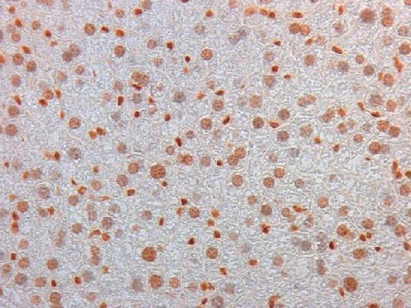
Click image to see more details
Immunohistochemistry of Mouse Monoclonal anti AKT3 Antibody in Mouse Embryonic Kidney. Tissue: Mouse Liver. Fixation: FFPE buffered formalin 10% conc. Ag Retrieval: Heat, Citrate pH 6.2. Pressure Cooker. Primary antibody: anti-AKT3 at 2ug/ml for 1.5 hour @ room Temp. Secondary Ab: MOUSE ON MOUSE HRP POLYMER 45” RT.

Click image to see more details
Western Blot of Mouse anti-AKT3 antibody. Lane 1: GST Tagged recombinant AKT1. Lane 2: GST Tagged recombinant AKT2. Lane 3: GST Tagged recombinant AKT3. Load: 25 ng per lane. Primary antibody: AKT3 antibody at 1:1,000 for overnight at 4°C. Secondary antibody: Peroxidase mouse secondary antibody at 1:40,000 for 30 min at RT. Block: 30 min at RT. Predicted/Observed size: 78 kDa for AKT3. Other band(s): none.

Click image to see more details
Western Blot of Mouse Anti-AKT3 antibody. Lane 1: C2C12. Lane 2: MEF#1. Lane 3: MEF#2. Lane 4: A549. Lane 5: Calu-1. Lane 6: PC3. Lane 7: HepG2. Lane 8: Jurkat. Lane 9: SKOV3. Lane 10: 293T. Load: 35 µg per lane. Primary antibody: AKT-3 antibody at 1:1000 for overnight at 4°C. Secondary antibody: Anti mouse secondary antibody at 1:20,000 for 1 h at RT. Block: 5% BLOTTO overnight at 4°C. Predicted/Observed size: 56 kDa for AKT3.

Click image to see more details
Western Blot of Mouse anti-AKT3 antibody. Lane 1: Control. Lane 2: Rapa. Lane 3: T50. Lane 4: T250. Lane 5: Control. Lane 6: Rapa. Lane 7: T50. Lane 8: T250. Lane 9: AKT3 null. Load: 35 µg per lane. Primary antibody: AKT-3 antibody at 1:1000 for overnight at 4°C. Secondary antibody: Anti mouse secondary antibody at 1:20,000 for 1 h at RT. Block: 5% BLOTTO overnight at 4°C. Predicted/Observed size: 56 kDa for AKT3.

Click image to see more details
Immunohistochemistry of Mouse Anti-AKT3 antibody. Tissue: human prostate carcinoma. A) AKT-3 antibody produced using CELLine, B) AKT-3 antibody produced using roller bottle. Fixation: formalin fixed paraffin embedded. Antigen retrieval: not required. Primary antibody: AKT-3 antibody at 10 µg/mL for 1 h at RT. Secondary antibody: Peroxidase mouse secondary antibody at 1:10,000 for 1 h at RT. Localization: AKT3 is nuclear and occasionally cytoplasmic. Staining: AKT3 as precipitated brown signal with hematoxylin purple nuclear counterstain.

Click image to see more details
Dot Blot of Mouse anti-AKT3 Monoclonal Antibody Fluorescein Conjugated. Antigen: His-tagged AKT3. Load: Lane 1 - 100 ng Lane 2 - 33.3 ng Lane 3 - 11.1 ng Lane 4 - 3.70 ng Lane 5 - 1.23 ng. Primary antibody: n/a. Secondary antibody: Mouse anti-AKT3 Monoclonal Antibody Fluorescein Conjugated at 1:1,000 for 60 min at RT. Block: 1 HR at RT.
Specific Publications For Anti-AKT3 FITC Monoclonal Antibody (F00520)
Loading publications
Recommended Resources
Here are featured tools and databases that you might find useful.
- Boster's Pathways Library
- Protein Databases
- Bioscience Research Protocol Resources
- Data Processing & Analysis Software
- Photo Editing Software
- Scientific Literature Resources
- Research Paper Management Tools
- Molecular Biology Software
- Primer Design Tools
- Bioinformatics Tools
- Phylogenetic Tree Analysis
Customer Reviews
Have you used Anti-AKT3 FITC Monoclonal Antibody?
Share your experimental results or join a short interview to earn up to $1,000 in product credits or other rewards.
0 Reviews For Anti-AKT3 FITC Monoclonal Antibody
Customer Q&As
Have a question?
Find answers in Q&As, reviews.
Can't find your answer?
Submit your question
2 Customer Q&As for Anti-AKT3 FITC Monoclonal Antibody
Question
I was wanting to use your anti-AKT3 FITC Monoclonal antibody for IHC for human liver on frozen tissues, but I want to know if it has been tested for this particular application. Has this antibody been tested and is this antibody a good choice for human liver identification?
Verified Customer
Verified customer
Asked: 2020-02-05
Answer
As indicated on the product datasheet, F00520 anti-AKT3 FITC Monoclonal antibody has been tested for IHC, WB on human tissues. We have an innovator award program that if you test this antibody and show it works in human liver in IHC-frozen, you can get your next antibody for free.
Boster Scientific Support
Answered: 2020-02-05
Question
My question regards to test anti-AKT3 FITC Monoclonal antibody F00520 on human liver for research purposes, then I may be interested in using anti-AKT3 FITC Monoclonal antibody F00520 for diagnostic purposes as well. Is the antibody suitable for diagnostic purposes?
C. Anderson
Verified customer
Asked: 2019-02-15
Answer
The products we sell, including anti-AKT3 FITC Monoclonal antibody F00520, are only intended for research use. They would not be suitable for use in diagnostic work. If you have the means to develop a product into diagnostic use, and are interested in collaborating with us and develop our product into an IVD product, please contact us for more discussions.
Boster Scientific Support
Answered: 2019-02-15
