Click image to see more details
-

-
-
-
-
+9
Product Info Summary
| SKU: | A00327-1 |
|---|---|
| Size: | 0.1 mg |
| Reactive Species: | Human, Mouse, Rat |
| Host: | Rabbit |
| Application: | ELISA, IF, IHC-P, ICC, WB |
Customers Who Bought This Also Bought

Product info
Product Name
Anti-Beclin-1 BECN1 Antibody
SKU/Catalog Number
A00327-1
Size
0.1 mg
Form
Liquid
Description
Boster Bio Anti-Beclin-1 BECN1 Antibody (Catalog # A00327-1). Tested in ELISA, WB, ICC/IF, IHC-P, IF applications. This antibody reacts with Human, Mouse, Rat.
Storage & Handling
Beclin-1 antibody can be stored at 4°C up to one year. Antibodies should not be exposed to prolonged high temperatures.
Cite This Product
Anti-Beclin-1 BECN1 Antibody (Boster Biological Technology, Pleasanton CA, USA, Catalog # A00327-1)
Host
Rabbit
Contents
Beclin-1 Antibody is supplied in PBS containing 0.02% sodium azide.
Clonality
Polyclonal
Isotype
IgG
Immunogen
Anti-Beclin-1 antibody was raised against a peptide corresponding to 17 amino acids near the center of human Beclin-1. The immunogen is located between 40-90 amino acids of Beclin-1.
Reactive Species
A00327-1 is reactive to BECN1 in Human, Mouse, Rat
Observed Molecular Weight
68 kDa
Calculated molecular weight
51.9 kDa
Background of BECN1
Autophagy, the process of bulk degradation of cellular proteins through an autophagosomic-lysosomal pathway is important for normal growth control and may be defective in tumor cells. Beclin-1, a coiled-coil Bcl-2-interacting protein homologous to the yeast autophagy gene apg6, is a mammalian autophagy gene that can inhibit tumorigenesis and is expressed at reduced levels in human breast carcinoma, suggesting that defects in autophagy proteins may contribute to the development or progression of tumors. Bcl-2 can bind to Beclin-1 and inhibit Beclin-1-dependent autophagy in yeast and mammalian cells, suggesting that Bcl-2 functions as an anti-autophagy protein as well as an anti-apoptotic protein, which helps maintain autophagy at levels that are more compatible with cell survival rather than cell death.
Antibody Validation
Boster validates all antibodies on WB, IHC, ICC, Immunofluorescence, and ELISA with known positive control and negative samples to ensure specificity and high affinity, including thorough antibody incubations.
Application & Images
Applications
A00327-1 is guaranteed for ELISA, IF, IHC-P, ICC, WB Boster Guarantee
Assay Dilutions Recommendation
The recommendations below provide a starting point for assay optimization. The actual working concentration varies and should be decided by the user.
WB: 1-2 μg/mL
IHC-P: 2-5 μg/mL
ICC/IF: 10-20 μg/mL
IHC-P/IF: 10-20 μg/mL.
Antibody validated: Western Blot in human, mouse and rat samples; Immunohistochemistry in human, mouse, and rat samples; Immunocytochemistry in human samples. All other applications and species not yet tested.
Validation Images & Assay Conditions
Click image to see more details
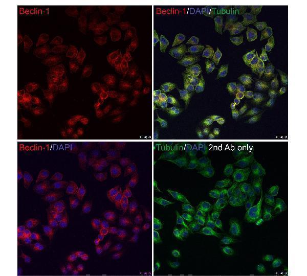
Immunofluorescence Validation of Beclin-1 in HeLa Cells
Immunofluorescent analysis of methanol-fixed HeLa cells labeling Beclin-1 with A00327-1 at 10 μg/mL, followed by goat anti-rabbit IgG secondary antibody at 1/1000 dilution (red) and DAPI staining (blue). Alpha tubulin was stained with anti-alpha tubulin antibody following by goat anti-mouse IgG secondary antibody (green). Images were captured with confocal microscopy.

Click image to see more details
WB Validation in Human Cell Lines
Loading: 15 μg of lysate
Antibodies: Beclin-1 A00327-1, 1 μg/mL , 1 h incubation at RT in 5% NFDM/TBST.
Secondary: Goat Anti-Rabbit IgG HRP conjugate at 1:10000 dilution.

Click image to see more details
WB Validation in Mouse Tissues
Loading: 15 μg of lysate
Antibodies: Beclin-1 A00327-1 , 1 μ g/mL , 1 h incubation at RT in 5% NFDM/TBST.
Secondary: Goat Anti-Rabbit IgG HRP conjugate at 1:10000 dilution.

Click image to see more details
WB Validation in Rat Tissues
Loading: 15 μg of lysate
Antibodies: Beclin-1 A00327-1, 1 μg/mL , 1 h incubation at RT in 5% NFDM/TBST.
Secondary: Goat Anti-Rabbit IgG HRP conjugate at 1:10000 dilution.

Click image to see more details
Immunohistochemistry Validation of Beclin-1 in Human kidney Tissue
Immunohistochemical analysis of paraffin-embedded human kidney tissue using anti-Beclin-1 antibody (A00327-1) at 2 μg/ml. Tissue was fixed with formaldehyde and blocked with 10% serum for 1 h at RT; antigen retrieval was by heat mediation with a citrate buffer (pH6). Samples were incubated with primary antibody overnight at 4˚C. A goat anti-rabbit IgG H&L (HRP) at 1/250 was used as secondary. Counter stained with Hematoxylin.

Click image to see more details
Immunohistochemistry Validation of Beclin-1 in Mouse Brain Tissue
Immunohistochemical analysis of paraffin-embedded mouse brain tissue using anti-Beclin-1 antibody (A00327-1) at 5 μg/ml. Tissue was fixed with formaldehyde and blocked with 10% serum for 1 h at RT; antigen retrieval was by heat mediation with a citrate buffer (pH6). Samples were incubated with primary antibody overnight at 4˚C. A goat anti-rabbit IgG H&L (HRP) at 1/250 was used as secondary. Counter stained with Hematoxylin.

Click image to see more details
Immunofluorescence Validation of Beclin-1 in Mouse Brain Tissue
Immunofluorescent analysis of 4% paraformaldehyde-fixed mouse brain labeling Beclin-1 with A00327-1 at 20 ?g/mL, followed by goat anti-rabbit IgG secondary antibody at 1/500 dilution (green) and DAPI antibody (blue).

Click image to see more details
Knockdown of Beclin-1 in Human T47D Cells (Thomas et al., 2011)
T47D cells were transfected with scrambled or Beclin-1 siRNA. The following day, these cells were treated with vehicle, C, 0.5 mM VPA, 10 lM 4OH-tamoxifen, T, or 0.5 mM VPA and 10 lM 4OH-tamoxifen, VT, for 48 h and viability was assayed by dye exclusion.

Click image to see more details
Immunohistochemistry Validation of Beclin-1 in Human HCC Cells (Qiu et al., 2014)
Beclin-1 exhibited cytoplasmic staining in adjacent non-tumor (ANT) tissues (A), bordeling site between HCC and ANT
(B: left, ANT; right: HCC), and HCC (C), respectively. Beclin-1 expression was stronger in ANT than in HCC. Magnification × 400.

Click image to see more details
Immunohistochemistry Validation of Beclin-1 in Rat Brain Tissue
Immunohistochemical analysis of paraffin-embedded rat brain tissue using anti-Beclin-1 antibody (A00327-1) at 2 μg/ml. Tissue was fixed with formaldehyde and blocked with 10% serum for 1 h at RT; antigen retrieval was by heat mediation with a citrate buffer (pH6). Samples were incubated with primary antibody overnight at 4˚C. A goat anti-rabbit IgG H&L (HRP) at 1/250 was used as secondary. Counter stained with Hematoxylin.

Click image to see more details
Immunohistochemistry Validation of Beclin-1 in Rat Liver Tissue
Immunohistochemical analysis of paraffin-embedded rat liver tissue using anti-Beclin-1 antibody (A00327-1) at 2 μg/ml. Tissue was fixed with formaldehyde and blocked with 10% serum for 1 h at RT; antigen retrieval was by heat mediation with a citrate buffer (pH6). Samples were incubated with primary antibody overnight at 4˚C. A goat anti-rabbit IgG H&L (HRP) at 1/250 was used as secondary. Counter stained with Hematoxylin.

Click image to see more details
Immunofluorescent Validation of Beclin-1 in Human Cortical Neurons and Rat Brain (Wang et al., 2017)
Cell specificity of Beclin 1 expression. Cortical neurons and glia co-cultures (upper panels) and cortex slices (lower panels) were immunostained
with anti-Beclin 1 (red) and NeuN (neuronal biomarker, green) or
GFAP (astrocyte biomarker, green). Neurons and astrocyte
in cultured cells were indicated by arrows and arrowhead,
respectively. Areas in white boxes were enlarged.

Click image to see more details
Regulated Expression of Beclin-1 in Human T47D Cells (Thomas et al., 2011)
Treatment with tamoxifen and an HDAC inhibitor alters the balance of autophagy and apoptosis inhibitors and drivers. T47D cells were treated with vehicle, C, 0.5 mM VPA, V, 10 lM 4OHtamoxifen, T, or 0.5 mM VPA and 10 lM 4OH-tamoxifen, VT, for 48 h and Beclin-1 protein and RNA
Specific Publications For Anti-Beclin-1 BECN1 Antibody (A00327-1)
Loading publications
Recommended Resources
Here are featured tools and databases that you might find useful.
- Boster's Pathways Library
- Protein Databases
- Bioscience Research Protocol Resources
- Data Processing & Analysis Software
- Photo Editing Software
- Scientific Literature Resources
- Research Paper Management Tools
- Molecular Biology Software
- Primer Design Tools
- Bioinformatics Tools
- Phylogenetic Tree Analysis
Customer Reviews
Have you used Anti-Beclin-1 BECN1 Antibody?
Share your experimental results or join a short interview to earn up to $1,000 in product credits or other rewards.
0 Reviews For Anti-Beclin-1 BECN1 Antibody
Customer Q&As
Have a question?
Find answers in Q&As, reviews.
Can't find your answer?
Submit your question
