Click image to see more details
-

-
-
-
-
+4
Product Info Summary
| SKU: | A00081-2 |
|---|---|
| Size: | 100ug |
| Reactive Species: | Human, Mouse |
| Host: | Rabbit |
| Application: | ELISA, IF, IHC, WB |
Customers Who Bought This Also Bought

Product info
Product Name
Anti-Beta Amyloid APP Antibody
SKU/Catalog Number
A00081-2
Size
100ug
Form
Liquid (sterile filtered)
Description
Boster Bio Anti-Beta Amyloid APP Antibody (Catalog # A00081-2). Tested in ELISA, IF, IHC, WB applications. This antibody reacts with Human, Mouse.
Storage & Handling
Store vial at -20°C prior to opening. Aliquot contents and freeze at -20°C or below for extended storage. Avoid cycles of freezing and thawing. Centrifuge product if not completely clear after standing at room temperature. This product is stable for several weeks at 4°C as an undiluted liquid. Dilute only prior to immediate use. Expiration date is one (1) year from date of opening. (Ship on dry ice.)
Cite This Product
Anti-Beta Amyloid APP Antibody (Boster Biological Technology, Pleasanton CA, USA, Catalog # A00081-2)
Host
Rabbit
Contents
0.02 M Potassium Phosphate, 0.15 M Sodium Chloride, pH 7.2, 0.01% (w/v) Sodium Azide
Clonality
Polyclonal
Isotype
IgG
Immunogen
This antibody was affinity purified from whole rabbit serum prepared by repeated immunizations with a synthetic peptide corresponding to an extracellular region of human beta amyloid conjugated to KLH using maleimide.
Cross-reactivity
No cross reactivity with other proteins.
Reactive Species
A00081-2 is reactive to APP in Human, Mouse
Calculated molecular weight
86.9 kDa
Background of APP
Beta amyloid, often abbreviated as A-beta, is a protein that builds up in the brains of persons with Alzheimer's disease, collecting in clumps called plaques or senile plaques. While some researchers question whether beta amyloid is the cause of the dementia, most agree that it is involved in the disruption of thinking that is a hallmark of the disease. In some cases of familial Alzheimer's disease, mutations in genes for the proteins called the presenilins lead to increased production of amyloid. Researchers have been looking at how presenilin-1 in particular contributes to the excess buildup of beta amyloid. Presenilin-1 apparently acts to increase the activity of gamma-secretase, an enzyme that changes a normal protein (amyloid precursor protein or APP) into beta amyloid itself. Furthermore, presenilin-1 might be gamma-secretase. Anti-Beta Amyloid Antibody is useful for researchers interested in TLR signaling and Alzheimer's research.
Antibody Validation
Boster validates all antibodies on WB, IHC, ICC, Immunofluorescence, and ELISA with known positive control and negative samples to ensure specificity and high affinity, including thorough antibody incubations.
Application & Images
Applications
A00081-2 is guaranteed for ELISA, IF, IHC, WB Boster Guarantee
Assay Dilutions Recommendation
The recommendations below provide a starting point for assay optimization. The actual working concentration varies and should be decided by the user.
ELISA: 1:10,000 - 1:30,000
IHC: 1:50-1:200
IF Microscopy: 1:50-1:200
WB: 1:1,000-1:5000
Affinity purified anti-beta amyloid has been tested by ELISA, IHC, WB, and IF. A 45.8kDa band is detected in western blot using whole tissue extracts and lysates from mouse and human. In general, we recommend the use of 4% PFA or 10% formalin for fixation of tissues with IHC-paraffin or IHC-frozen application of this antibody.
Validation Images & Assay Conditions
Click image to see more details
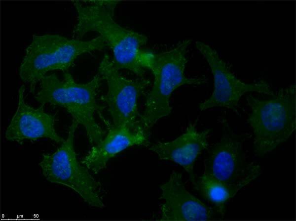
Immunofluorescence microscopy of Rabbit Anti-Beta Amyloid antibody using HeLa cells fixed with MeOH. Anti-Beta Amyloid Antibody was used at 1 µg/mL, O/N at 4°C. Secondary antibody: Anti-RABBIT IgG DyLight™ 488 Conjugated Preadsorbed at 2 ug/ml for 1 h at RT. Localization: APP is a cell surface protein that rapidly becomes internalized to endosomes and lysosomes. Some APP accumulates in secretory transport vesicles. Colocalizes with other proteins in a vesicular pattern in cytoplasm and perinuclear regions. Staining: Amyloid beta as green fluorescent signal with DAPI (blue) nuclear counterstain.

Click image to see more details
Immunohistochemistry with anti-beta amyloid antibody showing amyloid beta plaque staining in human Alzheimer’s disease brain at 10x and 20x (B & C). Staining was performed on Leica Bond system using the standard protocol. Formalin fixed/paraffin embedded tissue sections were subjected to antigen retrieval with E1 (Leica Microsystems) retrieval solution for 20 min and then incubated with rabbit anti-beta amyloid antibody 600-401-253 at 1:100 dilution for 60 minutes. Biotinylated Anti-rabbit secondary antibody was used at 1:200 dilution to detect primary antibody. The reaction was developed using streptavidin-HRP conjugated compact polymer system and visualized with chromogen substrate, 3’3-diamino-benzidine substrate (DAB). The sections were then counterstained with hematoxylin to detect cell nuclei.

Click image to see more details
Western Blot of Rabbit Anti-Beta Amyloid Antibody. Lane 1: Opal Prestained Molecular Weight Marker . Lane 2: HEK293T Whole Cell Lysate . Lane 3: Mouse Brain Whole Cell Lysate . Lane 4: A-172 Whole Cell Lysate . Load: 10µg/lane. Primary Antibody: Anti-Beta Amyloid at 1µg/mL overnight at 2-8°C. Secondary Antibody: Goat Anti-Rabbit IgG HRP Conjugated at 1:70,000 for 30min at RT. Block: BlockOut Buffer . Predicted MW: ~40-50kDa. Observed MW: ~48kDa.

Click image to see more details
Western Blot of Rabbit Anti-Beta Amyloid Antibody. Lane 1: Opal Prestained Molecular Weight Marker . Lane 2: HEK293T Whole Cell Lysate . Lane 3: Mouse Brain Whole Cell Lysate . Lane 4: A-172 Whole Cell Lysate . Lane 5: Daudi Whole Cell Lysate . Load: 10µg/lane. Primary Antibody: Anti-Beta Amyloid at 1:1000 overnight at 2-8°C. Secondary Antibody: Goat Anti-Rabbit IgG HRP Conjugated at 1:70,000 for 30min at RT. Block: BlockOut Buffer . Predicted MW: ~40-50kDa. Observed MW: ~48kDa.

Click image to see more details
Immunohistochemical detection of beta Amyloid using Anti-Beta Amyloid Antibody on TG APP23 mouse brain cortex frozen sections. Anti-Beta Amyloid Antibody used at 1:200 and incubated for 2 hours in TBS/BSA with Tween and azide. Fluorescent labelled anti rabbit IgG was then added. Carl Hobbs, King`s College London, United Kingdom.

Click image to see more details
Human Heart (formalin-fixed, paraffin-embedded) stained with Anti-Beta Amyloid Antibody at 5 ug/ml followed by biotinylated goat anti-rabbit IgG secondary antibody, alkaline phosphatase-streptavidin and chromogen.

Click image to see more details
Western Blot of Rabbit anti-Beta Amyloid antibody. Marker: Opal Pre-stained ladder . Lane 1: HEK293 lysate . Lane 2: HeLa Lysate . Lane 3: MCF-7 Lysate . Lane 4: Jurkat Lysate . Lane 5: A431 Lysate . Lane 6: LNCaP Lysate . Lane 7: A-172 Lysate . Lane 8: NIH/3T3 Lysate . Load: 35 µg per lane. Primary antibody: Beta Amyloid antibody at 1:5,000 for overnight at 4°C. Secondary antibody: Peroxidase rabbit secondary antibody at 1:30,000 for 60 min at RT. Blocking Buffer: 1% Casein-TTBS for 30 min at RT. Predicted MW: ~40-50 kDa. Observed size: ~48 kDa for Beta Amyloid.

Click image to see more details
ELISA results of purified Rabbit anti-Beta Amyloid Antibody tested against BSA-conjugated peptide of immunizing peptide. Each well was coated in duplicate with 0.1µg of conjugate. The starting dilution of antibody was 5μg/ml and the X-axis represents the Log10 of a 3-fold dilution. This titration is a 4-parameter curve fit where the IC50 is defined as the titer of the antibody. Assay performed using 3% fish gel, Goat anti-Rabbit IgG Antibody Peroxidase Conjugated (Min X Bv Ch Gt GP Ham Hs Hu Ms Rt & Sh Serum Proteins) and TMB ELISA Peroxidase Substrate .
Specific Publications For Anti-Beta Amyloid APP Antibody (A00081-2)
Loading publications
Recommended Resources
Here are featured tools and databases that you might find useful.
- Boster's Pathways Library
- Protein Databases
- Bioscience Research Protocol Resources
- Data Processing & Analysis Software
- Photo Editing Software
- Scientific Literature Resources
- Research Paper Management Tools
- Molecular Biology Software
- Primer Design Tools
- Bioinformatics Tools
- Phylogenetic Tree Analysis
Customer Reviews
Have you used Anti-Beta Amyloid APP Antibody?
Share your experimental results or join a short interview to earn up to $1,000 in product credits or other rewards.
0 Reviews For Anti-Beta Amyloid APP Antibody
Customer Q&As
Have a question?
Find answers in Q&As, reviews.
Can't find your answer?
Submit your question
16 Customer Q&As for Anti-Beta Amyloid APP Antibody
Question
Is this A00081-2 anti-Beta Amyloid antibody reactive to the isotypes of APP?
Verified Customer
Verified customer
Asked: 2020-04-17
Answer
The immunogen of A00081-2 anti-Beta Amyloid antibody is This affinity purified antibody was prepared from whole rabbit serum produced by repeated immunizations with a synthetic peptide corresponding to the amino terminus (aa 1-14) of human beta amyloid conjugated to KLH using maleimide. Could you tell me which isotype you are interested in so I can help see if the immunogen is part of this isotype?
Boster Scientific Support
Answered: 2020-04-17
Question
Would anti-Beta Amyloid antibody A00081-2 work for IHC with cerebellum hippocampus?
Verified Customer
Verified customer
Asked: 2020-03-04
Answer
According to the expression profile of cerebellum hippocampus, APP is highly expressed in cerebellum hippocampus. So, it is likely that anti-Beta Amyloid antibody A00081-2 will work for IHC with cerebellum hippocampus.
Boster Scientific Support
Answered: 2020-03-04
Question
See attached the WB image, lot number and protocol we used for cerebellum hippocampus using anti-Beta Amyloid antibody A00081-2. Please let me know if you require anything else.
Verified Customer
Verified customer
Asked: 2020-01-06
Answer
Thank you very much for the data. Our lab team are working to resolve this as quickly as possible, and we appreciate your patience and understanding! You have provided everything we needed. Please let me know if there is anything you need in the meantime.
Boster Scientific Support
Answered: 2020-01-06
Question
Our lab want to know about using your anti-Beta Amyloid antibody for modulation of excitatory postsynaptic potential studies. Has this antibody been tested with western blotting on jurkat cell lysates? We would like to see some validation images before ordering.
Verified Customer
Verified customer
Asked: 2019-12-26
Answer
We appreciate your inquiry. This A00081-2 anti-Beta Amyloid antibody is tested on mouse brain, jurkat cell lysates. It is guaranteed to work for ELISA, IF, IHC, WB in human, mouse. Our Boster guarantee will cover your intended experiment even if the sample type has not been be directly tested.
Boster Scientific Support
Answered: 2019-12-26
Question
Do you have a BSA free version of anti-Beta Amyloid antibody A00081-2 available?
Verified Customer
Verified customer
Asked: 2019-11-04
Answer
We appreciate your recent telephone inquiry. I can confirm that some lots of this anti-Beta Amyloid antibody A00081-2 are BSA free. For now, these lots are available and we can make a BSA free formula for you free of charge. It will take 3 extra days to prepare. If you require this antibody BSA free again in future, please do not hesitate to contact me and I will be pleased to check which lots we have in stock that are BSA free.
Boster Scientific Support
Answered: 2019-11-04
Question
My lab would like to test anti-Beta Amyloid antibody A00081-2 on human cerebellum hippocampus for research purposes, then I may be interested in using anti-Beta Amyloid antibody A00081-2 for diagnostic purposes as well. Is the antibody suitable for diagnostic purposes?
Verified Customer
Verified customer
Asked: 2019-10-11
Answer
The products we sell, including anti-Beta Amyloid antibody A00081-2, are only intended for research use. They would not be suitable for use in diagnostic work. If you have the means to develop a product into diagnostic use, and are interested in collaborating with us and develop our product into an IVD product, please contact us for more discussions.
Boster Scientific Support
Answered: 2019-10-11
Question
We are currently using anti-Beta Amyloid antibody A00081-2 for human tissue, and we are satisfied with the IHC results. The species of reactivity given in the datasheet says human, mouse. Is it likely that the antibody can work on dog tissues as well?
Verified Customer
Verified customer
Asked: 2019-07-05
Answer
The anti-Beta Amyloid antibody (A00081-2) has not been tested for cross reactivity specifically with dog tissues, though there is a good chance of cross reactivity. We have an innovator award program that if you test this antibody and show it works in dog you can get your next antibody for free. Please contact me if I can help you with anything.
Boster Scientific Support
Answered: 2019-07-05
Question
Would A00081-2 anti-Beta Amyloid antibody work on parafin embedded sections? If so, which fixation method do you recommend we use (PFA, paraformaldehyde, other)?
Verified Customer
Verified customer
Asked: 2019-05-28
Answer
It shows on the product datasheet, A00081-2 anti-Beta Amyloid antibody as been tested on IHC. It is best to use PFA for fixation because it has better tissue penetration ability. PFA needs to be prepared fresh before use. Long term stored PFA turns into formalin, as the PFA molecules congregate and become formalin.
Boster Scientific Support
Answered: 2019-05-28
Question
We purchased anti-Beta Amyloid antibody for IHC on eye pancreas in the past. I am using human, and We intend to use the antibody for IF next. We want examining eye pancreas as well as cerebellum hippocampus in our next experiment. Could you please give me some suggestion on which antibody would work the best for IF?
Verified Customer
Verified customer
Asked: 2019-02-27
Answer
I took a look at the website and datasheets of our anti-Beta Amyloid antibody and it seems that A00081-2 has been validated on human in both IHC and IF. Thus A00081-2 should work for your application. Our Boster satisfaction guarantee will cover this product for IF in human even if the specific tissue type has not been validated. We do have a comprehensive range of products for IF detection and you can check out our website bosterbio.com to find out more information about them.
Boster Scientific Support
Answered: 2019-02-27
Question
Can you help my question with product A00081-2, anti-Beta Amyloid antibody. I was wondering if it would be possible to conjugate this antibody with biotin. I would need it to be without BSA or sodium azide. I am planning on using a buffer exchange of sodium azide with PBS only. Would there be problems for me to conjugate the antibody and store it in -20 degrees in small aliquots?
D. Moore
Verified customer
Asked: 2018-05-29
Answer
We do not recommend storing this antibody with PBS buffer only in -20 degrees. If you want to store it in -20 degrees it is best to add some cryoprotectant like glycerol. If you want carrier free A00081-2 anti-Beta Amyloid antibody, we can provide it to you in a special formula with trehalose and/or glycerol. These molecules will not interfere with conjugation chemistry and provide a good level of protection for the antibody from degradation. Please be sure to specify this in your purchase order.
Boster Scientific Support
Answered: 2018-05-29
Question
We have observed staining in human platelet. Any tips? Is anti-Beta Amyloid antibody supposed to stain platelet positively?
Verified Customer
Verified customer
Asked: 2018-01-16
Answer
According to literature platelet does express APP. According to Uniprot.org, APP is expressed in frontal cortex, brain, leukocyte, cerebellum hippocampus, eye pancreas, liver, fibroblast, platelet, brain cortex, cerebrospinal fluid, blood vessel, fetal brain, plasma, among other tissues. Regarding which tissues have APP expression, here are a few articles citing expression in various tissues:
Blood vessel, Pubmed ID: 8248178
Brain, Pubmed ID: 2569763, 2881207, 2893289, 3035574, 3810169, 9338779
Brain cortex, Pubmed ID: 2893379, 3312495
Cerebrospinal fluid, Pubmed ID: 8229004, 22576872
Fetal brain, Pubmed ID: 2949367
Leukocyte, Pubmed ID: 1587857
Liver, Pubmed ID: 24275569
Plasma, Pubmed ID: 16335952
Platelet, Pubmed ID: 12665801
Boster Scientific Support
Answered: 2018-01-16
Question
Our team were satisfied with the WB result of your anti-Beta Amyloid antibody. However we have observed positive staining in cerebrospinal fluid cell membrane using this antibody. Is that expected? Could you tell me where is APP supposed to be expressed?
Z. Miller
Verified customer
Asked: 2016-01-27
Answer
According to literature, cerebrospinal fluid does express APP. Generally APP expresses in cell membrane. Regarding which tissues have APP expression, here are a few articles citing expression in various tissues:
Blood vessel, Pubmed ID: 8248178
Brain, Pubmed ID: 2569763, 2881207, 2893289, 3035574, 3810169, 9338779
Brain cortex, Pubmed ID: 2893379, 3312495
Cerebrospinal fluid, Pubmed ID: 8229004, 22576872
Fetal brain, Pubmed ID: 2949367
Leukocyte, Pubmed ID: 1587857
Liver, Pubmed ID: 24275569
Plasma, Pubmed ID: 16335952
Platelet, Pubmed ID: 12665801
Boster Scientific Support
Answered: 2016-01-27
Question
I was wanting to use your anti-Beta Amyloid antibody for IHC for human cerebellum hippocampus on frozen tissues, but I want to know if it has been tested for this particular application. Has this antibody been tested and is this antibody a good choice for human cerebellum hippocampus identification?
P. Krishna
Verified customer
Asked: 2015-03-23
Answer
You can see on the product datasheet, A00081-2 anti-Beta Amyloid antibody has been validated for ELISA, IF, IHC, WB on human, mouse tissues. We have an innovator award program that if you test this antibody and show it works in human cerebellum hippocampus in IHC-frozen, you can get your next antibody for free.
Boster Scientific Support
Answered: 2015-03-23
Question
I see that the anti-Beta Amyloid antibody A00081-2 works with IHC, what is the protocol used to produce the result images on the product page?
B. Johnson
Verified customer
Asked: 2013-12-31
Answer
You can find protocols for IHC on the "support/technical resources" section of our navigation menu. If you have any further questions, please send an email to support@bosterbio.com
Boster Scientific Support
Answered: 2013-12-31
Question
Thanks for helping with my inquiry over the phone. Here are the WB image, lot number and protocol we used for cerebellum hippocampus using anti-Beta Amyloid antibody A00081-2. Let me know if you need anything else.
B. Johnson
Verified customer
Asked: 2013-08-02
Answer
Thank you for the data. You have provided everything we needed. Our lab team are working to resolve your inquiry as quickly as possible, and we appreciate your patience and understanding! Please let me know if there is anything you need in the meantime.
Boster Scientific Support
Answered: 2013-08-02
Question
Is a blocking peptide available for product anti-Beta Amyloid antibody (A00081-2)?
A. Mangal
Verified customer
Asked: 2013-06-03
Answer
We do provide the blocking peptide for product anti-Beta Amyloid antibody (A00081-2). If you would like to place an order for it please contact support@bosterbio.com and make a special request.
Boster Scientific Support
Answered: 2013-06-03
