Click image to see more details
-

-
-
-
-
+9
Product Info Summary
| SKU: | A01857-1 |
|---|---|
| Size: | 100 μg/vial |
| Reactive Species: | Chicken, Human, Monkey, Mouse, Rat |
| Host: | Rabbit |
| Application: | Flow Cytometry, IF, IHC, ICC, WB |
Customers Who Bought This Also Bought

Product info
Product Name
Anti-Beta Tubulin/TUBB Antibody Picoband®
SKU/Catalog Number
A01857-1
Size
100 μg/vial
Form
Lyophilized
Description
Boster Bio Anti-Beta Tubulin/TUBB Antibody Picoband® catalog # A01857-1. Tested in Flow Cytometry, IF, IHC, ICC, WB applications. This antibody reacts with Human, Monkey, Mouse, Rat, Chicken. The brand Picoband indicates this is a premium antibody that guarantees superior quality, high affinity, and strong signals with minimal background in Western blot applications. Only our best-performing antibodies are designated as Picoband, ensuring unmatched performance.
Storage & Handling
Store at -20˚C for one year from date of receipt. After reconstitution, at 4˚C for one month. It can also be aliquotted and stored frozen at -20˚C for six months. Avoid repeated freeze-thaw cycles.
Cite This Product
Anti-Beta Tubulin/TUBB Antibody Picoband® (Boster Biological Technology, Pleasanton CA, USA, Catalog # A01857-1)
Host
Rabbit
Contents
Each vial contains 4 mg Trehalose, 0.9 mg NaCl and 0.2 mg Na2HPO4.
Clonality
Polyclonal
Isotype
Rabbit IgG
Immunogen
A synthetic peptide corresponding to a sequence at the C-terminus of human Beta Tubulin, identical to the related mouse and rat sequences.
Cross-reactivity
No cross-reactivity with other proteins
Reactive Species
A01857-1 is reactive to TUBB in Chicken, Human, Monkey, Mouse, Rat
Observed Molecular Weight
50 kDa
Calculated molecular weight
49.7 kDa
Background of TUBB
Tubulin beta chain is a protein that in humans is encoded by the TUBB gene. This gene encodes a beta tubulin protein. This protein forms a dimer with alpha tubulin and acts as a structural component of microtubules. Mutations in this gene cause cortical dysplasia, complex, with other brain malformations 6. Alternative splicing results in multiple splice variants. There are multiple pseudogenes for this gene on chromosomes 1, 6, 7, 8, 9, and 13.
Antibody Validation
Boster validates all antibodies on WB, IHC, ICC, Immunofluorescence, and ELISA with known positive control and negative samples to ensure specificity and high affinity, including thorough antibody incubations.
Application & Images
Applications
A01857-1 is guaranteed for Flow Cytometry, IF, IHC, ICC, WB Boster Guarantee
Recommend Dilution
| Application | Dilution | Species |
|---|---|---|
| Western blot | 0.1-0.5μg/ml | Human, Monkey, Mouse, Rat, Chicken |
| Immunohistochemistry (Paraffin-embedded Section) | 2-5μg/ml | Human |
| Immunocytochemistry/Immunofluorescence | 5μg/ml | Human |
| Flow Cytometry(Fixed) | 1-3μg/1x106 cells | Human |
Tested application
Suggested blocking solution with 5% non-fat milk or BSA; (*)Recommended protein loading: 20-40 µg per lane
Use TE buffer pH 9.0 for antigen retrieval; (*) citrate buffer pH 6.0 is an alternative.
Validation Images & Assay Conditions
Click image to see more details
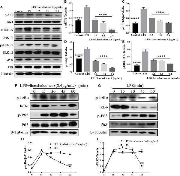
Effects of licochalcone A on LPS-induced activation of AKT/NF-κB and MAPK signaling pathways in mMECs. (A–E) Total protein in mMECs was collected after 4 h of LPS stimulation. Licochalcone A was added 1 h before LPS stimulation. Protein levels of p-AKT, p-JNK1/2, p-ERK1/2, and p-P38 were detected via western blot and quantitatively assessed via densitometry using β-tubulin as an internal control. Protein levels were measured using ImageJ software ( ) and normalized to that of β-tubulin. (F–I) MMECs were divided into LPS (1 μg/mL) or LPS + licochalcone A (2.4 μg/mL) groups. After adding LPS to serum-free medium for 1 h, licochalcone A was added. Protein was collected after 0, 15, 30, 45, and 60 min. (F) Western blot analysis of p-IκBα, IκBα, p-P65, and P65 in mMECs were treated with LPS + licochalcone A. (G) Western blot analysis of p-IκBα, IκBα, p-P65, and P65 in mMECs were treated with LPS only. (H–I) The phosphorylation of P65 and IκBα at different time-points under LPS and LPS + licochalcone A treatment were detected via western blot. Values are presented as means ± SD ( n = 3) (** p < 0.01 vs. LPS, **** p < 0.0001 vs. LPS).
Index in PubMed under a CC BY license. PMID: 30858849

Click image to see more details
Effects of licochalcone A on the protein levels of ZO-1, Occludin, and claudin3 in mMECs. mMECs were serum-starved for 3 h before treatment with Licochalcone A (1.8, 2.4 μg/mL) for 24 h (A) . The protein levels of ZO-1 (B) , claudin3 (C) , and Occludin (D) were determined by western blot, and the relative protein levels were quantified by scanning densitometry and normalized to β-tubulin. Values are presented as means ± SD ( n = 3) (** p < 0.01, *** p < 0.001 and **** p < 0.0001 vs. Control group).
Index in PubMed under a CC BY license. PMID: 30858849

Click image to see more details
Effects of Licochalcone A on LPS-induced inflammatory response in mouse mammary epithelial cells (mMECs). Cells were cultured with different concentrations of licochalcone A (1.2, 1.8, 2.4, and 3 μg/mL) for 4 h and viability determined with the CCK8 assay. (A) The effect of licochalcone A and licochalcone A + LPS were determined by CCK8 assay. mMECs were pretreated with Licochalcone A (1.2, 1.8, 2.4, and 3 μg/mL) for 1 h and then stimulated with LPS for 4 h, protein and mRNA levels were determined by qRT-PCR and western blot. The mRNA levels of IL-6 (B) , IL-1 β (C) , and TNF- α (D) , and the relative mRNA level was normalized to β -actin mRNA. The protein levels of COX-2 (E,G) and iNOS (E,F) , and the relative protein levels were quantified by scanning densitometry and normalized to β-tubulin. Values are presented as means ± SD ( n = 3) (* p < 0.05, ** p < 0.01, *** p < 0.001, and **** p < 0.0001 vs. LPS group).
Index in PubMed under a CC BY license. PMID: 30858849

Click image to see more details
Protein levels change in claudin-3, occluding, and ZO-1 after LPS or LPS + licochalcone A injection. Protein levels was measured using ImageJ software ( ) and normalized to that of β-tubulin. (A–D) Results of western blot analysis of claudin-3, occludin, ZO-1, and β-tubulin in mammary glands after LPS and LPS + licochalcone A injection. (B–D) Protein levels of claudin-3, occludin and ZO-1 normalized to that of β-tubulin. (E) Immunostaining images of ZO-1 (green) and nuclear staining with DAPI (blue) in mammary glands treated with LPS and licochalcone A. Values are presented as means ± SD ( n = 10 in each group) (* p < 0.05, ** p < 0.01, *** p < 0.001, and **** p < 0.0001 vs. LPS group).
Index in PubMed under a CC BY license. PMID: 30858849

Click image to see more details
Effects of licochalcone A on AKT/NF-κB signaling pathway in LPS-induced mice mastitis. Mammary gland tissues from different experimental groups were obtained 24 h after LPS administration and total protein. The tissue lysates were prepared and subjected to western blot by using p-AKT (A,B) , p-IκBα (A,C) , and p-P65 (A,D) antibodies, respectively. Each immunoreactive band was digitized and expressed as a ratio of the β-tubulin level. Values are presented as means ± SD (* p < 0.05, *** p < 0.001, and **** p < 0.0001 vs. LPS group).
Index in PubMed under a CC BY license. PMID: 30858849

Click image to see more details
Effects of licochalcone A on mitogen-activated protein kinase (MAPK) signaling pathway in LPS-induced mice mastitis. Mammary gland tissues from different experimental groups were obtained 24 h after LPS administration. The tissue lysates were prepared and subjected to western blot by using p-ERK1/2 (A,B) , p-JNK1/2 (A,C) , p-P38 (A,D) antibodies, respectively. Each immunoreactive band was digitized and expressed as a ratio of the β-tubulin level. Values are presented as means ± SD (**** p < 0.0001 vs. LPS group).
Index in PubMed under a CC BY license. PMID: 30858849

Click image to see more details
Effects of Licochalcone A on inflammatory response in LPS-induced mice mastitis. Mammary gland tissues from each experimental group ( n = 10) were obtained at 24 h after LPS administration. (A) Myeloperoxidase (MPO) activity assay. The protein levels of IL-1β (B) , TNF-α (C) , and IL-6 (D) were detected using ELISA. Western blot assay of inducible nitric oxide synthase (iNOS) (E,F) and cyclooxygenase-2 (COX-2) (E,G) , and the relative protein levels were quantified by scanning densitometry and normalized to β-tubulin. Values are presented as means ± SD (* p < 0.05, ** p < 0.01, and **** p < 0.0001 vs. LPS group).
Index in PubMed under a CC BY license. PMID: 30858849

Click image to see more details
Western blot analysis of Beta Tubulin using anti-Beta Tubulin antibody (A01857-1).
Electrophoresis was performed on a 5-20% SDS-PAGE gel at 70V (Stacking gel) / 90V (Resolving gel) for 2-3 hours. The sample well of each lane was loaded with 30 ug of sample under reducing conditions.
Lane 1: human SiHa whole cell lysates,
Lane 2: human 293T whole cell lysates,
Lane 3: human HepG2 whole cell lysates,
Lane 4: monkey COS-7 whole cell lysates,
Lane 5: chicken heart tissue lysates,
Lane 6: rat brain tissue lysates,
Lane 7: rat PC-12 whole cell lysates,
Lane 8: mouse brain tissue lysates,
Lane 9: mouse NIH/3T3 whole cell lysates.
After electrophoresis, proteins were transferred to a nitrocellulose membrane at 150 mA for 50-90 minutes. Blocked the membrane with 5% non-fat milk/TBS for 1.5 hour at RT. The membrane was incubated with rabbit anti-Beta Tubulin antigen affinity purified polyclonal antibody (Catalog # A01857-1) at 0.5 μg/mL overnight at 4°C, then washed with TBS-0.1%Tween 3 times with 5 minutes each and probed with a goat anti-rabbit IgG-HRP secondary antibody at a dilution of 1:5000 for 1.5 hour at RT. The signal is developed using an Enhanced Chemiluminescent detection (ECL) kit (Catalog # EK1002) with Tanon 5200 system. A specific band was detected for Beta Tubulin at approximately 50 kDa. The expected band size for Beta Tubulin is at 50 kDa.

Click image to see more details
Western blot analysis of TUBB using anti-TUBB antibody (A01857-1).
Electrophoresis was performed on a 5-20% SDS-PAGE gel at 80V (Stacking gel) / 120V (Resolving gel) for 2 hours. The sample well of each lane was loaded with 30 ug of sample under reducing conditions.
Lane 2-3: human OCI-LY1-SRPK1 KO whole cell lysates.
After electrophoresis, proteins were transferred to a nitrocellulose membrane at 150 mA for 50-90 minutes. Blocked the membrane with 5% non-fat milk/TBS for 1.5 hour at RT. The membrane was incubated with rabbit anti-TUBB antigen affinity purified polyclonal antibody (A04887-1) at 1:3000 overnight at 4°C, then washed with TBS-0.1%Tween 3 times with 5 minutes each and probed with a HRP-conjugated Anti-Rabbit IgG Secondary Antibodyat for 1 hour at RT. The signal is developed using an ECL Plus Western Blotting Substrate (Catalog # AR1196-200) with Tanon 5200 system. A specific band was detected for TUBB at approximately 55 kDa. The expected band size for TUBB is at 50 kDa.

Click image to see more details
IHC analysis of Beta Tubulin using anti-Beta Tubulin antibody (A01857-1).
Beta Tubulin was detected in a paraffin-embedded section of human liver cancer tissue. Heat mediated antigen retrieval was performed in EDTA buffer (pH 8.0, epitope retrieval solution). The tissue section was blocked with 10% goat serum. The tissue section was then incubated with 2 μg/ml rabbit anti-Beta Tubulin Antibody (A01857-1) overnight at 4°C. Peroxidase Conjugated Goat Anti-rabbit IgG was used as secondary antibody and incubated for 30 minutes at 37°C. The tissue section was developed using HRP Conjugated Rabbit IgG Super Vision Assay Kit (Catalog # SV0002) with DAB as the chromogen.

Click image to see more details
IHC analysis of Beta Tubulin using anti-Beta Tubulin antibody (A01857-1).
Beta Tubulin was detected in a paraffin-embedded section of human testicular germ cell tumor tissue. Heat mediated antigen retrieval was performed in EDTA buffer (pH 8.0, epitope retrieval solution). The tissue section was blocked with 10% goat serum. The tissue section was then incubated with 2 μg/ml rabbit anti-Beta Tubulin Antibody (A01857-1) overnight at 4°C. Peroxidase Conjugated Goat Anti-rabbit IgG was used as secondary antibody and incubated for 30 minutes at 37°C. The tissue section was developed using HRP Conjugated Rabbit IgG Super Vision Assay Kit (Catalog # SV0002) with DAB as the chromogen.

Click image to see more details
IF analysis of Beta Tubulin using anti-Beta Tubulin antibody (A01857-1).
Beta Tubulin was detected in an immunocytochemical section of U2OS cells. Enzyme antigen retrieval was performed using IHC enzyme antigen retrieval reagent (AR0022) for 15 mins. The cells were blocked with 10% goat serum. And then incubated with 5 μg/mL rabbit anti-Beta Tubulin Antibody (A01857-1) overnight at 4°C. Cy3 Conjugated Goat Anti-Rabbit IgG (BA1032) was used as secondary antibody at 1:500 dilution and incubated for 30 minutes at 37°C. The section was counterstained with DAPI. Visualize using a fluorescence microscope and filter sets appropriate for the label used.

Click image to see more details
Flow Cytometry analysis of SiHa cells using anti-Beta Tubulin antibody (A01857-1).
Overlay histogram showing SiHa cells stained with A01857-1 (Blue line). To facilitate intracellular staining, cells were fixed with 4% paraformaldehyde and permeabilized with permeabilization buffer. The cells were blocked with 10% normal goat serum. And then incubated with rabbit anti-Beta Tubulin Antibody (A01857-1, 1 μg/1x106 cells) for 30 min at 20°C. DyLight®488 conjugated goat anti-rabbit IgG (BA1127, 5-10 μg/1x106 cells) was used as secondary antibody for 30 minutes at 20°C. Isotype control antibody (Green line) was rabbit IgG (1 μg/1x106) used under the same conditions. Unlabelled sample without incubation with primary antibody and secondary antibody (Red line) was used as a blank control.
Specific Publications For Anti-Beta Tubulin/TUBB Antibody Picoband® (A01857-1)
Loading publications
Recommended Resources
Here are featured tools and databases that you might find useful.
- Boster's Pathways Library
- Protein Databases
- Bioscience Research Protocol Resources
- Data Processing & Analysis Software
- Photo Editing Software
- Scientific Literature Resources
- Research Paper Management Tools
- Molecular Biology Software
- Primer Design Tools
- Bioinformatics Tools
- Phylogenetic Tree Analysis
Customer Reviews
Have you used Anti-Beta Tubulin/TUBB Antibody Picoband®?
Share your experimental results or join a short interview to earn up to $1,000 in product credits or other rewards.
1 Reviews For Anti-Beta Tubulin/TUBB Antibody Picoband®
In WB using β-tubulin antibody (Cat# A01857-1), a clear, specific band was observed in OCI-LY8 cells with clean background, showing stable expression after SRPK1 knockdown.
Excellent

| SKU | A01857-1 |
|---|---|
| Application | Western Blot |
| Sample | human OCI-LY1 cells |
| Sample Processing Description | Cell samples were lysed by sonication in RIPA buffer containing protease and phosphatase inhibitors, followed by centrifugation for 10 minutes. The supernatant was mixed with loading buffer at a 4:1 ratio and boiled for 10 minutes. Fifteen microliters of each sample were loaded per well. |
| Other Reagents | 5% Non-fat milk |
| Primary Antibody | Beta Tubulin/TUBB Antibody |
| Primary Incubation | 1:3000, overnight at 4 ℃ |
| Secondary Antibody | HRP-conjugated Anti-Rabbit IgG Secondary Antibody |
| Secondary Incubation | 1 hour in room temperature |
| Detection | Substrate: ECL luminescent reagent, Imaging system:ChemiDoc MP |
| Results Summary | The antibody is highly specific and efficient, with a clean background and no nonspecific bands. The target band has clear and well-defined edges. |
Maolin Yao, Fujian Medical University
Verified customer
Submitted 2025-11-11
Customer Q&As
Have a question?
Find answers in Q&As, reviews.
Can't find your answer?
Submit your question
7 Customer Q&As for Anti-Beta Tubulin/TUBB Antibody Picoband®
Question
Is this A01857-1 anti-Beta Tubulin/TUBB antibody reactive to the isotypes of TUBB?
Verified Customer
Verified customer
Asked: 2020-04-06
Answer
The immunogen of A01857-1 anti-Beta Tubulin/TUBB antibody is A synthetic peptide corresponding to a sequence at the C-terminus of human Beta Tubulin (383-412aa EQFTAMFRRKAFLHWYTGEGMDEMEFTEAE), identical to the related mouse and rat sequences. Could you tell me which isotype you are interested in so I can help see if the immunogen is part of this isotype?
Boster Scientific Support
Answered: 2020-04-06
Question
We appreciate helping with my inquiry over the phone. Here are the WB image, lot number and protocol we used for lung using anti-Beta Tubulin/TUBB antibody A01857-1. Let me know if you need anything else.
Verified Customer
Verified customer
Asked: 2019-12-31
Answer
Thank you for the data. You have provided everything we needed. Our lab team are working to resolve your inquiry as quickly as possible, and we appreciate your patience and understanding! Please let me know if there is anything you need in the meantime.
Boster Scientific Support
Answered: 2019-12-31
Question
Is a blocking peptide available for product anti-Beta Tubulin/TUBB antibody (A01857-1)?
Verified Customer
Verified customer
Asked: 2019-12-09
Answer
We do provide the blocking peptide for product anti-Beta Tubulin/TUBB antibody (A01857-1). If you would like to place an order for it please contact support@bosterbio.com and make a special request.
Boster Scientific Support
Answered: 2019-12-09
Question
Would anti-Beta Tubulin/TUBB antibody A01857-1 work for WB with lung?
Verified Customer
Verified customer
Asked: 2019-09-19
Answer
According to the expression profile of lung, TUBB is highly expressed in lung. So, it is likely that anti-Beta Tubulin/TUBB antibody A01857-1 will work for WB with lung.
Boster Scientific Support
Answered: 2019-09-19
Question
I have attached the WB image, lot number and protocol we used for lung using anti-Beta Tubulin/TUBB antibody A01857-1. Please let me know if you require anything else.
Verified Customer
Verified customer
Asked: 2019-09-12
Answer
Thank you very much for the data. Our lab team are working to resolve this as quickly as possible, and we appreciate your patience and understanding! You have provided everything we needed. Please let me know if there is anything you need in the meantime.
Boster Scientific Support
Answered: 2019-09-12
Question
I have a question about product A01857-1, anti-Beta Tubulin/TUBB antibody. I was wondering if it would be possible to conjugate this antibody with biotin. I would need it to be without BSA or sodium azide. I am planning on using a buffer exchange of sodium azide with PBS only. Would there be problems for me to conjugate the antibody and store it in -20 degrees in small aliquots?
Verified Customer
Verified customer
Asked: 2019-08-12
Answer
We do not recommend storing this antibody with PBS buffer only in -20 degrees. If you want to store it in -20 degrees it is best to add some cryoprotectant like glycerol. If you want carrier free A01857-1 anti-Beta Tubulin/TUBB antibody, we can provide it to you in a special formula with trehalose and/or glycerol. These molecules will not interfere with conjugation chemistry and provide a good level of protection for the antibody from degradation. Please be sure to specify this in your purchase order.
Boster Scientific Support
Answered: 2019-08-12
Question
Would A01857-1 anti-Beta Tubulin/TUBB antibody work on parafin embedded sections? If so, which fixation method do you recommend we use (PFA, paraformaldehyde, other)?
A. Carter
Verified customer
Asked: 2014-02-24
Answer
You can see on the product datasheet, A01857-1 anti-Beta Tubulin/TUBB antibody as been tested on WB. It is best to use PFA for fixation because it has better tissue penetration ability. PFA needs to be prepared fresh before use. Long term stored PFA turns into formalin, as the PFA molecules congregate and become formalin.
Boster Scientific Support
Answered: 2014-02-24
