Click image to see more details
-

-
-
-
-
+3
Product Info Summary
| SKU: | M05613-4 |
|---|---|
| Size: | 100 μg/vial |
| Reactive Species: | Human, Mouse, Rat |
| Host: | Mouse |
| Application: | Flow Cytometry, IF, ICC, WB |
Customers Who Bought This Also Bought

Product info
Product Name
Anti-Beta Tubulin TUBB Antibody Picoband® (monoclonal, 2E11)
SKU/Catalog Number
M05613-4
Size
100 μg/vial
Form
Lyophilized
Description
Boster Bio Anti-Beta Tubulin TUBB Antibody Picoband® (monoclonal, 2E11) catalog # M05613-4. Tested in Flow Cytometry, IF, ICC, WB applications. This antibody reacts with Human, Mouse, Rat. The brand Picoband indicates this is a premium antibody that guarantees superior quality, high affinity, and strong signals with minimal background in Western blot applications. Only our best-performing antibodies are designated as Picoband, ensuring unmatched performance.
Storage & Handling
Store at -20˚C for one year from date of receipt. After reconstitution, at 4˚C for one month. It can also be aliquotted and stored frozen at -20˚C for six months. Avoid repeated freeze-thaw cycles.
Cite This Product
Anti-Beta Tubulin TUBB Antibody Picoband® (monoclonal, 2E11) (Boster Biological Technology, Pleasanton CA, USA, Catalog # M05613-4)
Host
Mouse
Contents
Each vial contains 4mg Trehalose, 0.9mg NaCl, 0.2mg Na2HPO4, 0.05mg NaN3.
Clonality
Monoclonal
Clone Number
2E11
Isotype
Mouse IgG2a
Immunogen
A synthetic peptide corresponding to a sequence at the C-terminus of human Beta Tubulin, identical to the related mouse and rat sequences.
Cross-reactivity
No cross-reactivity with other proteins.
Reactive Species
M05613-4 is reactive to TUBB in Human, Mouse, Rat
Observed Molecular Weight
55 kDa
Calculated molecular weight
49.7 kDa
Background of TUBB
Tubulin beta chain is a protein that in humans is encoded by the TUBB gene. This gene encodes a beta tubulin protein. This protein forms a dimer with alpha tubulin and acts as a structural component of microtubules. Mutations in this gene cause cortical dysplasia, complex, with other brain malformations 6. Alternative splicing results in multiple splice variants. There are multiple pseudogenes for this gene on chromosomes 1, 6, 7, 8, 9, and 13.
Antibody Validation
Boster validates all antibodies on WB, IHC, ICC, Immunofluorescence, and ELISA with known positive control and negative samples to ensure specificity and high affinity, including thorough antibody incubations.
Application & Images
Applications
M05613-4 is guaranteed for Flow Cytometry, IF, ICC, WB Boster Guarantee
Assay Dilutions Recommendation
The recommendations below provide a starting point for assay optimization. The actual working concentration varies and should be decided by the user.
Western blot, 0.1-0.5μg/ml, Human, Mouse, Rat
Immunocytochemistry/Immunofluorescence,2μg/ml,Human
Flow Cytometry (Fixed), 1-3μg/1x106 cells, Human
Positive Control
WB: human Hela whole cell, human HepG2 whole cell, human K562 whole cell, human Raji whole cell, rat brain tissue, rat lung tissue, mouse brain tissue, mouse lung tissue
ICC/IF: U2OS cell, U2OS cell, CACO-2 cell
FCM: U937 cell, HEPA1-6 cell
Validation Images & Assay Conditions
Click image to see more details
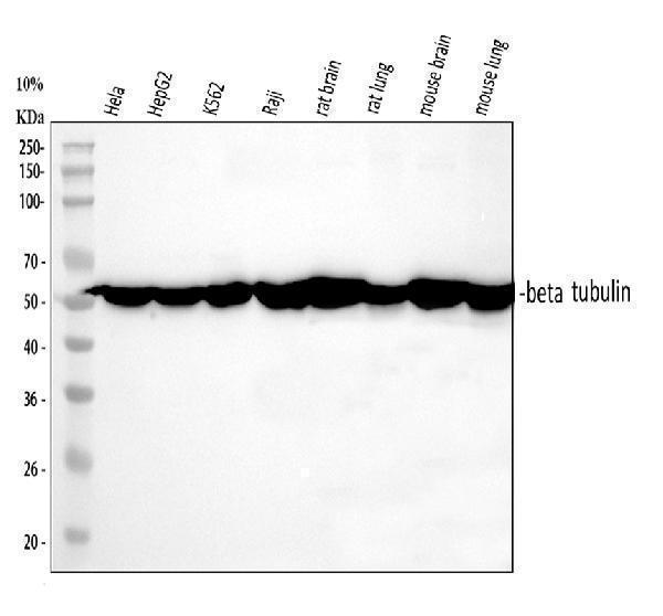
Western blot analysis of Tubulin beta using anti-Tubulin beta antibody (M05613-4).
Electrophoresis was performed on a 5-20% SDS-PAGE gel at 70V (Stacking gel) / 90V (Resolving gel) for 2-3 hours. The sample well of each lane was loaded with 30 ug of sample under reducing conditions.
Lane 1: human Hela whole cell lysates,
Lane 2: human HepG2 whole cell lysates,
Lane 3: human K562 whole cell lysates,
Lane 4: human Raji whole cell lysates,
Lane 5: rat brain tissue lysates,
Lane 6: rat lung tissue lysates,
Lane 6: mouse brain tissue lysates,
Lane 6: mouse lung tissue lysates.
After electrophoresis, proteins were transferred to a nitrocellulose membrane at 150 mA for 50-90 minutes. Blocked the membrane with 5% non-fat milk/TBS for 1.5 hour at RT. The membrane was incubated with mouse anti-Tubulin beta antigen affinity purified monoclonal antibody (Catalog # M05613-4) at 0.5 μg/mL overnight at 4°C, then washed with TBS-0.1%Tween 3 times with 5 minutes each and probed with a goat anti-mouse IgG-HRP secondary antibody at a dilution of 1:10000 for 1.5 hour at RT. The signal is developed using an Enhanced Chemiluminescent detection (ECL) kit (Catalog # EK1001) with Tanon 5200 system. A specific band was detected for Tubulin beta at approximately 55 kDa. The expected band size for Tubulin beta is at 50 kDa.

Click image to see more details
IF analysis of Tubulin beta using anti-Tubulin beta antibody (M05613-4) and anti-FOSB antibody (A01569-1).
Tubulin beta was detected in immunocytochemical section of CACO-2 cells. Enzyme antigen retrieval was performed using IHC enzyme antigen retrieval reagent (AR0022) for 15 mins. The cells were blocked with 10% goat serum. And then incubated with 2μg/mL mouse anti-Tubulin beta Antibody (M05613-4) and rabbit anti-FOSB antibody (A01569-1) overnight at 4°C. DyLight®594 Conjugated Goat Anti-Mouse IgG (BA1141) and DyLight®488 Conjugated Goat Anti-Rabbit IgG (BA1127) were used as secondary antibody at 1:100 dilution and incubated for 30 minutes at 37°C. Visualize using a fluorescence microscope and filter sets appropriate for the label used.

Click image to see more details
IF analysis of Tubulin beta using anti-Tubulin beta antibody (M05613-4).
Tubulin beta was detected in immunocytochemical section of U2OS cell. Enzyme antigen retrieval was performed using IHC enzyme antigen retrieval reagent (AR0022) for 15 mins. The cells were blocked with 10% goat serum. And then incubated with 2μg/mL mouse anti-Tubulin beta Antibody (M05613-4) overnight at 4°C. DyLight®488 Conjugated Goat Anti-Mouse IgG (BA1126) was used as secondary antibody at 1:100 dilution and incubated for 30 minutes at 37°C. The section was counterstained with DAPI. Visualize using a fluorescence microscope and filter sets appropriate for the label used.

Click image to see more details
Flow Cytometry analysis of U937 cells using anti-Tubulin beta antibody M05613-4).
Overlay histogram showing U937 cells stained with M05613-4 (Blue line). To facilitate intracellular staining, cells were fixed with 4% paraformaldehyde and permeabilized with permeabilization buffer. The cells were blocked with 10% normal goat serum. And then incubated with mouse anti-Tubulin beta Antibody (M05613-4, 1μg/1x106 cells) for 30 min at 20°C. DyLight®488 conjugated goat anti-mouse IgG (BA1126, 5-10μg/1x106 cells) was used as secondary antibody for 30 minutes at 20°C. Isotype control antibody (Green line) was mouse IgG (1μg/1x106) used under the same conditions. Unlabelled sample without incubation with primary antibody and secondary antibody (Red line) was used as a blank control.

Click image to see more details
Flow Cytometry analysis of HEPA1-6 cells using anti-Tubulin beta antibody M05613-4).
Overlay histogram showing HEPA1-6 cells stained with M05613-4 (Blue line). To facilitate intracellular staining, cells were fixed with 4% paraformaldehyde and permeabilized with permeabilization buffer. The cells were blocked with 10% normal goat serum. And then incubated with mouse anti-Tubulin beta Antibody (M05613-4, 1μg/1x106 cells) for 30 min at 20°C. DyLight®488 conjugated goat anti-mouse IgG (BA1126, 5-10μg/1x106 cells) was used as secondary antibody for 30 minutes at 20°C. Isotype control antibody (Green line) was mouse IgG (1μg/1x106) used under the same conditions. Unlabelled sample without incubation with primary antibody and secondary antibody (Red line) was used as a blank control.

Click image to see more details
Western blot analysis of Tubulin beta using anti-Tubulin beta antibody (M05613-4).
Electrophoresis was performed on a 5-20% SDS-PAGE gel at 70V (Stacking gel) / 90V (Resolving gel) for 2-3 hours. The sample well of each lane was loaded with 30 ug of sample under reducing conditions.
Lane 1: human Hela whole cell lysates,
Lane 2: human HepG2 whole cell lysates,
Lane 3: human K562 whole cell lysates,
Lane 4: human Raji whole cell lysates,
Lane 5: rat brain tissue lysates,
Lane 6: rat lung tissue lysates,
Lane 7: mouse brain tissue lysates,
Lane 8: mouse lung tissue lysates.
After electrophoresis, proteins were transferred to a nitrocellulose membrane at 150 mA for 50-90 minutes. Blocked the membrane with 5% non-fat milk/TBS for 1.5 hour at RT. The membrane was incubated with mouse anti-Tubulin beta antigen affinity purified monoclonal antibody (M05613-4) at 0.5 μg/mL overnight at 4°C, then washed with TBS-0.1%Tween 3 times with 5 minutes each and probed with a goat anti-mouse IgG-DyLight 647 Conjugated secondary antibody at a dilution of 1:2000 for 1.5 hour at RT. A specific band was detected for Tubulin beta at approximately 50 kDa. The expected band size for Tubulin beta is at 50 kDa.
Specific Publications For Anti-Beta Tubulin TUBB Antibody Picoband® (monoclonal, 2E11) (M05613-4)
Loading publications
Recommended Resources
Here are featured tools and databases that you might find useful.
- Boster's Pathways Library
- Protein Databases
- Bioscience Research Protocol Resources
- Data Processing & Analysis Software
- Photo Editing Software
- Scientific Literature Resources
- Research Paper Management Tools
- Molecular Biology Software
- Primer Design Tools
- Bioinformatics Tools
- Phylogenetic Tree Analysis
Customer Reviews
Have you used Anti-Beta Tubulin TUBB Antibody Picoband® (monoclonal, 2E11)?
Share your experimental results or join a short interview to earn up to $1,000 in product credits or other rewards.
0 Reviews For Anti-Beta Tubulin TUBB Antibody Picoband® (monoclonal, 2E11)
Customer Q&As
Have a question?
Find answers in Q&As, reviews.
Can't find your answer?
Submit your question
1 Customer Q&As for Anti-Beta Tubulin TUBB Antibody Picoband® (monoclonal, 2E11)
Question
We are currently using anti-Beta Tubulin antibody (monoclonal, 2E11) M05613-4 for human tissue, and we are well pleased with the WB results. The species of reactivity given in the datasheet says human, mouse, rat. Is it likely that the antibody can work on pig tissues as well?
Verified Customer
Verified customer
Asked: 2018-09-04
Answer
The anti-Beta Tubulin antibody (monoclonal, 2E11) (M05613-4) has not been tested for cross reactivity specifically with pig tissues, though there is a good chance of cross reactivity. We have an innovator award program that if you test this antibody and show it works in pig you can get your next antibody for free. Please contact me if I can help you with anything.
Boster Scientific Support
Answered: 2018-09-04
