Click image to see more details
-

-
-
-
-
+5
Product Info Summary
| SKU: | M00894 |
|---|---|
| Size: | 100 μl |
| Reactive Species: | Human, Mouse, Rat |
| Host: | Rabbit |
| Application: | Flow Cytometry, IP, IF, IHC, ICC, WB |
Customers Who Bought This Also Bought

Product info
Product Name
Anti-Calreticulin Rabbit Monoclonal Antibody
SKU/Catalog Number
M00894
BM4228 is an alternative SKU for this antibody, used in previous lots.
Size
100 μl
Form
Liquid
Description
Boster Bio Anti-Calreticulin Rabbit Monoclonal Antibody catalog # M00894. Tested in WB, IHC, ICC/IF, IP, Flow Cytometry applications. This antibody reacts with Human, Mouse, Rat.
Storage & Handling
Store at -20°C for one year. For short term storage and frequent use, store at 4°C for up to one month. Avoid repeated freeze-thaw cycles.
Cite This Product
Anti-Calreticulin Rabbit Monoclonal Antibody (Boster Biological Technology, Pleasanton CA, USA, Catalog # M00894)
Host
Rabbit
Contents
Rabbit IgG in stabilizing components, phosphate buffered saline, pH 7.4, 150mM NaCl, 0.02% sodium azide and 50% glycerol.
*This antibody is supplied in a stabilized formulation.
Compatibility with conjugation reactions depends on the chemistry of the conjugation method used.
For conjugation methods that are not compatible with the stabilizing components present in this formulation, a carrier-free antibody format is required.
Clonality
Monoclonal
Clone Number
CGO-3
Isotype
Rabbit IgG
Immunogen
A synthesized peptide derived from human Calreticulin - ER Marker
Reactive Species
M00894 is reactive to CALR in Human, Mouse, Rat
Observed Molecular Weight
60 kDa
Calculated molecular weight
48.1 kDa
Antibody Validation
Boster validates all antibodies on WB, IHC, ICC, Immunofluorescence, and ELISA with known positive control and negative samples to ensure specificity and high affinity, including thorough antibody incubations.
Application & Images
Applications
M00894 is guaranteed for Flow Cytometry, IP, IF, IHC, ICC, WB Boster Guarantee
Recommend Dilution
WB 1:500-2000
IHC 1:50-200
ICC/IF 1:50-200
IP 1:20
FC 1:20
Tested application
Suggested blocking solution with 5% non-fat milk or BSA; (*)Recommended protein loading: 20-40 µg per lane
Use TE buffer pH 9.0 for antigen retrieval; (*) citrate buffer pH 6.0 is an alternative.
Validation Images & Assay Conditions
Click image to see more details
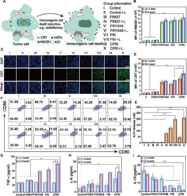
(A) CFRI-mediated ICD and DCs matured in vitro. Mechanism scheme of PTT-induced immunogenic death; 1,064-nm laser irradiation (1.0 W/cm 2 ) with or without treatment with DMEM, FR837, FIR1048, FRI, and CFRI (2 μg/ml). (B) The efflux of HMGB1 was quantitatively analyzed by flow cytometry. (C) CLSM images of CRT expression on the 4T1 cell surface after different treatments (scale bar: 100 μm). (D) Mean fluorescence intensity of CRT expression in 4T1 cells after different treatments. (E) The average fluorescence intensity of ATP exposure in 4T1 cells after different treatments. (F) The corresponding quantification (CD80 + CD86 + ) of mature DCs was quantitatively analyzed by flow cytometry. (G) After different treatments by enzyme-linked immunosorbent assay (ELISA), secretion levels of TNF-ɑ in the cell supernatant. (H) After different treatments by ELISA, the secretion level of IL-6 in the cell supernatant. (I) After different treatments by ELISA, the secretion level of IL-10 in the cell supernatant. Information of each group: I, control; II, control + L; III, FR837; IV, FR837 + L; V, FIR1048; VI, FIR1048 + L; VII, FRI; VII, FRI + L; IX, CFRI; X, CFRI + L. Significance is determined using one-way analysis of variance (* P < 0.05, ** P < 0.01, and *** P < 0.001). HSPs, heat shock proteins.
Index in PubMed under a CC BY license. PMID: 40040955

Click image to see more details
In vivo CFRI-mediated PTT-enhanced immunotherapy elicited an immune response after 16 d of treatment. (A) Schematic diagram of immunity induced by damage-associated molecular patterns (DAMPs). (B and C) Analysis of DC maturity (CD11 + CD80 + CD86 + ) using flow cytometry in the primary tumor of 4T1 tumor-bearing mice. (D and E) Regulatory T cells (Tregs) (CD4 + CD25 + ) in the spleen. (F and G) T cell proliferation in the blood (CD4 + CD8 + ). (H) The content of TNF-α in the spleen of 4T1 tumor-bearing mice. (I) The content of IL-6 in the spleen of 4T1 tumor-bearing mice. (J) The content of IL-10 in the spleen of 4T1 tumor-bearing mice. (K) Heat map of TNF-α and IL-10 content in the blood of 4T1 tumor-bearing mice. (L) CRT and HMGB1 staining images of 4T1 tumor-bearing mice after different treatments. Scale bar: 100 μm. Group information: I, PBS; II, PBS + L; III, FR837; IV, FR837 + L; V, FIR1048; VI, FIR1048 + L; VII, FRI; VIII, FRI + L; IX, CFRI; X, CFRI + L (* P < 0.05, ** P < 0.01, and *** P < 0.001). CTL, cytotoxic T lymphocyte; TCR, T cell receptor.
Index in PubMed under a CC BY license. PMID: 40040955

Click image to see more details
Western blot analysis of Calreticulin using anti-Calreticulin antibody (M00894).
Electrophoresis was performed on a 5-20% SDS-PAGE gel at 70V (Stacking gel) / 90V (Resolving gel) for 2-3 hours. The sample well of each lane was loaded with 30 ug of sample under reducing conditions.
Lane 1: human COLO320 whole cell lysates,
Lane 2: human HL-60 whole cell lysates,
Lane 3: human Hela whole cell lysates,
Lane 4: human MCF-7 whole cell lysates,
Lane 5: rat brain tissue lysates,
Lane 6: rat liver tissue lysates,
Lane 7: mouse brain tissue lysates,
Lane 8: mouse liver tissue lysates.
After electrophoresis, proteins were transferred to a nitrocellulose membrane at 150 mA for 50-90 minutes. Blocked the membrane with 5% non-fat milk/TBS for 1.5 hour at RT. The membrane was incubated with rabbit anti-Calreticulin antigen affinity purified monoclonal antibody (Catalog # M00894) at 1:500 overnight at 4°C, then washed with TBS-0.1%Tween 3 times with 5 minutes each and probed with a goat anti-rabbit IgG-HRP secondary antibody at a dilution of 1:5000 for 1.5 hour at RT. The signal is developed using an Enhanced Chemiluminescent detection (ECL) kit (Catalog # EK1002) with Tanon 5200 system. A specific band was detected for Calreticulin at approximately 60 kDa. The expected band size for Calreticulin is at 48 kDa.

Click image to see more details
Immunohistochemical analysis of paraffin-embedded Rat cerebral cortex, using the Antibody at 1:150 dilution.

Click image to see more details
Immunohistochemical analysis of paraffin-embedded Human renal cancer, using the Antibody at 1:150 dilution.

Click image to see more details
Immunohistochemical analysis of paraffin-embedded Human placenta, using the Antibody at 1:150 dilution.

Click image to see more details
Immunohistochemical analysis of paraffin-embedded Mouse testis, using the Antibody at 1:150 dilution.

Click image to see more details
Immunohistochemical analysis of paraffin-embedded Mouse cerebellum, using the Antibody at 1:150 dilution.

Click image to see more details
Immunofluorescent analysis using the Antibody at 1:50 dilution.
Specific Publications For Anti-Calreticulin Rabbit Monoclonal Antibody (M00894)
Loading publications
Recommended Resources
Here are featured tools and databases that you might find useful.
- Boster's Pathways Library
- Protein Databases
- Bioscience Research Protocol Resources
- Data Processing & Analysis Software
- Photo Editing Software
- Scientific Literature Resources
- Research Paper Management Tools
- Molecular Biology Software
- Primer Design Tools
- Bioinformatics Tools
- Phylogenetic Tree Analysis
Customer Reviews
Have you used Anti-Calreticulin Rabbit Monoclonal Antibody?
Share your experimental results or join a short interview to earn up to $1,000 in product credits or other rewards.
0 Reviews For Anti-Calreticulin Rabbit Monoclonal Antibody
Customer Q&As
Have a question?
Find answers in Q&As, reviews.
Can't find your answer?
Submit your question
15 Customer Q&As for Anti-Calreticulin Rabbit Monoclonal Antibody
Question
Will M00894 anti-Calreticulin Rabbit Monoclonal antibody work on parafin embedded sections? If so, which fixation method do you recommend we use (PFA, paraformaldehyde, other)?
Verified Customer
Verified customer
Asked: 2020-04-30
Answer
It shows on the product datasheet, M00894 anti-Calreticulin Rabbit Monoclonal antibody as been tested on IF. It is best to use PFA for fixation because it has better tissue penetration ability. PFA needs to be prepared fresh before use. Long term stored PFA turns into formalin, as the PFA molecules congregate and become formalin.
Boster Scientific Support
Answered: 2020-04-30
Question
I am looking for to test anti-Calreticulin Rabbit Monoclonal antibody M00894 on mouse cajal-retzius cell fetal brain cortex for research purposes, then I may be interested in using anti-Calreticulin Rabbit Monoclonal antibody M00894 for diagnostic purposes as well. Is the antibody suitable for diagnostic purposes?
Verified Customer
Verified customer
Asked: 2019-12-16
Answer
The products we sell, including anti-Calreticulin Rabbit Monoclonal antibody M00894, are only intended for research use. They would not be suitable for use in diagnostic work. If you have the means to develop a product into diagnostic use, and are interested in collaborating with us and develop our product into an IVD product, please contact us for more discussions.
Boster Scientific Support
Answered: 2019-12-16
Question
I was wanting to use your anti-Calreticulin Rabbit Monoclonal antibody for IF for mouse cajal-retzius cell fetal brain cortex on frozen tissues, but I want to know if it has been tested for this particular application. Has this antibody been tested and is this antibody a good choice for mouse cajal-retzius cell fetal brain cortex identification?
Verified Customer
Verified customer
Asked: 2019-12-05
Answer
As indicated on the product datasheet, M00894 anti-Calreticulin Rabbit Monoclonal antibody has been validated for IP, IF, WB on human, mouse, rat tissues. We have an innovator award program that if you test this antibody and show it works in mouse cajal-retzius cell fetal brain cortex in IHC-frozen, you can get your next antibody for free.
Boster Scientific Support
Answered: 2019-12-05
Question
We purchased anti-Calreticulin Rabbit Monoclonal antibody for WB on placenta a few years ago. I am using rat, and We are going to use the antibody for IF next. We want examining placenta as well as colon carcinoma in our next experiment. Do you have any suggestion on which antibody would work the best for IF?
Verified Customer
Verified customer
Asked: 2019-12-04
Answer
I have checked the website and datasheets of our anti-Calreticulin Rabbit Monoclonal antibody and it seems that M00894 has been validated on rat in both WB and IF. Thus M00894 should work for your application. Our Boster satisfaction guarantee will cover this product for IF in rat even if the specific tissue type has not been validated. We do have a comprehensive range of products for IF detection and you can check out our website bosterbio.com to find out more information about them.
Boster Scientific Support
Answered: 2019-12-04
Question
Thank you for helping with my inquiry over the phone. Here are the WB image, lot number and protocol we used for cajal-retzius cell fetal brain cortex using anti-Calreticulin Rabbit Monoclonal antibody M00894. Let me know if you need anything else.
Verified Customer
Verified customer
Asked: 2019-11-04
Answer
Thank you for the data. You have provided everything we needed. Our lab team are working to resolve your inquiry as quickly as possible, and we appreciate your patience and understanding! Please let me know if there is anything you need in the meantime.
Boster Scientific Support
Answered: 2019-11-04
Question
Do you have a BSA free version of anti-Calreticulin Rabbit Monoclonal antibody M00894 available?
Verified Customer
Verified customer
Asked: 2019-10-09
Answer
Thank you for your recent telephone inquiry. I can confirm that some lots of this anti-Calreticulin Rabbit Monoclonal antibody M00894 are BSA free. For now, these lots are available and we can make a BSA free formula for you free of charge. It will take 3 extra days to prepare. If you require this antibody BSA free again in future, please do not hesitate to contact me and I will be pleased to check which lots we have in stock that are BSA free.
Boster Scientific Support
Answered: 2019-10-09
Question
Is this M00894 anti-Calreticulin Rabbit Monoclonal antibody reactive to the isotypes of CALR?
G. Huang
Verified customer
Asked: 2019-09-16
Answer
The immunogen of M00894 anti-Calreticulin Rabbit Monoclonal antibody is A synthesized peptide derived from human Calreticulin - ER Marker. Could you tell me which isotype you are interested in so I can help see if the immunogen is part of this isotype?
Boster Scientific Support
Answered: 2019-09-16
Question
See below the WB image, lot number and protocol we used for cajal-retzius cell fetal brain cortex using anti-Calreticulin Rabbit Monoclonal antibody M00894. Please let me know if you require anything else.
Verified Customer
Verified customer
Asked: 2019-08-23
Answer
Thank you very much for the data. Our lab team are working to resolve this as quickly as possible, and we appreciate your patience and understanding! You have provided everything we needed. Please let me know if there is anything you need in the meantime.
Boster Scientific Support
Answered: 2019-08-23
Question
Is a blocking peptide available for product anti-Calreticulin Rabbit Monoclonal antibody (M00894)?
D. Zhang
Verified customer
Asked: 2019-07-16
Answer
We do provide the blocking peptide for product anti-Calreticulin Rabbit Monoclonal antibody (M00894). If you would like to place an order for it please contact support@bosterbio.com and make a special request.
Boster Scientific Support
Answered: 2019-07-16
Question
We have observed staining in human pancreas skin. Any tips? Is anti-Calreticulin Rabbit Monoclonal antibody supposed to stain pancreas skin positively?
Verified Customer
Verified customer
Asked: 2019-02-05
Answer
According to literature pancreas skin does express CALR. According to Uniprot.org, CALR is expressed in thyroid gland, eye, pancreas skin, placenta, liver, brain, cajal-retzius cell fetal brain cortex, keratinocyte, colon carcinoma, among other tissues. Regarding which tissues have CALR expression, here are a few articles citing expression in various tissues:
Colon carcinoma, Pubmed ID: 9150948
Eye, Pancreas, and Skin, Pubmed ID: 15489334
Keratinocyte, Pubmed ID: 1286667
Liver, Pubmed ID: 19159218, 24275569
Placenta, Pubmed ID: 7841019, 11322874
Boster Scientific Support
Answered: 2019-02-05
Question
My team were content with the WB result of your anti-Calreticulin Rabbit Monoclonal antibody. However we have been able to see positive staining in placenta endoplasmic reticulum lumen using this antibody. Is that expected? Could you tell me where is CALR supposed to be expressed?
Verified Customer
Verified customer
Asked: 2019-02-04
Answer
From literature, placenta does express CALR. Generally CALR expresses in endoplasmic reticulum lumen. Regarding which tissues have CALR expression, here are a few articles citing expression in various tissues:
Colon carcinoma, Pubmed ID: 9150948
Eye, Pancreas, and Skin, Pubmed ID: 15489334
Keratinocyte, Pubmed ID: 1286667
Liver, Pubmed ID: 19159218, 24275569
Placenta, Pubmed ID: 7841019, 11322874
Boster Scientific Support
Answered: 2019-02-04
Question
Can you help my question with product M00894, anti-Calreticulin Rabbit Monoclonal antibody. I was wondering if it would be possible to conjugate this antibody with biotin. I would need it to be without BSA or sodium azide. I am planning on using a buffer exchange of sodium azide with PBS only. Would there be problems for me to conjugate the antibody and store it in -20 degrees in small aliquots?
Verified Customer
Verified customer
Asked: 2018-10-17
Answer
It is not recommended storing this antibody with PBS buffer only in -20 degrees. If you want to store it in -20 degrees it is best to add some cryoprotectant like glycerol. If you want carrier free M00894 anti-Calreticulin Rabbit Monoclonal antibody, we can provide it to you in a special formula with trehalose and/or glycerol. These molecules will not interfere with conjugation chemistry and provide a good level of protection for the antibody from degradation. Please be sure to specify this in your purchase order.
Boster Scientific Support
Answered: 2018-10-17
Question
Would anti-Calreticulin Rabbit Monoclonal antibody M00894 work for IF with cajal-retzius cell fetal brain cortex?
P. Li
Verified customer
Asked: 2015-06-18
Answer
According to the expression profile of cajal-retzius cell fetal brain cortex, CALR is highly expressed in cajal-retzius cell fetal brain cortex. So, it is likely that anti-Calreticulin Rabbit Monoclonal antibody M00894 will work for IF with cajal-retzius cell fetal brain cortex.
Boster Scientific Support
Answered: 2015-06-18
Question
I see that the anti-Calreticulin Rabbit Monoclonal antibody M00894 works with IF, what is the protocol used to produce the result images on the product page?
O. Krishna
Verified customer
Asked: 2014-10-24
Answer
You can find protocols for IF on the "support/technical resources" section of our navigation menu. If you have any further questions, please send an email to support@bosterbio.com
Boster Scientific Support
Answered: 2014-10-24
Question
We are currently using anti-Calreticulin Rabbit Monoclonal antibody M00894 for human tissue, and we are happy with the IP results. The species of reactivity given in the datasheet says human, mouse, rat. Is it true that the antibody can work on feline tissues as well?
Z. Patel
Verified customer
Asked: 2014-04-10
Answer
The anti-Calreticulin Rabbit Monoclonal antibody (M00894) has not been tested for cross reactivity specifically with feline tissues, though there is a good chance of cross reactivity. We have an innovator award program that if you test this antibody and show it works in feline you can get your next antibody for free. Please contact me if I can help you with anything.
Boster Scientific Support
Answered: 2014-04-10
