Click image to see more details
Product Info Summary
| SKU: | M01943-1 |
|---|---|
| Size: | 80 µl |
| Reactive Species: | Human |
| Host: | Mouse |
| Application: | Flow Cytometry, IHC-P, WB |
Customers Who Bought This Also Bought

Product info
Product Name
Anti-CAPN1 Antibody
SKU/Catalog Number
M01943-1
Size
80 µl
Description
Boster Bio Anti-CAPN1 Antibody (Catalog # M01943-1). Tested in IHC-P, Flow Cytometry, WB application(s). This antibody reacts with Human.
Storage & Handling
Maintain refrigerated at 2-8°C for up to 2 weeks. For long-term storage, store at -20°C in small aliquots to prevent freeze-thaw cycles.
Cite This Product
Anti-CAPN1 Antibody (Boster Biological Technology, Pleasanton CA, USA, Catalog # M01943-1)
Host
Mouse
Contents
Purified monoclonal antibody supplied in PBS with 0.09% (W/V) sodium azide.
Clonality
Monoclonal
Clone Number
1376CT809.24.85
Isotype
IgG1,k
Immunogen
This CAPN1 antibody is generated from a mouse immunized with a KLH conjugated synthetic peptide between amino acids from the human region of human CAPN1.
Reactive Species
M01943-1 is reactive to CAPN1 in Human
Calculated molecular weight
81.9 kDa
Background of CAPN1
Calcium-regulated non-lysosomal thiol-protease which catalyze limited proteolysis of substrates involved in cytoskeletal remodeling and signal transduction.
Antibody Validation
Boster validates all antibodies on WB, IHC, ICC, Immunofluorescence, and ELISA with known positive control and negative samples to ensure specificity and high affinity, including thorough antibody incubations.
Application & Images
Applications
M01943-1 is guaranteed for Flow Cytometry, IHC-P, WB Boster Guarantee
Recommend Dilution
WB: 1:1000
IHC-P: 1:25
FC: 1:25
Validation Images & Assay Conditions
Click image to see more details
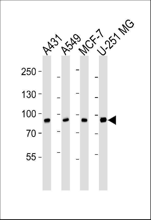
Western blot analysis of lysates from A431, A549, MCF-7, U-251 MG cell line (from left to right), using CAPN1 Antibody. M01943-1 was diluted at 1:1000 at each lane. A goat anti-mouse IgG H&L (HRP) at 1:3000 dilution was used as the secondary antibody. Lysates at 35μg per lane.

Click image to see more details
Immunohistochemical analysis of paraffin-embedded H. kidney section using CAPN1 Antibody (Cat#M01943-1). M01943-1 was diluted at 1:25 dilution. A peroxidase-conjugated goat anti-mouse IgG at 1:400 dilution was used as the secondary antibody, followed by DAB staining.

Click image to see more details
Flow cytometric analysis of Hela cells using CAPN1 Antibody (green, Cat#M01943-1) compared to an isotype control of mouse IgG1 (blue). M01943-1 was diluted at 1:25 dilution. An Alexa Fluor® 488 goat anti-mouse lgG at 1:400 dilution was used as the secondary antibody.
Specific Publications For Anti-CAPN1 Antibody (M01943-1)
Loading publications
Recommended Resources
Here are featured tools and databases that you might find useful.
- Boster's Pathways Library
- Protein Databases
- Bioscience Research Protocol Resources
- Data Processing & Analysis Software
- Photo Editing Software
- Scientific Literature Resources
- Research Paper Management Tools
- Molecular Biology Software
- Primer Design Tools
- Bioinformatics Tools
- Phylogenetic Tree Analysis
Customer Reviews
Have you used Anti-CAPN1 Antibody?
Share your experimental results or join a short interview to earn up to $1,000 in product credits or other rewards.
0 Reviews For Anti-CAPN1 Antibody
Customer Q&As
Have a question?
Find answers in Q&As, reviews.
Can't find your answer?
Submit your question
