Click image to see more details
Product Info Summary
| SKU: | PA1849 |
|---|---|
| Size: | 100 μg/vial |
| Reactive Species: | Human, Mouse, Rat |
| Host: | Rabbit |
| Application: | WB |
Customers Who Bought This Also Bought

Product info
Product Name
Anti-Caspase 3/CASP3 Antibody Picoband®
SKU/Catalog Number
PA1849
Size
100 μg/vial
Form
Lyophilized
Description
Boster Bio Anti-Caspase 3/CASP3 Antibody catalog # PA1849. Tested in WB applications. This antibody reacts with Human, Mouse, Rat. The brand Picoband indicates this is a premium antibody that guarantees superior quality, high affinity, and strong signals with minimal background in Western blot applications. Only our best-performing antibodies are designated as Picoband, ensuring unmatched performance.
Storage & Handling
Store at -20˚C for one year from date of receipt. After reconstitution, at 4˚C for one month. It can also be aliquotted and stored frozen at -20˚C for six months. Avoid repeated freeze-thaw cycles.
Cite This Product
Anti-Caspase 3/CASP3 Antibody Picoband® (Boster Biological Technology, Pleasanton CA, USA, Catalog # PA1849)
Host
Rabbit
Contents
Each vial contains 4 mg Trehalose, 0.9 mg NaCl and 0.2mg Na2HPO4.
Clonality
Polyclonal
Isotype
Rabbit IgG
Immunogen
A synthetic peptide corresponding to a sequence at the C-terminal of human Caspase 3, identical to the related mouse sequence, and different from the related rat sequence by one amino acid.
Cross-reactivity
No cross-reactivity with other proteins
Reactive Species
PA1849 is reactive to CASP3 in Human, Mouse, Rat
Observed Molecular Weight
31 kDa
Calculated molecular weight
31.6 kDa
Background of CASP3
Caspase 3 (caspase 3, apoptosis-related cysteine peptidase) is a caspase protein that interacts with caspase 8 and caspase 9, also known as Caspase-3, PARP CLEAVAGE PROTEASE, APOPAIN, CPP32, CPP32B, YAMA. It is a member of the cysteine-aspartic acid protease (caspase) family. PCR analysis of 16 human tissues revealed expression of full-length CASP3, as well as CASP3s at somewhat lower levels, in all tissues tested. Western blot analysis of 3 cell lines revealed the prominent CASP3 band at 32 kD and CASP3s at 20 kD. Several human cancer cell lines showed coexpression of both variants at the mRNA and protein levels. Overexpression of the catalytically inactive CASP3s by human kidney cells offered some resistance to inducers of apoptosis, and CASP3s accumulation could be enhanced with addition of proteasome inhibitors. Sequential activation of caspases plays a central role in the execution-phase of cell apoptosis. Alternative splicing of this gene results in two transcript variants that encode the same protein. Encoded by the CASP3 gene, CASP3 orthologs have been identified in numerous mammals for which complete genome data are available. Unique orthologs are also present in birds, lizards, lissamphibians, and teleosts. Nicholson et al. developed a potent peptide aldehyde inhibitor and showed that it prevented apoptotic events in vitro, suggesting that apopain/CPP32 is important for the initiation of apoptotic cell death.
Antibody Validation
Boster validates all antibodies on WB, IHC, ICC, Immunofluorescence, and ELISA with known positive control and negative samples to ensure specificity and high affinity, including thorough antibody incubations.
Application & Images
Applications
PA1849 is guaranteed for WB Boster Guarantee
Assay Dilutions Recommendation
The recommendations below provide a starting point for assay optimization. The actual working concentration varies and should be decided by the user.
Western blot, 0.1-0.5μg/ml, Human, Rat, Mouse
Positive Control
WB: Rat Cardiac Muscle Tissue, Rat Liver Tissue, Rat Thymus Tissue, MCF-7 Whole Cell, SMMC Whole Cell, HT1080 Whole Cell
Validation Images & Assay Conditions
Click image to see more details
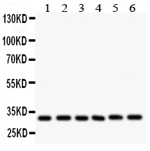
Anti-CASP3 antibody, PA1849, Western blotting
All lanes: Anti CASP3 (PA1849) at 0.5ug/ml
Lane 1: Rat Cardiac Muscle Tissue Lysate at 50ug
Lane 2: Rat Liver Tissue Lysate at 50ug
Lane 3: Rat Thymus Tissue Lysate at 50ug
Lane 4: MCF-7 Whole Cell Lysate at 40ug
Lane 5: SMMC Whole Cell Lysate at 40ug
Lane 6: HT1080 Whole Cell Lysate at 40ug
Predicted bind size: 31KD
Observed bind size: 31KD
Specific Publications For Anti-Caspase 3/CASP3 Antibody Picoband® (PA1849)
Loading publications
Recommended Resources
Here are featured tools and databases that you might find useful.
- Boster's Pathways Library
- Protein Databases
- Bioscience Research Protocol Resources
- Data Processing & Analysis Software
- Photo Editing Software
- Scientific Literature Resources
- Research Paper Management Tools
- Molecular Biology Software
- Primer Design Tools
- Bioinformatics Tools
- Phylogenetic Tree Analysis
Customer Reviews
Have you used Anti-Caspase 3/CASP3 Antibody Picoband®?
Share your experimental results or join a short interview to earn up to $1,000 in product credits or other rewards.
0 Reviews For Anti-Caspase 3/CASP3 Antibody Picoband®
Customer Q&As
Have a question?
Find answers in Q&As, reviews.
Can't find your answer?
Submit your question
4 Customer Q&As for Anti-Caspase 3/CASP3 Antibody Picoband®
Question
We have seen staining in rat lymph. Are there any suggestions? Is anti-Caspase 3/CASP3 antibody supposed to stain lymph positively?
Verified Customer
Verified customer
Asked: 2019-12-11
Answer
Based on literature lymph does express CASP3. Based on Uniprot.org, CASP3 is expressed in jejunal mucosa, t-cell, tongue, lymph, cervix carcinoma erythroleukemia, among other tissues. Regarding which tissues have CASP3 expression, here are a few articles citing expression in various tissues:
Cervix carcinoma, and Erythroleukemia, Pubmed ID: 23186163
Lymph, Pubmed ID: 15489334
T-cell, Pubmed ID: 7983002, 7774019
Tongue, Pubmed ID: 14702039
Boster Scientific Support
Answered: 2019-12-11
Question
Our lab were happy with the WB result of your anti-Caspase 3/CASP3 antibody. However we have been able to see positive staining in t-cell cytoplasm. using this antibody. Is that expected? Could you tell me where is CASP3 supposed to be expressed?
D. Jones
Verified customer
Asked: 2019-12-11
Answer
According to literature, t-cell does express CASP3. Generally CASP3 expresses in cytoplasm. Regarding which tissues have CASP3 expression, here are a few articles citing expression in various tissues:
Cervix carcinoma, and Erythroleukemia, Pubmed ID: 23186163
Lymph, Pubmed ID: 15489334
T-cell, Pubmed ID: 7983002, 7774019
Tongue, Pubmed ID: 14702039
Boster Scientific Support
Answered: 2019-12-11
Question
We need using your anti-Caspase 3/CASP3 antibody for wound healing studies. Has this antibody been tested with western blotting on ht1080 whole cell lysate? We would like to see some validation images before ordering.
Verified Customer
Verified customer
Asked: 2019-06-13
Answer
Thank you for your inquiry. This PA1849 anti-Caspase 3/CASP3 antibody is validated on rat thymus tissue, cardiac muscle tissue, tissue lysate, liver tissue, smmc whole cell lysate, ht1080 whole cell lysate. It is guaranteed to work for WB in human, mouse, rat. Our Boster guarantee will cover your intended experiment even if the sample type has not been be directly tested.
Boster Scientific Support
Answered: 2019-06-13
Question
We are currently using anti-Caspase 3/CASP3 antibody PA1849 for mouse tissue, and we are happy with the WB results. The species of reactivity given in the datasheet says human, mouse, rat. Is it possible that the antibody can work on bovine tissues as well?
P. Krishna
Verified customer
Asked: 2019-05-10
Answer
The anti-Caspase 3/CASP3 antibody (PA1849) has not been validated for cross reactivity specifically with bovine tissues, though there is a good chance of cross reactivity. We have an innovator award program that if you test this antibody and show it works in bovine you can get your next antibody for free. Please contact me if I can help you with anything.
Boster Scientific Support
Answered: 2019-05-10
