Click image to see more details
-

-
-
-
-
+1
Product Info Summary
| SKU: | PB9188 |
|---|---|
| Size: | 100 μg/vial |
| Reactive Species: | Human, Mouse, Rat |
| Host: | Rabbit |
| Application: | IF, IHC, ICC, WB |
Customers Who Bought This Also Bought

Product info
Product Name
Anti-Caspase-3/CASP3 Antibody Picoband®
SKU/Catalog Number
PB9188
Size
100 μg/vial
Form
Lyophilized
Description
Boster Bio Anti-Caspase-3/CASP3 Antibody Picoband® catalog # PB9188. Tested in IF, IHC, ICC, WB applications. This antibody reacts with Human, Mouse, Rat. The brand Picoband indicates this is a premium antibody that guarantees superior quality, high affinity, and strong signals with minimal background in Western blot applications. Only our best-performing antibodies are designated as Picoband, ensuring unmatched performance.
Storage & Handling
Store at -20˚C for one year from date of receipt. After reconstitution, at 4˚C for one month. It can also be aliquotted and stored frozen at -20˚C for six months. Avoid repeated freeze-thaw cycles.
Cite This Product
Anti-Caspase-3/CASP3 Antibody Picoband® (Boster Biological Technology, Pleasanton CA, USA, Catalog # PB9188)
Host
Rabbit
Contents
Each vial contains 4mg Trehalose, 0.9mg NaCl, 0.2mg Na2HPO4, 0.01mg NaN3.
Clonality
Polyclonal
Isotype
Rabbit IgG
Immunogen
E.coli-derived human Caspase 3 recombinant protein (Position: T67-D175). Human Caspase 3 shares 86% and 90% amino acid (aa) sequences identity with mouse and rat Caspase 3, respectively.
Cross-reactivity
No cross-reactivity with other proteins
Reactive Species
PB9188 is reactive to CASP3 in Human, Mouse, Rat
Observed Molecular Weight
35 kDa, 17 kDa (Cleaved)
Calculated molecular weight
31.6 kDa
Background of CASP3
Caspase 3 is a caspase protein which interacts with Survivin, XIAP, CFLAR, Caspase 8, HCLS1, Deleted in Colorectal Cancer, TRAF3 and GroEL. This gene which is located on 4q35 encodes a protein that is a member of the cysteine-aspartic acid protease (caspase) family. Sequential activation of caspases plays a central role in the execution-phase of cell apoptosis. Caspases exist as inactive proenzymes that undergo proteolytic processing at conserved aspartic residues to produce two subunits, large and small, that dimerize to form the active enzyme. It is the predominant caspase involved in the cleavage of amyloid-beta 4A precursor protein, which is associated with neuronal death in Alzheimer's disease. And the caspase-3 activation in heart failure sequentially cleaves SRF and generates a truncated SRF that appears to function as a dominant-negative transcription factor. Additionally, the caspase-3 influence on bone mineral density should be considered in any in vivo application of caspase-3 inhibitors to the treatment of human disease. In erythroid precursors undergoing terminal differentiation, Hsp70 prevents active CASP3 from cleaving GATA1 and inducing apoptosis.
Antibody Validation
Boster validates all antibodies on WB, IHC, ICC, Immunofluorescence, and ELISA with known positive control and negative samples to ensure specificity and high affinity, including thorough antibody incubations.
Application & Images
Applications
PB9188 is guaranteed for IF, IHC, ICC, WB Boster Guarantee
Recommend Dilution
| Application | Dilution | Species |
|---|---|---|
| Western blot | 0.1-0.5μg/ml | Human, Mouse, Rat |
| Immunohistochemistry (Paraffin-embedded Section) | 2-5μg/ml | Human |
| Immunocytochemistry/Immunofluorescence | 5μg/ml | Human |
Tested application
Suggested blocking solution with 5% non-fat milk or BSA; (*)Recommended protein loading: 20-40 µg per lane
Use TE buffer pH 9.0 for antigen retrieval; (*) citrate buffer pH 6.0 is an alternative.
Validation Images & Assay Conditions
Click image to see more details
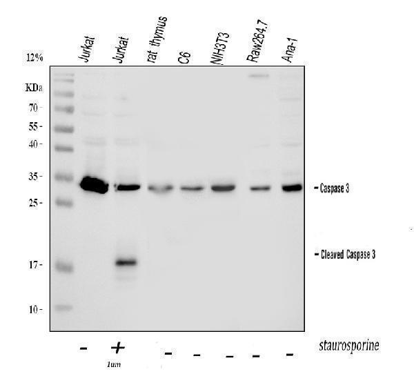
Western blot analysis of Caspase-3 using anti-Caspase-3 antibody (PB9188).
Electrophoresis was performed on a 5-20% SDS-PAGE gel at 70V (Stacking gel) / 90V (Resolving gel) for 2-3 hours. The sample well of each lane was loaded with 30 ug of sample under reducing conditions.
Lane 1: human Jurkat whole cell lysates,
Lane 2: human Jurkat whole cell lysates,
Lane 3: rat thymus tissue lysates,
Lane 4: rat C6 whole cell lysates,
Lane 5: mouse NIH/3T3 whole cell lysates,
Lane 6: mouse RAW264.7 whole cell lysates,
Lane 7: mouse ANA-1 whole cell lysates.
After electrophoresis, proteins were transferred to a nitrocellulose membrane at 150 mA for 50-90 minutes. Blocked the membrane with 5% non-fat milk/TBS for 1.5 hour at RT. The membrane was incubated with rabbit anti-Caspase-3 antigen affinity purified polyclonal antibody (Catalog # PB9188) at 0.5 μg/mL overnight at 4°C, then washed with TBS-0.1%Tween 3 times with 5 minutes each and probed with a goat anti-rabbit IgG-HRP secondary antibody at a dilution of 1:5000 for 1.5 hour at RT. The signal is developed using an Enhanced Chemiluminescent detection (ECL) kit (Catalog # EK1002) with Tanon 5200 system. A specific band was detected for Caspase-3 at approximately 35 kDa, 17 kDa (Cleaved). The expected band size for Caspase-3 is at 32 kDa.

Click image to see more details
Western blot analysis of Caspase-3 using anti-Caspase-3 antibody (PB9188).
Electrophoresis was performed on a 5-20% SDS-PAGE gel at 70V (Stacking gel) / 90V (Resolving gel) for 2-3 hours. The sample well of each lane was loaded with 30 ug of sample under reducing conditions.
Lane 1: human Jurkat whole cell lysates,
Lane 2: human Jurkat whole cell lysates,
Lane 3: human Jurkat whole cell lysates,
Lane 4: human HepG2 whole cell lysates.
After electrophoresis, proteins were transferred to a nitrocellulose membrane at 150 mA for 50-90 minutes. Blocked the membrane with 5% non-fat milk/TBS for 1.5 hour at RT. The membrane was incubated with rabbit anti-Caspase-3 antigen affinity purified polyclonal antibody (Catalog # PB9188) at 0.5 μg/mL overnight at 4°C, then washed with TBS-0.1%Tween 3 times with 5 minutes each and probed with a goat anti-rabbit IgG-HRP secondary antibody at a dilution of 1:5000 for 1.5 hour at RT. The signal is developed using an Enhanced Chemiluminescent detection (ECL) kit (Catalog # EK1002) with Tanon 5200 system. A specific band was detected for Caspase-3 at approximately 35 kDa, 17 kDa (Cleaved). The expected band size for Caspase-3 is at 32 kDa.

Click image to see more details
IHC analysis of Caspase-3 using anti-Caspase-3 antibody (PB9188).
Caspase-3 was detected in a paraffin-embedded section of human tonsil tissue. Heat mediated antigen retrieval was performed in EDTA buffer (pH 8.0, epitope retrieval solution). The tissue section was blocked with 10% goat serum. The tissue section was then incubated with 2 μg/ml rabbit anti-Caspase-3 Antibody (PB9188) overnight at 4°C. Peroxidase Conjugated Goat Anti-rabbit IgG was used as secondary antibody and incubated for 30 minutes at 37°C. The tissue section was developed using HRP Conjugated Rabbit IgG Super Vision Assay Kit (Catalog # SV0002) with DAB as the chromogen.

Click image to see more details
IF analysis of Caspase-3 using anti-Caspase-3 antibody (PB9188).
Caspase-3 was detected in an immunocytochemical section of U20S cells. Enzyme antigen retrieval was performed using IHC enzyme antigen retrieval reagent (AR0022) for 15 mins. The cells were blocked with 10% goat serum. And then incubated with 5 μg/mL rabbit anti-Caspase-3 Antibody (PB9188) overnight at 4°C. DyLight®488 Conjugated Goat Anti-Rabbit IgG (BA1127) was used as secondary antibody at 1:500 dilution and incubated for 30 minutes at 37°C. Visualize using a fluorescence microscope and filter sets appropriate for the label used.

Click image to see more details
Regulatory effects of KSG on the expression of cytokines and apoptosis-related factors in Aβ-induced PC12 cells. (A) Expression of IL-1β (n = 3). (B) Expression of IL-18 (n = 3). (C) Expression of TNF-α (n = 3). (D) Protein expression of apoptosis-related factors in PC12 cells of each group (n = 3). (E) Expression of Caspase-3 (n = 3). (F) Expression of Bax (n = 3). Data were expressed as mean ± SD. * P < 0.05, ** P < 0.01, and *** P < 0.001 vs. Con. # P < 0.05, ## P < 0.01, and ### P < 0.001 vs. AD.
Index in PubMed under a CC BY license. PMID: 41050397
Specific Publications For Anti-Caspase-3/CASP3 Antibody Picoband® (PB9188)
Loading publications
Recommended Resources
Here are featured tools and databases that you might find useful.
- Boster's Pathways Library
- Protein Databases
- Bioscience Research Protocol Resources
- Data Processing & Analysis Software
- Photo Editing Software
- Scientific Literature Resources
- Research Paper Management Tools
- Molecular Biology Software
- Primer Design Tools
- Bioinformatics Tools
- Phylogenetic Tree Analysis
Customer Reviews
Have you used Anti-Caspase-3/CASP3 Antibody Picoband®?
Share your experimental results or join a short interview to earn up to $1,000 in product credits or other rewards.
0 Reviews For Anti-Caspase-3/CASP3 Antibody Picoband®
Customer Q&As
Have a question?
Find answers in Q&As, reviews.
Can't find your answer?
Submit your question
16 Customer Q&As for Anti-Caspase-3/CASP3 Antibody Picoband®
Question
I see that the anti-Caspase-3/CASP3 antibody PB9188 works with IHC-P, what is the protocol used to produce the result images on the product page?
Verified Customer
Verified customer
Asked: 2020-04-03
Answer
You can find protocols for IHC-P on the "support/technical resources" section of our navigation menu. If you have any further questions, please send an email to support@bosterbio.com
Boster Scientific Support
Answered: 2020-04-03
Question
I am interested in using your anti-Caspase-3/CASP3 antibody for t cell homeostasis studies. Has this antibody been tested with western blotting on smmc whole cell lysate? We would like to see some validation images before ordering.
Verified Customer
Verified customer
Asked: 2019-12-25
Answer
I appreciate your inquiry. This PB9188 anti-Caspase-3/CASP3 antibody is tested on mouse intestine tissue, tissue lysate, rat thymus tissue, lung cancer tissue, liver tissue, smmc whole cell lysate, k562 cells. It is guaranteed to work for Flow Cytometry, IF, IHC-P, ICC, WB in human, mouse, rat. Our Boster guarantee will cover your intended experiment even if the sample type has not been be directly tested.
Boster Scientific Support
Answered: 2019-12-25
Question
See attached the WB image, lot number and protocol we used for tongue using anti-Caspase-3/CASP3 antibody PB9188. Please let me know if you require anything else.
Verified Customer
Verified customer
Asked: 2019-10-09
Answer
Thank you very much for the data. Our lab team are working to resolve this as quickly as possible, and we appreciate your patience and understanding! You have provided everything we needed. Please let me know if there is anything you need in the meantime.
Boster Scientific Support
Answered: 2019-10-09
Question
Would PB9188 anti-Caspase-3/CASP3 antibody work on parafin embedded sections? If so, which fixation method do you recommend we use (PFA, paraformaldehyde, other)?
Verified Customer
Verified customer
Asked: 2019-09-25
Answer
You can see on the product datasheet, PB9188 anti-Caspase-3/CASP3 antibody as been tested on IHC-P. It is best to use PFA for fixation because it has better tissue penetration ability. PFA needs to be prepared fresh before use. Long term stored PFA turns into formalin, as the PFA molecules congregate and become formalin.
Boster Scientific Support
Answered: 2019-09-25
Question
We appreciate helping with my inquiry over the phone. Here are the WB image, lot number and protocol we used for tongue using anti-Caspase-3/CASP3 antibody PB9188. Let me know if you need anything else.
Verified Customer
Verified customer
Asked: 2019-09-20
Answer
I appreciate the data. You have provided everything we needed. Our lab team are working to resolve your inquiry as quickly as possible, and we appreciate your patience and understanding! Please let me know if there is anything you need in the meantime.
Boster Scientific Support
Answered: 2019-09-20
Question
My colleagues were content with the WB result of your anti-Caspase-3/CASP3 antibody. However we have been able to see positive staining in jejunal mucosa cytoplasm. using this antibody. Is that expected? Could you tell me where is CASP3 supposed to be expressed?
Verified Customer
Verified customer
Asked: 2019-08-20
Answer
From literature, jejunal mucosa does express CASP3. Generally CASP3 expresses in cytoplasm. Regarding which tissues have CASP3 expression, here are a few articles citing expression in various tissues:
Cervix carcinoma, and Erythroleukemia, Pubmed ID: 23186163
Lymph, Pubmed ID: 15489334
T-cell, Pubmed ID: 7983002, 7774019
Tongue, Pubmed ID: 14702039
Boster Scientific Support
Answered: 2019-08-20
Question
My question regarding product PB9188, anti-Caspase-3/CASP3 antibody. I was wondering if it would be possible to conjugate this antibody with biotin. I would need it to be without BSA or sodium azide. I am planning on using a buffer exchange of sodium azide with PBS only. Would there be problems for me to conjugate the antibody and store it in -20 degrees in small aliquots?
Verified Customer
Verified customer
Asked: 2019-08-20
Answer
It is not recommended storing this antibody with PBS buffer only in -20 degrees. If you want to store it in -20 degrees it is best to add some cryoprotectant like glycerol. If you want carrier free PB9188 anti-Caspase-3/CASP3 antibody, we can provide it to you in a special formula with trehalose and/or glycerol. These molecules will not interfere with conjugation chemistry and provide a good level of protection for the antibody from degradation. Please be sure to specify this in your purchase order.
Boster Scientific Support
Answered: 2019-08-20
Question
Does anti-Caspase-3/CASP3 antibody PB9188 work for IHC-P with tongue?
Verified Customer
Verified customer
Asked: 2019-07-25
Answer
According to the expression profile of tongue, CASP3 is highly expressed in tongue. So, it is likely that anti-Caspase-3/CASP3 antibody PB9188 will work for IHC-P with tongue.
Boster Scientific Support
Answered: 2019-07-25
Question
Do you have a BSA free version of anti-Caspase-3/CASP3 antibody PB9188 available?
Verified Customer
Verified customer
Asked: 2019-07-18
Answer
Thanks for your recent telephone inquiry. I can confirm that some lots of this anti-Caspase-3/CASP3 antibody PB9188 are BSA free. For now, these lots are available and we can make a BSA free formula for you free of charge. It will take 3 extra days to prepare. If you require this antibody BSA free again in future, please do not hesitate to contact me and I will be pleased to check which lots we have in stock that are BSA free.
Boster Scientific Support
Answered: 2019-07-18
Question
Is this PB9188 anti-Caspase-3/CASP3 antibody reactive to the isotypes of CASP3?
Verified Customer
Verified customer
Asked: 2019-06-05
Answer
The immunogen of PB9188 anti-Caspase-3/CASP3 antibody is E.coli-derived human Caspase 3 recombinant protein (Position: T67-D175). Human Caspase 3 shares 86% and 90% amino acid (aa) sequences identity with mouse and rat Caspase 3, respectively. Could you tell me which isotype you are interested in so I can help see if the immunogen is part of this isotype?
Boster Scientific Support
Answered: 2019-06-05
Question
I was wanting to use your anti-Caspase-3/CASP3 antibody for IHC-P for rat tongue on frozen tissues, but I want to know if it has been validated for this particular application. Has this antibody been validated and is this antibody a good choice for rat tongue identification?
H. Mitchell
Verified customer
Asked: 2019-02-22
Answer
You can see on the product datasheet, PB9188 anti-Caspase-3/CASP3 antibody has been validated for Flow Cytometry, IF, IHC-P, ICC, WB on human, mouse, rat tissues. We have an innovator award program that if you test this antibody and show it works in rat tongue in IHC-frozen, you can get your next antibody for free.
Boster Scientific Support
Answered: 2019-02-22
Question
Is a blocking peptide available for product anti-Caspase-3/CASP3 antibody (PB9188)?
Verified Customer
Verified customer
Asked: 2018-07-06
Answer
We do provide the blocking peptide for product anti-Caspase-3/CASP3 antibody (PB9188). If you would like to place an order for it please contact support@bosterbio.com and make a special request.
Boster Scientific Support
Answered: 2018-07-06
Question
We are currently using anti-Caspase-3/CASP3 antibody PB9188 for human tissue, and we are content with the IHC-P results. The species of reactivity given in the datasheet says human, mouse, rat. Is it true that the antibody can work on horse tissues as well?
Verified Customer
Verified customer
Asked: 2017-12-18
Answer
The anti-Caspase-3/CASP3 antibody (PB9188) has not been tested for cross reactivity specifically with horse tissues, though there is a good chance of cross reactivity. We have an innovator award program that if you test this antibody and show it works in horse you can get your next antibody for free. Please contact me if I can help you with anything.
Boster Scientific Support
Answered: 2017-12-18
Question
We ordered your anti-Caspase-3/CASP3 antibody for IF on t-cell a few months ago. I am using mouse, and We intend to use the antibody for IHC-P next. I am interested in examining t-cell as well as lymph in our next experiment. Could you please give me some suggestion on which antibody would work the best for IHC-P?
Verified Customer
Verified customer
Asked: 2017-08-09
Answer
I viewed the website and datasheets of our anti-Caspase-3/CASP3 antibody and it seems that PB9188 has been validated on mouse in both IF and IHC-P. Thus PB9188 should work for your application. Our Boster satisfaction guarantee will cover this product for IHC-P in mouse even if the specific tissue type has not been validated. We do have a comprehensive range of products for IHC-P detection and you can check out our website bosterbio.com to find out more information about them.
Boster Scientific Support
Answered: 2017-08-09
Question
We have been able to see staining in human cervix carcinoma erythroleukemia. Do you have any suggestions? Is anti-Caspase-3/CASP3 antibody supposed to stain cervix carcinoma erythroleukemia positively?
Verified Customer
Verified customer
Asked: 2017-05-16
Answer
From literature cervix carcinoma erythroleukemia does express CASP3. From Uniprot.org, CASP3 is expressed in jejunal mucosa, t-cell, tongue, lymph, cervix carcinoma erythroleukemia, among other tissues. Regarding which tissues have CASP3 expression, here are a few articles citing expression in various tissues:
Cervix carcinoma, and Erythroleukemia, Pubmed ID: 23186163
Lymph, Pubmed ID: 15489334
T-cell, Pubmed ID: 7983002, 7774019
Tongue, Pubmed ID: 14702039
Boster Scientific Support
Answered: 2017-05-16
Question
you antibody to test anti-Caspase-3/CASP3 antibody PB9188 on rat tongue for research purposes, then I may be interested in using anti-Caspase-3/CASP3 antibody PB9188 for diagnostic purposes as well. Is the antibody suitable for diagnostic purposes?
S. Johnson
Verified customer
Asked: 2016-05-30
Answer
The products we sell, including anti-Caspase-3/CASP3 antibody PB9188, are only intended for research use. They would not be suitable for use in diagnostic work. If you have the means to develop a product into diagnostic use, and are interested in collaborating with us and develop our product into an IVD product, please contact us for more discussions.
Boster Scientific Support
Answered: 2016-05-30
