This website uses cookies to ensure you get the best experience on our website.
- Table of Contents
4 Citations 6 Q&As

103 Citations 17 Q&As
117 Citations 5 Q&As


148 Citations 16 Q&As
46 Citations 15 Q&As
32 Citations 16 Q&As


26 Citations 16 Q&As
56 Citations 15 Q&As
18 Citations 5 Q&As
58 Citations 16 Q&As
49 Citations 4 Q&As
12 Citations 14 Q&As

16 Citations 16 Q&As
17 Citations 15 Q&As
12 Citations 16 Q&As


1 Citations

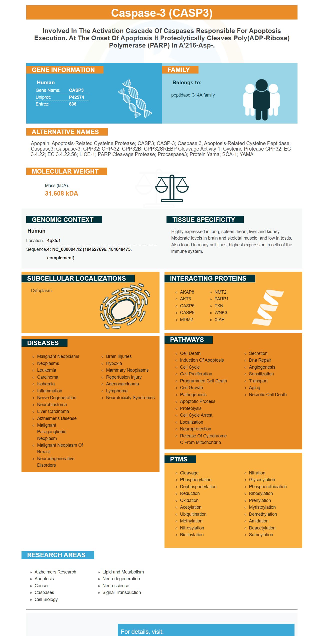
Facts about Caspase-3.

| Human | |
|---|---|
| Gene Name: | CASP3 |
| Uniprot: | P42574 |
| Entrez: | 836 |

| Belongs to: |
|---|
| peptidase C14A family |

Apopain; apoptosis-related cysteine protease; CASP3; CASP-3; caspase 3, apoptosis-related cysteine peptidase; Caspase3; Caspase-3; CPP32; CPP-32; CPP32B; CPP32SREBP cleavage activity 1; Cysteine protease CPP32; EC 3.4.22; EC 3.4.22.56; LICE-1; PARP cleavage protease; procaspase3; Protein Yama; SCA-1; YAMA
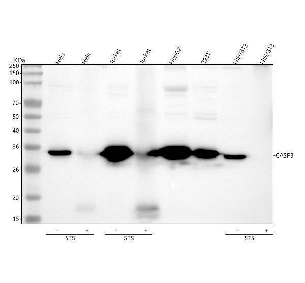
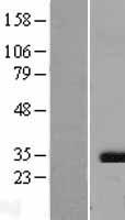
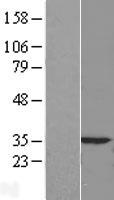
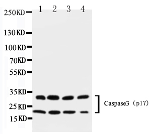
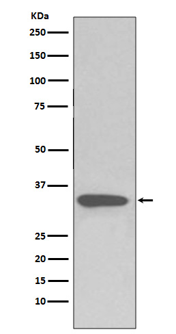
Mass (kDA):
31.608 kDA

| Human | |
|---|---|
| Location: | 4q35.1 |
| Sequence: | 4; NC_000004.12 (184627696..184649475, complement) |
Highly expressed in lung, spleen, heart, liver and kidney. Moderate levels in brain and skeletal muscle, and low in testis. Also found in many cell lines, highest expression in cells of the immune system.
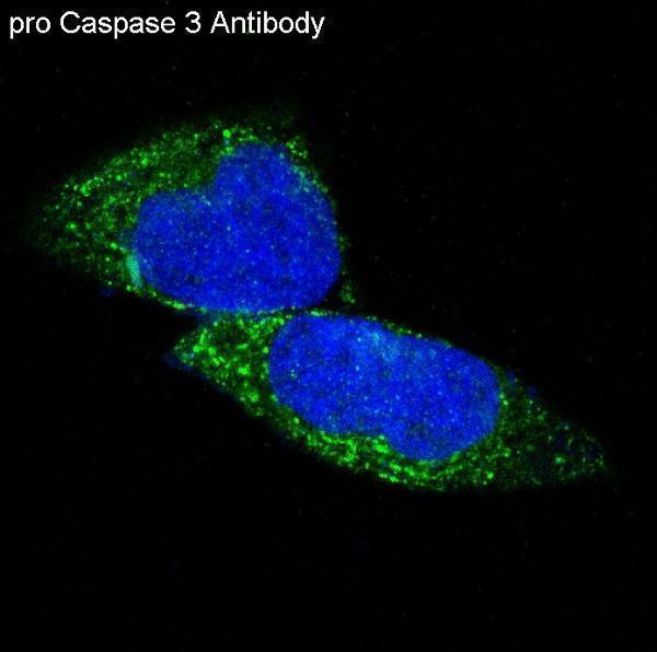
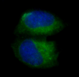
Cytoplasm.


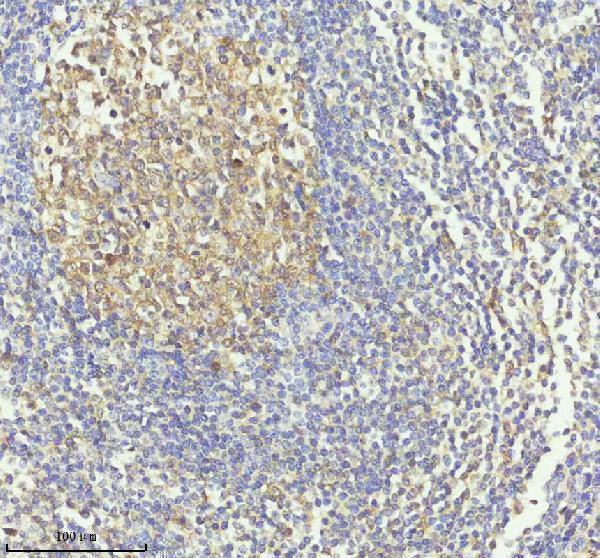
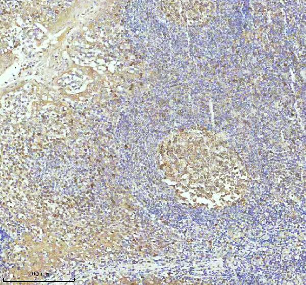
This article discusses the IHC protocol and the biological significance of the CASP3 marker. It also examines Molecular biology as well as clinical applications. It also describes the immunohistochemistry (IHC) protocol for this new marker. CASP3 is a crucial indicator for detecting cancer. Boster Bio is a company that offers a wide range of antibodies that can be used in a variety of applications. For more information, go to the Boster Bio website.
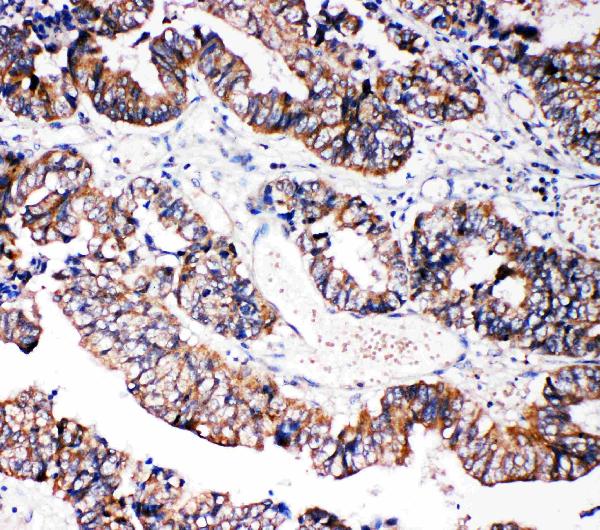
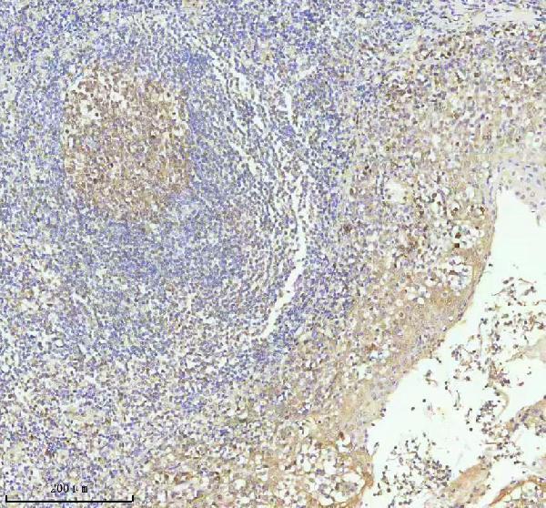
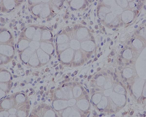
IHC protocols are widely used for visualizing the distribution of antigens and haptens inside cells and tissue context. There are a variety of ways to identify antigens. But, it is important to look at the quality of the sample preparation. The quality of the sample can affect the display of antigens. Therefore, IHC methods should be chosen with care. Here are some guidelines to follow when performing this assay. This protocol is based on the binding chemical properties of CASP3.
New slides should be soaked in 95 percent alcohol for 12-24hrs prior to beginning the IHC protocol. Slides should then be thoroughly dried by wiping or by baking in the infrared. It is vital not to scratch the slides during this process. Slides for microscopic examinations should be five millimeters in diameter, whereas the nervous tissue should be 20-100 mm in size. This will allow for better tracking of the never fiber direction. For coverslips, the pre-treatment procedure is similar to that for microscopically-sized slides, but is different from the procedures mentioned above.
IHC protocols for the caspase-3 enzyme use the ABC immunoperoxidase method. The second antibody that is biotin-labeled attaches to the primary antibody, CASP3, and binds to the protein of interest. After the primary antibody has bound to the protein it attaches to the CASP3 marker and allows it to be identified in the same image. The secondary antibody can be dilute to make the resulting staining more precise.
The next step of the IHC procedure is to prepare the samples for staining. The type of sample will determine the preparation. Tissues are either obtained by biopsy, surgery or animal model. In the latter the autopsy is conducted after the animal has died for two hours. This is like a postmortem examination. As a result, antigens denature after a few hours.
The CASP3 marker is an epigenetic molecule that has recently been identified as a biomarker for the development of gastrointestinal cancer. Despite the complexity of GI cancer's development and its multi-stage progression, it's unclear what the molecular cause was. However the molecular biology behind this cancer has led to a better understanding of the disease as well as the use of biomarkers for diagnostic and prognostication testing. In addition to its diagnostic value CASP3 is also a CASP3 marker has many possibilities for clinical applications.
The CASP3 gene encodes the caspase-3 enzyme that interacts with caspases 8 , and 9. Many mammals have orthologs to the CASP3 marker in their genomes including humans. Some animals have their own orthologs such as birds, teleosts and Lizards. The CASP3 gene is present only in a few tissues in humans.
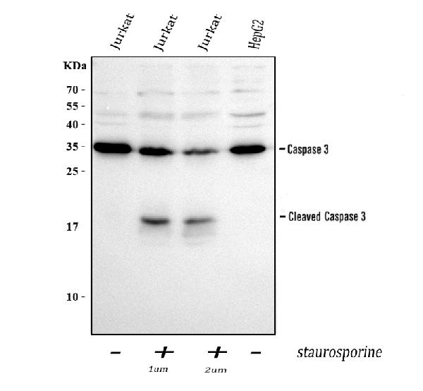
Molecular biology and the CASP3 marker: In addition to its importance in determining whether cancerous cells are cancerous, it is important to understand how this gene works. This enzyme is responsible for the death of cells in MCF-7 mammalian cell line. This gene is present in several cancer cell lines, including MCF-7 cells and SKOV-90 cells. Several clones of these cancer cells harbor the same deletion.
The CASP3 gene encodes the caspase-3 enzyme, which interacts with caspase-8 or caspase-9. Many mammals have orthologs of the CASP3 gene. Several species have full genome data. Teleosts, lizards, and birds all have distinct orthologs to the CASP3 gene. These results support the hypothesis that cell death is caused by the CASP3 gene.
PMID: 7983002 by Fernandes-Alnemri T., et al. CPP32, a novel human apoptotic protein with homology to Caenorhabditis elegans cell death protein Ced-3 and mammalian interleukin-1 beta-converting enzyme.
PMID: 7774019 by Tewari M., et al. Yama/CPP32 beta, a mammalian homolog of CED-3, is a CrmA-inhibitable protease that cleaves the death substrate poly(ADP-ribose) polymerase.
*Showing only the more recent 20. More publications can be found for each product on its corresponding product page