Click image to see more details
-

-
-
-
-
+2
Product Info Summary
| SKU: | PA1961-1 |
|---|---|
| Size: | 100 μg/vial |
| Reactive Species: | Mouse, Rat |
| Host: | Rabbit |
| Application: | IHC, WB |
Customers Who Bought This Also Bought

Product info
Product Name
Anti-Caspase-3(P17)/CASP3 Antibody Picoband®
SKU/Catalog Number
PA1961-1
BA3257 is an alternative SKU for this antibody, used in previous lots.
Size
100 μg/vial
Form
Lyophilized
Description
Boster Bio Anti-Caspase-3 (P17)/CASP3 Antibody catalog # PA1961-1. Tested in IHC, WB applications. This antibody reacts with Mouse, Rat. The brand Picoband indicates this is a premium antibody that guarantees superior quality, high affinity, and strong signals with minimal background in Western blot applications. Only our best-performing antibodies are designated as Picoband, ensuring unmatched performance.
Storage & Handling
Store at -20˚C for one year from date of receipt. After reconstitution, at 4˚C for one month. It can also be aliquotted and stored frozen at -20˚C for six months. Avoid repeated freeze-thaw cycles.
Cite This Product
Anti-Caspase-3(P17)/CASP3 Antibody Picoband® (Boster Biological Technology, Pleasanton CA, USA, Catalog # PA1961-1)
Host
Rabbit
Contents
Each vial contains antibody formulated with stabilizing components, 0.9mg NaCl, 0.2mg Na2HPO4, 0.05mg Thimerosal, 0.05mg NaN3.
*This antibody is supplied in a stabilized formulation.
Compatibility with conjugation reactions depends on the chemistry of the conjugation method used.
For conjugation methods that are not compatible with the stabilizing components present in this formulation, a carrier-free antibody format is required.
Clonality
Polyclonal
Isotype
Rabbit IgG
Immunogen
A synthetic peptide corresponding to a sequence at the N-terminus of mouse Caspase-3(P17), different from the related rat sequence by one amino acid.
Cross-reactivity
No cross-reactivity with other proteins
Reactive Species
PA1961-1 is reactive to Casp3 in Mouse, Rat
Observed Molecular Weight
32 kDa, 19 kDa
Calculated molecular weight
31.5 kDa
Background of Casp3
CASP3 (Caspase3 Apoptosis-Related Cysteine Protease), also known as YAMA, CPP32 or APOPAIN, is a caspase protein that interacts with caspase 8 and caspase 9. It is encoded by the CASP3 gene. The CASP3 protein is a member of the cysteine-aspartic acid protease (caspase) family. Tiso et al. (1996) used radiation hybrid mapping to localize the CPP32 gene to human chromosome 4q33-q35.1. Fernandes-Alnemri et al. (1994) found that overexpression of CPP32 in insect cells induced apoptosis. Coexpression of the 2 CPP32 subunits in insect cells also resulted in apoptosis. Tewari et al. (1995) showed that purified human ICE cleaved the Yama proenzyme into a proteolytically active form and that activated Yama cleaved PARP into the 85-kD apoptotic form.
Antibody Validation
Boster validates all antibodies on WB, IHC, ICC, Immunofluorescence, and ELISA with known positive control and negative samples to ensure specificity and high affinity, including thorough antibody incubations.
Application & Images
Applications
PA1961-1 is guaranteed for IHC, WB Boster Guarantee
Recommend Dilution
| Application | Dilution | Species |
|---|---|---|
| Immunohistochemistry (Paraffin-embedded Section) | 0.5-1μg/ml | Rat, Mouse |
| Western blot | 0.1-0.5μg/ml | Mouse, Rat |
Tested application
Suggested blocking solution with 5% non-fat milk or BSA; (*)Recommended protein loading: 20-40 µg per lane
Use TE buffer pH 9.0 for antigen retrieval; (*) citrate buffer pH 6.0 is an alternative.
Validation Images & Assay Conditions
Click image to see more details
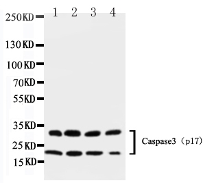
Western blot analysis of Caspase-3 (P17) using anti-Caspase-3 (P17) antibody (PA1961-1).
Electrophoresis was performed on a 5-20% SDS-PAGE gel at 70V (Stacking gel) / 90V (Resolving gel) for 2-3 hours. The sample well of each lane was loaded with 50ug of sample under reducing conditions.
Lane 1: Rat Heart Tissue Lysate
Lane 2: Rat Liver Tissue Lysate
Lane 3: Rat Thymus Tissue Lysate
Lane 4: Rat Spleen Tissue Lysate.
After Electrophoresis, proteins were transferred to a Nitrocellulose membrane at 150mA for 50-90 minutes. Blocked the membrane with 5% Non-fat Milk/ TBS for 1.5 hour at RT. The membrane was incubated with rabbit anti-Caspase-3 (P17) antigen affinity purified polyclonal antibody (Catalog # PA1961-1) at 0.5 μg/mL overnight at 4°C, then washed with TBS-0.1%Tween 3 times with 5 minutes each and probed with a goat anti-rabbit IgG-HRP secondary antibody at a dilution of 1:10000 for 1.5 hour at RT. The signal is developed using an Enhanced Chemiluminescent detection (ECL) kit (Catalog # EK1002) with Tanon 5200 system. A specific band was detected for Caspase-3 (P17) at approximately 32KD,19KD. The expected band size for Caspase-3 (P17) is at 32KD,17KD.

Click image to see more details
Mean gray value analyzed according to western blotting results of IL-6, caspase-3, and cleaved-caspase-3.
Index in PubMed under a CC BY license. PMID: 34975719

Click image to see more details
Experimental workflow. One hundred four rats were randomly divided into five groups: group S (sham, n = 20), group M (middle cerebral artery occlusion [MCAO], n = 28), group H2M (intermittent hypobaric hypoxia preconditioned MCAO group, 2 h/day, n = 20), group H6M (intermittent hypobaric hypoxia preconditioned MCAO group, 6 h/day, n = 28), and group HpM (persistent hypobaric hypoxia preconditioned MCAO group, n = 28). Behavioral tests and morphological staining (TTC staining) were used to analyze the severity of infarction. Total protein expression of NeuN (a specific marker of mature neurons), caspase-3, cleaved-caspase-3, and IL-6 was estimated using western blotting, which explained the severity of injury from different perspectives. Ultrastructural changes were observed under a transmission electron microscope. The most effective pretreatment group was selected for further label-free proteomic study and provided a reliable direction for mechanism exploration. Western blotting was used to verify the expression of the target protein, and key markers for the biological process were detected using immunofluorescence. caspase-3, cysteinyl aspartate specific proteinase 3; IL-6, interleukin 6; NeuN, neuron-specific nuclear protein; TTC, 2,3,5-triphenyl tetrazolium chloride.
Index in PubMed under a CC BY license. PMID: 34975719

Click image to see more details
Relative expression of IL-6, caspase-3, and cleaved-caspase-3. (A) Immunoblot results of IL-6, caspase-3, and cleaved-caspase-3. (B) Analysis of IL-6 relative expression. One-way ANOVA showed differences among the five groups, F = 10.86, p < 0.0001. (C) Analysis of caspase-3 relative expression. One-way ANOVA showed differences among the five groups, F = 8.50, p = 0.0004. (D) Analysis of cleaved-caspase-3 relative expression. One-way ANOVA showed differences among the five groups, F = 17.36, p < 0.0001. ANOVA, analysis of variance; caspase-3, cysteinyl aspartate specific proteinase 3; IL-6, interleukin 6.
Index in PubMed under a CC BY license. PMID: 34975719

Click image to see more details
IHC analysis of Caspase-3 (P17)using anti-Caspase-3 (P17)antibody (PA1961-1).
Caspase-3 (P17)was detected in paraffin-embedded section of rat intestine tissues. Heat mediated antigen retrieval was performed in citrate buffer (pH6, epitope retrieval solution) for 20 mins. The tissue section was blocked with 10% goat serum. The tissue section was then incubated with 1μg/ml rabbit anti-Caspase-3 (P17)Antibody (PA1961-1) overnight at 4°C. Biotinylated goat anti-rabbit IgG was used as secondary antibody and incubated for 30 minutes at 37°C. The tissue section was developed using Strepavidin-Biotin-Complex (SABC)(Catalog # SA1022) with DAB as the chromogen.

Click image to see more details
Western blot analysis of Caspase-3 (P17) using anti-Caspase-3 (P17) antibody (PA1961-1).
Electrophoresis was performed on a 5-20% SDS-PAGE gel at 70V (Stacking gel) / 90V (Resolving gel) for 2-3 hours. The sample well of each lane was loaded with 50ug of sample under reducing conditions.
Lane 1: mouse thymus tissue lysates,
Lane 2: mouse spleen tissue lysates,
Lane 3: mouse lung tissue lysates,
Lane 4: mouse brain tissue lysates,
Lane 5: mouse testis tissue lysates.
After Electrophoresis, proteins were transferred to a Nitrocellulose membrane at 150mA for 50-90 minutes. Blocked the membrane with 5% Non-fat Milk/ TBS for 1.5 hour at RT. The membrane was incubated with rabbit anti-Caspase-3 (P17) antigen affinity purified polyclonal antibody (Catalog # PA1961-1) at 0.5 μg/mL overnight at 4°C, then washed with TBS-0.1%Tween 3 times with 5 minutes each and probed with a goat anti-rabbit IgG-HRP secondary antibody at a dilution of 1:10000 for 1.5 hour at RT. The signal is developed using an Enhanced Chemiluminescent detection (ECL) kit (Catalog # EK1002) with Tanon 5200 system. A specific band was detected for Caspase-3 (P17) at approximately 32KD,19KD. The expected band size for Caspase-3 (P17) is at 32KD,17KD.
Specific Publications For Anti-Caspase-3(P17)/CASP3 Antibody Picoband® (PA1961-1)
Loading publications
Recommended Resources
Here are featured tools and databases that you might find useful.
- Boster's Pathways Library
- Protein Databases
- Bioscience Research Protocol Resources
- Data Processing & Analysis Software
- Photo Editing Software
- Scientific Literature Resources
- Research Paper Management Tools
- Molecular Biology Software
- Primer Design Tools
- Bioinformatics Tools
- Phylogenetic Tree Analysis
Customer Reviews
Have you used Anti-Caspase-3(P17)/CASP3 Antibody Picoband®?
Share your experimental results or join a short interview to earn up to $1,000 in product credits or other rewards.
0 Reviews For Anti-Caspase-3(P17)/CASP3 Antibody Picoband®
Customer Q&As
Have a question?
Find answers in Q&As, reviews.
Can't find your answer?
Submit your question
5 Customer Q&As for Anti-Caspase-3(P17)/CASP3 Antibody Picoband®
Question
I am interested in using your anti-Caspase-3(P17)/CASP3 antibody for response to lipopolysaccharide studies. Has this antibody been tested with western blotting on spleen tissue? We would like to see some validation images before ordering.
Verified Customer
Verified customer
Asked: 2019-08-19
Answer
Thank you for your inquiry. This PA1961-1 anti-Caspase-3(P17)/CASP3 antibody is validated on rat thymus tissue, heart tissue, tissue lysate, liver tissue, spleen tissue, mouse thymus tissue, lung tissue, brain tissue, testis tissue. It is guaranteed to work for IHC, WB in mouse, rat. Our Boster guarantee will cover your intended experiment even if the sample type has not been be directly tested.
Boster Scientific Support
Answered: 2019-08-19
Question
We have seen staining in mouse lymph. Any tips? Is anti-Caspase-3(P17)/CASP3 antibody supposed to stain lymph positively?
Verified Customer
Verified customer
Asked: 2018-12-25
Answer
From what I have seen in literature lymph does express CASP3. From what I have seen in Uniprot.org, CASP3 is expressed in jejunal mucosa, t-cell, tongue, lymph, cervix carcinoma erythroleukemia, among other tissues. Regarding which tissues have CASP3 expression, here are a few articles citing expression in various tissues:
Cervix carcinoma, and Erythroleukemia, Pubmed ID: 23186163
Lymph, Pubmed ID: 15489334
T-cell, Pubmed ID: 7983002, 7774019
Tongue, Pubmed ID: 14702039
Boster Scientific Support
Answered: 2018-12-25
Question
We purchased anti-Caspase-3(P17)/CASP3 antibody for IHC on t-cell last year. I am using rat, and We intend to use the antibody for WB next. I was wanting to use examining t-cell as well as cervix carcinoma erythroleukemia in our next experiment. Could give a recommendation on which antibody would work the best for WB?
Verified Customer
Verified customer
Asked: 2017-06-19
Answer
I have checked the website and datasheets of our anti-Caspase-3(P17)/CASP3 antibody and it seems that PA1961-1 has been tested on rat in both IHC and WB. Thus PA1961-1 should work for your application. Our Boster satisfaction guarantee will cover this product for WB in rat even if the specific tissue type has not been validated. We do have a comprehensive range of products for WB detection and you can check out our website bosterbio.com to find out more information about them.
Boster Scientific Support
Answered: 2017-06-19
Question
We were content with the WB result of your anti-Caspase-3(P17)/CASP3 antibody. However we have been able to see positive staining in jejunal mucosa cytoplasm. using this antibody. Is that expected? Could you tell me where is CASP3 supposed to be expressed?
D. Zhao
Verified customer
Asked: 2017-02-22
Answer
From literature, jejunal mucosa does express CASP3. Generally CASP3 expresses in cytoplasm. Regarding which tissues have CASP3 expression, here are a few articles citing expression in various tissues:
Cervix carcinoma, and Erythroleukemia, Pubmed ID: 23186163
Lymph, Pubmed ID: 15489334
T-cell, Pubmed ID: 7983002, 7774019
Tongue, Pubmed ID: 14702039
Boster Scientific Support
Answered: 2017-02-22
Question
We are currently using anti-Caspase-3(P17)/CASP3 antibody PA1961-1 for mouse tissue, and we are well pleased with the WB results. The species of reactivity given in the datasheet says mouse, rat. Is it true that the antibody can work on dog tissues as well?
J. Zhao
Verified customer
Asked: 2014-10-31
Answer
The anti-Caspase-3(P17)/CASP3 antibody (PA1961-1) has not been tested for cross reactivity specifically with dog tissues, though there is a good chance of cross reactivity. We have an innovator award program that if you test this antibody and show it works in dog you can get your next antibody for free. Please contact me if I can help you with anything.
Boster Scientific Support
Answered: 2014-10-31
