Click image to see more details
-

-
-
-
-
+7
Product Info Summary
| SKU: | A00248-1 |
|---|---|
| Size: | 100 μg/vial |
| Reactive Species: | Human |
| Host: | Rabbit |
| Application: | ELISA, Flow Cytometry, IF, IHC, ICC, WB |
Customers Who Bought This Also Bought

Product info
Product Name
Anti-CD147/Emmprin/BSG Antibody Picoband®
SKU/Catalog Number
A00248-1
Size
100 μg/vial
Form
Lyophilized
Description
Boster Bio Anti-CD147/Emmprin/BSG Antibody Picoband® catalog # A00248-1. Tested in ELISA, Flow Cytometry, IF, IHC, ICC, WB applications. This antibody reacts with Human. The brand Picoband indicates this is a premium antibody that guarantees superior quality, high affinity, and strong signals with minimal background in Western blot applications. Only our best-performing antibodies are designated as Picoband, ensuring unmatched performance.
Storage & Handling
Store at -20˚C for one year from date of receipt. After reconstitution, at 4˚C for one month. It can also be aliquotted and stored frozen at -20˚C for six months. Avoid repeated freeze-thaw cycles.
Cite This Product
Anti-CD147/Emmprin/BSG Antibody Picoband® (Boster Biological Technology, Pleasanton CA, USA, Catalog # A00248-1)
Host
Rabbit
Contents
Each vial contains antibody formulated with stabilizing components, 0.9 mg NaCl, 0.2 mg Na2HPO4, and 0.05 mg NaN3.
*This antibody is supplied in a stabilized formulation.
Compatibility with conjugation reactions depends on the chemistry of the conjugation method used.
For conjugation methods that are not compatible with the stabilizing components present in this formulation, a carrier-free antibody format is required.
Clonality
Polyclonal
Isotype
Rabbit IgG
Immunogen
E.coli-derived human CD147/Emmprin recombinant protein (Position: E138-A323). Human CD147/Emmprin shares 51.1% and 51.9% amino acid (aa) sequence identity with mouse and rat CD147/Emmprin, respectively.
Cross-reactivity
No cross-reactivity with other proteins
Reactive Species
A00248-1 is reactive to BSG in Human
Observed Molecular Weight
38-55 kDa
Calculated molecular weight
42.2 kDa
Background of BSG
Emmprin, extracellular matrix metalloproteinase inducer, also known as Emmprin (BSG) or cluster of differentiation 147 (CD147) is a protein that in humans is encoded by the Emmprin gene. The human BSG gene is mapped to 19p13.3. This protein is a determinant for the Ok blood group system. BSG has been shown to be an essential receptor on red blood cells for the malaria parasite. It is a member of the immunoglobulin superfamily, with a structure related to the putative primordial form of the family. As members of the immunoglobulin superfamily, it plays fundamental roles in intercellular recognition involved in various immunologic phenomena, differentiation, and development. BSG is thought also to play a role in intercellular recognition. It also regulates several distinct functions, such as spermatogenesis, expression of the monocarboxylate transporter and the responsiveness of lymphocytes. BSG is a type I integral membrane receptor that has many ligands, including the cyclophilin (CyP) proteins Cyp-A and CyP-B and certain integrins. It is expressed by many cell types, including epithelial cells, endothelial cells and leukocytes.
Antibody Validation
Boster validates all antibodies on WB, IHC, ICC, Immunofluorescence, and ELISA with known positive control and negative samples to ensure specificity and high affinity, including thorough antibody incubations.
Application & Images
Applications
A00248-1 is guaranteed for ELISA, Flow Cytometry, IF, IHC, ICC, WB Boster Guarantee
Recommend Dilution
| Application | Dilution | Species |
|---|---|---|
| Western blot | 0.1-0.5μg/ml | Human |
| Immunohistochemistry (Paraffin-embedded Section) | 0.5-1μg/ml | Human |
| Immunocytochemistry/Immunofluorescence | 2μg/ml | Human |
| Immunofluorescence | 2μg/ml | Human |
| Flow Cytometry | 1-3μg/1x106 cells | Human |
| ELISA | 0.1-0.5μg/ml | - |
Tested application
Suggested blocking solution with 5% non-fat milk or BSA; (*)Recommended protein loading: 20-40 µg per lane
Use TE buffer pH 9.0 for antigen retrieval; (*) citrate buffer pH 6.0 is an alternative.
Validation Images & Assay Conditions
Click image to see more details
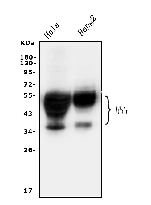
Western blot analysis of CD147/Emmprin using anti-CD147/Emmprin antibody (A00248-1).
Electrophoresis was performed on a 5-20% SDS-PAGE gel at 70V (Stacking gel) / 90V (Resolving gel) for 2-3 hours. The sample well of each lane was loaded with 30 ug of sample under reducing conditions.
Lane 1: human Hela whole cell lysates,
Lane 2: human HepG2 whole cell lysates.
After electrophoresis, proteins were transferred to a nitrocellulose membrane at 150 mA for 50-90 minutes. Blocked the membrane with 5% non-fat milk/TBS for 1.5 hour at RT. The membrane was incubated with rabbit anti-CD147/Emmprin antigen affinity purified polyclonal antibody (Catalog # A00248-1) at 0.5 μg/mL overnight at 4°C, then washed with TBS-0.1%Tween 3 times with 5 minutes each and probed with a goat anti-rabbit IgG-HRP secondary antibody at a dilution of 1:5000 for 1.5 hour at RT. The signal is developed using an Enhanced Chemiluminescent detection (ECL) kit (Catalog # EK1002) with Tanon 5200 system. A specific band was detected for CD147/Emmprin at approximately 38-55 kDa. The expected band size for CD147/Emmprin is at 42 kDa.

Click image to see more details
IHC analysis of CD147/Emmprin using anti-CD147/Emmprin antibody (A00248-1).
CD147/Emmprin was detected in paraffin-embedded section of human lung cancer tissues. Heat mediated antigen retrieval was performed in citrate buffer (pH6, epitope retrieval solution) for 20 mins. The tissue section was blocked with 10% goat serum. The tissue section was then incubated with 1μg/ml rabbit anti-CD147/Emmprin Antibody (A00248-1) overnight at 4°C. Biotinylated goat anti-rabbit IgG was used as secondary antibody and incubated for 30 minutes at 37°C. The tissue section was developed using Strepavidin-Biotin-Complex (SABC)(Catalog # SA1022) with DAB as the chromogen.

Click image to see more details
IHC analysis of CD147/Emmprin using anti-CD147/Emmprin antibody (A00248-1).
CD147/Emmprin was detected in paraffin-embedded section of human tonsil tissues. Heat mediated antigen retrieval was performed in citrate buffer (pH6, epitope retrieval solution) for 20 mins. The tissue section was blocked with 10% goat serum. The tissue section was then incubated with 1μg/ml rabbit anti-CD147/Emmprin Antibody (A00248-1) overnight at 4°C. Biotinylated goat anti-rabbit IgG was used as secondary antibody and incubated for 30 minutes at 37°C. The tissue section was developed using Strepavidin-Biotin-Complex (SABC)(Catalog # SA1022) with DAB as the chromogen.

Click image to see more details
IHC analysis of CD147/Emmprin using anti-CD147/Emmprin antibody (A00248-1).
CD147/Emmprin was detected in paraffin-embedded section of human intestinal cancer tissues. Heat mediated antigen retrieval was performed in citrate buffer (pH6, epitope retrieval solution) for 20 mins. The tissue section was blocked with 10% goat serum. The tissue section was then incubated with 1μg/ml rabbit anti-CD147/Emmprin Antibody (A00248-1) overnight at 4°C. Biotinylated goat anti-rabbit IgG was used as secondary antibody and incubated for 30 minutes at 37°C. The tissue section was developed using Strepavidin-Biotin-Complex (SABC)(Catalog # SA1022) with DAB as the chromogen.

Click image to see more details
Flow Cytometry analysis of A549 cells using anti-CD147/Emmprin antibody (A00248-1).
Overlay histogram showing A549 cells stained with A00248-1 (Blue line).The cells were blocked with 10% normal goat serum. And then incubated with rabbit anti-CD147/Emmprin Antibody (A00248-1,1μg/1x106 cells) for 30 min at 20°C. DyLight488 conjugated goat anti-rabbit IgG (BA1127, 5-10μg/1x106 cells) was used as secondary antibody for 30 minutes at 20°C. Isotype control antibody (Green line) was rabbit IgG (1μg/1x106) used under the same conditions. Unlabelled sample (Red line) was also used as a control.

Click image to see more details
Flow Cytometry analysis of Hela cells using anti-CD147/Emmprin antibody (A00248-1).
Overlay histogram showing Hela cells stained with A00248-1 (Blue line).The cells were blocked with 10% normal goat serum. And then incubated with rabbit anti-CD147/Emmprin Antibody (A00248-1,1μg/1x106 cells) for 30 min at 20°C. DyLight488 conjugated goat anti-rabbit IgG (BA1127, 5-10μg/1x106 cells) was used as secondary antibody for 30 minutes at 20°C. Isotype control antibody (Green line) was rabbit IgG (1μg/1x106) used under the same conditions. Unlabelled sample (Red line) was also used as a control.

Click image to see more details
IF analysis of CD147/Emmprin using anti-CD147/Emmprin antibody (A00248-1)
CD147/Emmprin was detected in immunocytochemical section of A549 cell. Enzyme antigen retrieval was performed using IHC enzyme antigen retrieval reagent (AR0022) for 15 mins. The cells were blocked with 10% goat serum. And then incubated with 2μg/ml rabbit anti-CD147/Emmprin Antibody (A00248-1) overnight at 4°C. Biotin conjugated goat anti-rabbit IgG (BA1003) was used as secondary antibody and incubated for 30 minutes at 37°C. The section was developed using DyLight®488 Conjugated Avidin (BA1128). Visualize using a fluorescence microscope and filter sets appropriate for the label used.

Click image to see more details
IF analysis of CD147/Emmprin using anti-CD147/Emmprin antibody (A00248-1)
CD147/Emmprin was detected in immunocytochemical section of HELA cell. Enzyme antigen retrieval was performed using IHC enzyme antigen retrieval reagent (AR0022) for 15 mins. The cells were blocked with 10% goat serum. And then incubated with 2μg/ml rabbit anti-CD147/Emmprin Antibody (A00248-1) overnight at 4°C. Biotin conjugated goat anti-rabbit IgG (BA1003) was used as secondary antibody and incubated for 30 minutes at 37°C. The section was developed using DyLight®488 Conjugated Avidin (BA1128). Visualize using a fluorescence microscope and filter sets appropriate for the label used.

Click image to see more details
IF analysis of CD147/Emmprin using anti-CD147/Emmprin antibody (A00248-1)
CD147/Emmprin was detected in paraffin-embedded section of human intestinal cancer tissues. Heat mediated antigen retrieval was performed in citrate buffer (pH6, epitope retrieval solution ) for 20 mins. The tissue section was blocked with 10% goat serum. The tissue section was then incubated with 1μg/mL rabbit anti-CD147/Emmprin Antibody (A00248-1) overnight at 4°C. DyLight®488 Conjugated Goat Anti-Rabbit IgG (BA1127) was used as secondary antibody at 1:100 dilution and incubated for 30 minutes at 37°C. The section was counterstained with DAPI. Visualize using a fluorescence microscope and filter sets appropriate for the label used.

Click image to see more details
Sandwich ELISA - Recombinant human Emmprin protein standard curve.
Use in combination with reagents from Human Emmprin ELISA Kit EZ-Set (DIY Antibody Pairs) (EZ0751).
Specific Publications For Anti-CD147/Emmprin/BSG Antibody Picoband® (A00248-1)
Loading publications
Recommended Resources
Here are featured tools and databases that you might find useful.
- Boster's Pathways Library
- Protein Databases
- Bioscience Research Protocol Resources
- Data Processing & Analysis Software
- Photo Editing Software
- Scientific Literature Resources
- Research Paper Management Tools
- Molecular Biology Software
- Primer Design Tools
- Bioinformatics Tools
- Phylogenetic Tree Analysis
Customer Reviews
Have you used Anti-CD147/Emmprin/BSG Antibody Picoband®?
Share your experimental results or join a short interview to earn up to $1,000 in product credits or other rewards.
2 Reviews For Anti-CD147/Emmprin/BSG Antibody Picoband®
Immunohistochemistry review for CD147 Emmprin BSG antibody
Excellent
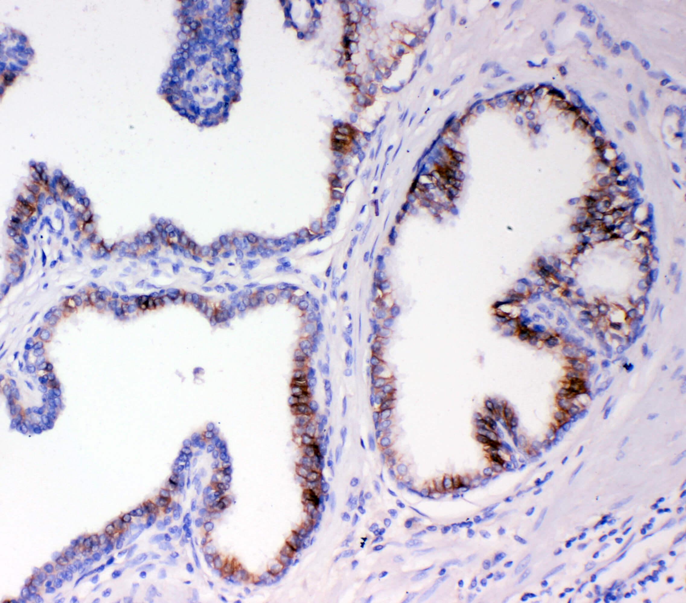
| SKU | A00248-1 |
|---|---|
| Application | Immunohistochemistry (paraffin embedded) |
| Blocking step | 5% BSA as blocking agent for 30 min at 37°C |
| Sample | Prostate cancer |
| Fixative | Fixed with 4% paraformaldehyde |
| Primary Incubation | 4°C overnight |
| Primary Incubation diluent | 5% BSA in TBS |
| Primary Concentration | 1ug/ml |
| Secondary Antibody | SABC kit from Boster Bio, (SA1022) |
| Secondary Dilution | The kit was ready to use, no dilution needed |
| Secondary Incubation | at 37°C for 30 min |
E. Taylor
Verified customer
Submitted 2020-07-22
Immunohistochemistry Review For Anti-CD147 Emmprin BSG Antibody
Excellent
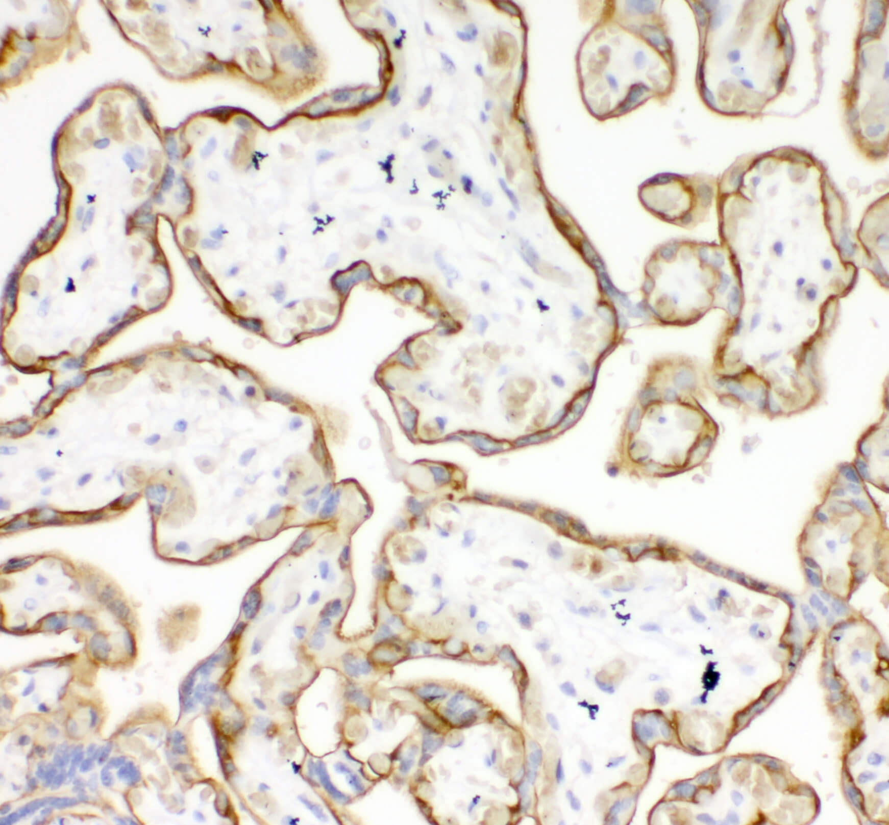
| SKU | A00248-1 |
|---|---|
| Application | Immunohistochemistry (paraffin embedded) |
| Blocking step | 5% BSA as blocking agent for 30 min at 37°C |
| Sample | Placenta |
| Fixative | Fixed with 4% paraformaldehyde |
| Primary Ab Incubation | 4°C overnight |
| Primary Ab Incubation diluent | 5% BSA in TBS |
| Primary Ab Concentration | 1ug/ml |
| Secondary Antibody | SABC kit from Boster Bio, (SA1022) |
| Secondary Ab Dilution | The kit was ready to use, no dilution needed |
| Secondary Ab Incubation | at 37°C for 30 min |
Verified customer
Verified customer
Submitted 2020-04-13
Customer Q&As
Have a question?
Find answers in Q&As, reviews.
Can't find your answer?
Submit your question
