Click image to see more details
-

-
-
-
-
+4
Product Info Summary
| SKU: | A01756-2 |
|---|---|
| Size: | 100 μg/vial |
| Reactive Species: | Human, Mouse, Rat |
| Host: | Rabbit |
| Application: | ELISA, Flow Cytometry, IP, IF, IHC, ICC, WB |
Customers Who Bought This Also Bought

Product info
Product Name
Anti-CD2AP Antibody Picoband®
SKU/Catalog Number
A01756-2
Size
100 μg/vial
Form
Lyophilized
Description
Boster Bio Anti-CD2AP Antibody Picoband® catalog # A01756-2. Tested in ELISA, Flow Cytometry, IP, IF, IHC, ICC, WB applications. This antibody reacts with Human, Mouse, Rat. The brand Picoband indicates this is a premium antibody that guarantees superior quality, high affinity, and strong signals with minimal background in Western blot applications. Only our best-performing antibodies are designated as Picoband, ensuring unmatched performance.
Storage & Handling
Store at -20˚C for one year from date of receipt. After reconstitution, at 4˚C for one month. It can also be aliquotted and stored frozen at -20˚C for six months. Avoid repeated freeze-thaw cycles.
Cite This Product
Anti-CD2AP Antibody Picoband® (Boster Biological Technology, Pleasanton CA, USA, Catalog # A01756-2)
Host
Rabbit
Contents
Each vial contains 4 mg Trehalose, 0.9 mg NaCl and 0.2 mg Na2HPO4.
Clonality
Polyclonal
Isotype
Rabbit IgG
Immunogen
E. coli-derived human CD2AP recombinant protein (Position: K253-K337).
Cross-reactivity
No cross-reactivity with other proteins.
Reactive Species
A01756-2 is reactive to CD2AP in Human, Mouse, Rat
Observed Molecular Weight
80 kDa
Calculated molecular weight
71.5 kDa
Background of CD2AP
CD2-associated protein is a protein that in humans is encoded by the CD2AP gene. This gene encodes a scaffolding molecule that regulates the actin cytoskeleton. The protein directly interacts with filamentous actin and a variety of cell membrane proteins through multiple actin binding sites, SH3 domains, and a proline-rich region containing binding sites for SH3 domains. The cytoplasmic protein localizes to membrane ruffles, lipid rafts, and the leading edges of cells. It is implicated in dynamic actin remodeling and membrane trafficking that occurs during receptor endocytosis and cytokinesis. Haploinsufficiency of this gene is implicated in susceptibility to glomerular disease.
Antibody Validation
Boster validates all antibodies on WB, IHC, ICC, Immunofluorescence, and ELISA with known positive control and negative samples to ensure specificity and high affinity, including thorough antibody incubations.
Application & Images
Applications
A01756-2 is guaranteed for ELISA, Flow Cytometry, IP, IF, IHC, ICC, WB Boster Guarantee
Recommend Dilution
| Application | Dilution | Species |
|---|---|---|
| Western blot | 0.1-0.5μg/ml | |
| Immunohistochemistry (Paraffin-embedded Section) | 2-5μg/ml | |
| Immunocytochemistry/Immunofluorescence | 5μg/ml | |
| Immunoprecipitation | 0.5-2 μg/ml | Human |
| Flow Cytometry (Fixed) | 1-3μg/1x106 cells | |
| ELISA | 0.1-0.5μg/ml |
Tested application
Suggested blocking solution with 5% non-fat milk or BSA; (*)Recommended protein loading: 20-40 µg per lane
Use TE buffer pH 9.0 for antigen retrieval; (*) citrate buffer pH 6.0 is an alternative.
Validation Images & Assay Conditions
Click image to see more details
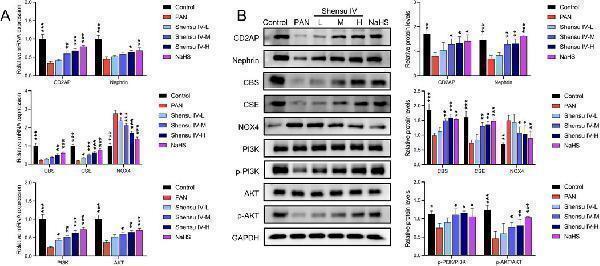
Shensu IV regulates the PI3K/AKT signaling pathway through H2S. (A) The effects of Shensu IV and NaHS on the mRNA expression of CD2AP, nephrin, CBS, CSE, NOX4, PI3K, and AKT in renal tissue of PAN rats were analyzed by RT-qPCR. (B) Western blot analysis of the effects of Shensu IV and NaHS on the protein levels of CD2AP, nephrin, CBS, CSE, NOX4, PI3K, p-PI3K,AKT,p-AKT in renal tissue of PAN rats. * P < 0.05, ** P < 0.01, *** P < 0.001. Abbreviations: CD2AP, CD2-associated protein; CBS, Cystathionine β-synthase; CSE, Cystathionine γ-lyase; PI3K, Phosphoinositide 3-Kinase; AKT, Protein Kinase B.
Index in PubMed under a CC BY license. PMID: 39720588

Click image to see more details
Shensu IV regulates the PI3K/AKT signaling pathway through H2S in podocytes. (A) The effects of Shensu IV and NaHS on the mRNA expression of CD2AP, nephrin, CBS, CSE, NOX4, PI3K, and AKT in podocytes were analyzed by RT-qPCR. (B) Western blot analysis of the effects of Shensu IV and NaHS on the protein levels of CD2AP, nephrin, CBS, CSE, NOX4, PI3K, p-PI3K,AKT,p-AKT in PAN-induced podocyocytes. * P < 0.05, ** P < 0.01, *** P < 0.001. Abbreviations: CD2AP, CD2-associated protein; CBS, Cystathionine β-synthase; CSE, Cystathionine γ-lyase; PI3K, Phosphoinositide 3-Kinase; AKT, Protein Kinase B.
Index in PubMed under a CC BY license. PMID: 39720588

Click image to see more details
Western blot analysis of CD2AP using anti-CD2AP antibody (A01756-2).
Electrophoresis was performed on a 10% SDS-PAGE gel at 80V (Stacking gel) / 120V (Resolving gel) for 2 hours. The sample well of each lane was loaded with 30 ug of sample under reducing conditions.
Lane 1: human Jurkat whole cell lysates,
Lane 2: human A431 whole cell lysates,
Lane 3: human Hela whole cell lysates,
Lane 4: human K562 whole cell lysates.
After electrophoresis, proteins were transferred to a nitrocellulose membrane at 150 mA for 50-90 minutes. Blocked the membrane with 5% non-fat milk/TBS for 1.5 hour at RT. The membrane was incubated with rabbit anti-CD2AP antigen affinity purified polyclonal antibody (A01756-2) at 0.5 μg/mL overnight at 4°C, then washed with TBS-0.1%Tween 3 times with 5 minutes each and probed with a goat anti-rabbit IgG-HRP secondary antibody (Catalog # BA1054) at a dilution of 1:5000 for 1.5 hour at RT. The signal is developed using an ECL Plus Western Blotting Substrate (Catalog # AR1196-200) with Tanon 5200 system. A specific band was detected for CD2AP at approximately 80 kDa. The expected band size for CD2AP is at 71 kDa.

Click image to see more details
IHC analysis of CD2AP using anti-CD2AP antibody (A01756-2).
CD2AP was detected in a paraffin-embedded section of mouse kidney tissue. Heat mediated antigen retrieval was performed in EDTA buffer (pH 8.0, epitope retrieval solution). The tissue section was blocked with 10% goat serum. The tissue section was then incubated with 2 μg/ml rabbit anti-CD2AP Antibody (A01756-2) overnight at 4°C. Biotinylated goat anti-rabbit IgG was used as secondary antibody and incubated for 30 minutes at 37°C. The tissue section was developed using HRP Conjugated Rabbit IgG Super Vision Assay Kit (Catalog # SV0002) with DAB as the chromogen.

Click image to see more details
IHC analysis of CD2AP using anti-CD2AP antibody (A01756-2).
CD2AP was detected in a paraffin-embedded section of rat kidney tissue. Heat mediated antigen retrieval was performed in EDTA buffer (pH 8.0, epitope retrieval solution). The tissue section was blocked with 10% goat serum. The tissue section was then incubated with 2 μg/ml rabbit anti-CD2AP Antibody (A01756-2) overnight at 4°C. Biotinylated goat anti-rabbit IgG was used as secondary antibody and incubated for 30 minutes at 37°C. The tissue section was developed using HRP Conjugated Rabbit IgG Super Vision Assay Kit (Catalog # SV0002) with DAB as the chromogen.

Click image to see more details
IF analysis of CD2AP using anti-CD2AP antibody (A01756-2).
CD2AP was detected in an immunocytochemical section of Hela cells. Enzyme antigen retrieval was performed using IHC enzyme antigen retrieval reagent (AR0022) for 15 mins. The cells were blocked with 10% goat serum. And then incubated with 5 μg/mL rabbit anti-CD2AP Antibody (A01756-2) overnight at 4°C. DyLight®488 Conjugated Goat Anti-Rabbit IgG (BA1127) was used as secondary antibody at 1:500 dilution and incubated for 30 minutes at 37°C. The section was counterstained with DAPI. Visualize using a fluorescence microscope and filter sets appropriate for the label used.

Click image to see more details
Immunoprecipitating CD2AP in Jurkat whole cell lysate .
Western blot analysis of CD2AP using anti-CD2AP antibody (A01756-2).
Lane 1: Jurkat whole cell lysates (30ug),
Lane 2: Rabbit control IgG instead of anti-CD2AP antibody in Jurkat whole cell lysate,
Lane 3: anti-CD2AP antibody (2μg) + Jurkat whole cell lysate (500μg).
After electrophoresis, proteins were transferred to a membrane. Then the membrane was incubated with rabbit anti-CD2AP antigen affinity purified polyclonal antibody (A01756-2) at a dilution of 0.5 μg/mL and probed with a mouse anti-rabbit IgG-HRP secondary antibody (Catalog # BA1054). The signal is developed using ECL Plus Western Blotting Substrate (Catalog # AR1196-200). A specific band was detected for CD2AP at approximately 80 kDa. The expected band size for CD2AP is at 71 kDa.

Click image to see more details
Flow Cytometry analysis of K562 cells using anti-CD2AP antibody (A01756-2).
Overlay histogram showing K562 cells stained with A01756-2 (Blue line). To facilitate intracellular staining, cells were fixed with 4% paraformaldehyde and permeabilized with permeabilization buffer. The cells were blocked with 10% normal goat serum. And then incubated with rabbit anti-CD2AP Antibody (A01756-2, 1 μg/1x106 cells) for 30 min at 20°C. DyLight®488 conjugated goat anti-rabbit IgG (BA1127, 5-10 μg/1x106 cells) was used as secondary antibody for 30 minutes at 20°C. Isotype control antibody (Green line) was rabbit IgG (1 μg/1x106) used under the same conditions. Unlabelled sample without incubation with primary antibody and secondary antibody (Red line) was used as a blank control.
Specific Publications For Anti-CD2AP Antibody Picoband® (A01756-2)
Loading publications
Recommended Resources
Here are featured tools and databases that you might find useful.
- Boster's Pathways Library
- Protein Databases
- Bioscience Research Protocol Resources
- Data Processing & Analysis Software
- Photo Editing Software
- Scientific Literature Resources
- Research Paper Management Tools
- Molecular Biology Software
- Primer Design Tools
- Bioinformatics Tools
- Phylogenetic Tree Analysis
Customer Reviews
Have you used Anti-CD2AP Antibody Picoband®?
Share your experimental results or join a short interview to earn up to $1,000 in product credits or other rewards.
0 Reviews For Anti-CD2AP Antibody Picoband®
Customer Q&As
Have a question?
Find answers in Q&As, reviews.
Can't find your answer?
Submit your question
